

Brain Aging in Patients With Cardiovascular Disease From the UK Biobank
- PMID: 40489382
- PMCID: PMC12147859
- DOI: 10.1002/hbm.70252
Brain Aging in Patients With Cardiovascular Disease From the UK Biobank
Abstract
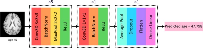
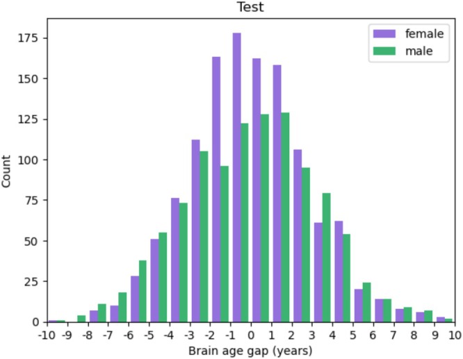
The brain undergoes complex but normal structural changes during the aging process in healthy adults, whereas deviations from the normal aging patterns of the brain can be indicative of various conditions as well as an increased risk for the development of diseases. The brain age gap (BAG), which is defined as the difference between the chronological age and the machine learning-predicted biological age of an individual, is a promising biomarker for determining whether an individual deviates from normal brain aging patterns. While the BAG has shown promise for various neurological diseases and cardiovascular risk factors, its utility to quantify brain changes associated with diagnosed cardiovascular diseases has not been investigated to date, which is the aim of this study. T1-weighted MRI scans from healthy participants in the UK Biobank were used to train a convolutional neural network (CNN) model for biological brain age prediction. The trained model was then used to quantify and compare the BAGs for all participants in the UK Biobank with known cardiovascular diseases, as well as healthy controls and patients with known neurological diseases for benchmark comparisons. Saliency maps were computed for each individual to investigate whether brain regions used for biological brain age prediction by the CNN differ between groups. The analyses revealed significant differences in BAG distributions for 10 of the 42 sex-specific cardiovascular disease groups investigated compared to healthy participants, indicating disease-specific variations in brain aging. However, no significant differences were found regarding the brain regions used for brain age prediction as determined by saliency maps, indicating that the model mostly relied on healthy brain aging patterns, even in the presence of cardiovascular diseases. Overall, the findings of this work demonstrate that the BAG is a sensitive imaging biomarker to detect differences in brain aging associated with specific cardiovascular diseases. This further supports the theory of the heart-brain axis by exemplifying that many cardiovascular diseases are associated with atypical brain aging.
© 2025 The Author(s). Human Brain Mapping published by Wiley Periodicals LLC.
Conflict of interest statement
The authors declare no conflicts of interest.
Figures


Similar articles
-
UK Biobank MRI data can power the development of generalizable brain clocks: A study of standard ML/DL methodologies and performance analysis on external databases.Neuroimage. 2025 Mar;308:121064. doi: 10.1016/j.neuroimage.2025.121064. Epub 2025 Jan 30. Neuroimage. 2025. PMID: 39892529
-
Learning patterns of the ageing brain in MRI using deep convolutional networks.Neuroimage. 2021 Jan 1;224:117401. doi: 10.1016/j.neuroimage.2020.117401. Epub 2020 Sep 24. Neuroimage. 2021. PMID: 32979523
-
Multimodal brain age prediction fusing morphometric and imaging data and association with cardiovascular risk factors.Front Neurol. 2022 Dec 14;13:979774. doi: 10.3389/fneur.2022.979774. eCollection 2022. Front Neurol. 2022. PMID: 36588902 Free PMC article.
-
Multimodal brain age estimates relate to Alzheimer disease biomarkers and cognition in early stages: a cross-sectional observational study.Elife. 2023 Jan 6;12:e81869. doi: 10.7554/eLife.81869. Elife. 2023. PMID: 36607335 Free PMC article.
-
Predicting brain age with deep learning from raw imaging data results in a reliable and heritable biomarker.Neuroimage. 2017 Dec;163:115-124. doi: 10.1016/j.neuroimage.2017.07.059. Epub 2017 Jul 29. Neuroimage. 2017. PMID: 28765056
Cited by
-
Biological heart and brain ageing in subjects with cardiovascular diseases.Front Cardiovasc Med. 2025 Jul 7;12:1569423. doi: 10.3389/fcvm.2025.1569423. eCollection 2025. Front Cardiovasc Med. 2025. PMID: 40693220 Free PMC article.
References
-
- Benjamini, Y. , and Hochberg Y.. 1995. “Controlling the False Discovery Rate: A Practical and Powerful Approach to Multiple Testing.” Journal of the Royal Statistical Society: Series B: Methodological 57, no. 1: 289–300. 10.1111/j.2517-6161.1995.tb02031.x. - DOI
MeSH terms
Grants and funding
LinkOut - more resources
Full Text Sources
Medical
Miscellaneous
