. 2025 Sep:99:102179.
doi: 10.1016/j.molmet.2025.102179. Epub 2025 Jun 7.

Adiponectin-receptor agonism prevents right ventricular tissue pathology in a mouse model of Duchenne muscular dystrophy

Affiliations

Adiponectin-receptor agonism prevents right ventricular tissue pathology in a mouse model of Duchenne muscular dystrophy

Shivam Gandhi et al. Mol Metab. 2025 Sep.

Abstract

Objective: Cardiac fibrosis during Duchenne muscular dystrophy (DMD) arises from cellular damage and inflammation and is associated with metabolic dysfunction. The extent to which these relationships develop across all 4 cardiac chambers, particularly during early-stage disease, remains unknown.

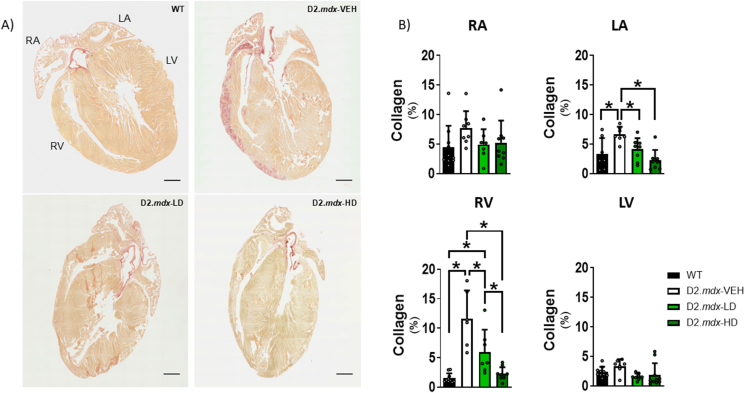
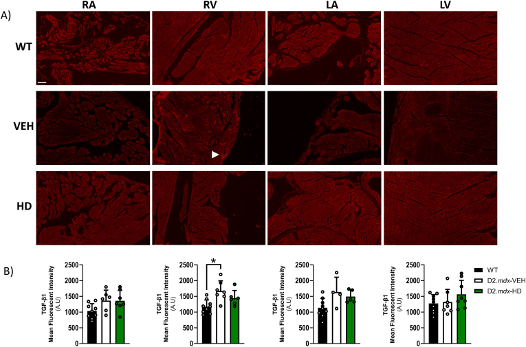
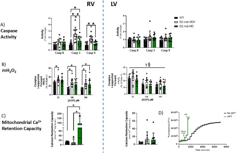
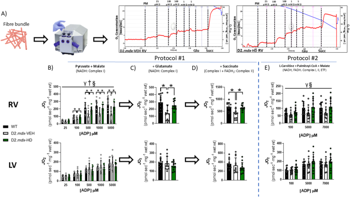
Methods and results: We discovered that very young D2.mdx mice exhibit fibrosis exclusively in the right ventricle (RV) and left atrium. Concurrent myocardial disorganization in the RV was related to a highly specific inflammatory signature of increased infiltrating pro-inflammatory macrophages (CD11b+CD45+CD64+F4/80+CCR2+), myofibre mitochondrial-linked apoptosis, and reduced carbohydrate and fat oxidation. This relationship did not occur in the left ventricle. Short-term daily administration of a peptidomimetic adiponectin receptor agonist, ALY688, prevented RV fibrosis, infiltrating macrophages, and mitochondrial stress as well as left atrial fibrosis.

Conclusions: Our discoveries demonstrate early-stage cardiac tissue pathology occurs in a chamber-specific manner and is prevented by adiponectin receptor agonism, thereby opening a new direction for developing therapies that prevent tissue remodeling during DMD.

Keywords: Cardiomyopathy; Duchenne muscular dystrophy; Fibrosis; Inflammation; Mitochondria.


Conflict of interest statement

Declaration of competing interest Authors declare that they have no competing interests. HHH is an employee of Allysta and GS and AAS consult for Allysta.

Figures

Figure 1
Figure 1
4-week-old D2.mdx mice exhibit robust, chamber-specific cardiac fibrosis, which is protected by daily ALY688 administration. A) 5 μM thick sagittal sections of paraffin-embedded cardiac tissue were stained with picrosirius red, which selectively binds collagen fibres. B) All four chambers were assessed for collagen deposition. Images were taken with EVOS M7000 Imaging System at 4x magnification. Dark red segments denote collagen. Results represent mean ± SD; n = 6–10. Scale bars are 1 mm. All p values are FDR-adjusted by Benjamini, Krieger, and Yekutieli post-hoc analyses. ∗p < 0.05 denotes significance. WT = Wildtype; D2.mdx-VEH = vehicle (saline)-treated mdx; D2.mdx-HD = high dose (ALY688)-treated mdx; RA = right atrium; LA = left atrium; RV = right ventricle; LV = left ventricle. (For interpretation of the references to colour in this figure legend, the reader is referred to the Web version of this article.)
Figure 2
Figure 2
D2.mdx-VEH mice have smaller hearts and overall body weights compared to WT, and this is not prevented by ALY688. A) Morphometric data comparing heart weight (mg), body weight (g), heart weight-to-tibia length (mg/mm) and ventricle-specific weights (phase 4 only) between WT, D2.mdx-VEH, and D2.mdx-HD, with B) representative images of perfused whole-heart with the RV oriented frontward. Results represent mean ± SD; n = 23–35 (combined phases 1 + 4). All p values are FDR-adjusted by Benjamini, Krieger, and Yekutieli post-hoc analyses. ∗p < 0.05 denotes significance. WT = Wildtype mouse; VEH = vehicle (saline)-treated mdx; HD = high dose (ALY688)-treated mdx; RV = right ventricle; LV = left ventricle.
Figure 3
Figure 3
Chamber-specific immunofluorescent staining of TGF-β1. A) TGF-β1 (denoted by white arrow) was quantified by Mean Fluorescent Intensity (A.U.). (B) Whole atria and free-wall segments of the ventricles were analyzed. Stains were conducted on 5 μm thick paraffin-embedded sagittal sections and imaged by confocal microscopy at 20x magnification. Results represent mean ± SD; n = 4–9. Scale bars are 100 μm. All p values are FDR-adjusted by Benjamini, Krieger, and Yekutieli post-hoc analyses. ∗p < 0.05 denotes significance. WT = Wildtype; VEH = vehicle (saline)-treated mdx; HD = high dose (ALY688)-treated mdx; RA = right atrium; LA = left atrium; RV = right ventricle; LV = left ventricle.
Figure 4
Figure 4
Ventricle-specific spectral flow cytometry analysis of infiltrating and resident macrophage populations in D2.mdx. A) Representative gating scheme of CCR2+/− cells in all three groups between ventricles. B) Ventricle-specific comparison of CD11b+CD45+CD64+F4/80+CCR2+ cells (representative of an infiltrating macrophage population), normalized to milligram tissue weight. C) Ventricle-specific comparison of CD11b+CD45+CD64+F4/80+CCR2- cells (representative of a resident macrophage population), normalized to milligram tissue weight. Results represent mean ± SD; n = 7. All p values are FDR-adjusted by Benjamini, Krieger, and Yekutieli post-hoc analyses. ∗p < 0.05 denotes significance. WT = Wildtype mouse; VEH = vehicle (saline)-treated mdx; HD = high dose (ALY688)-treated mdx; RV = right ventricle; LV = left ventricle.
Figure 5
Figure 5
D2.mdx exhibit indices of mitochondrial stress primarily in the RV that are partially restored by ALY688 administration. A) Detection of caspase 3 and 9 activity - representative of potentially mitochondrial-linked apoptotic pathways. Caspase 8 (death receptor) activity was also examined. Results represent mean ± SD; n = 10–12. ∗p < 0.05 denotes significance. B) Analysis of mitochondrial complex I-supported (pyruvate + malate) forward electron transfer mH2O2 (represented as % of State II) in the presence of creatine in both ventricles across metabolic demands (25, 100, 500 μM ADP). Results represent mean ± SD; n = 10–12. RV: ∗p < 0.05 denotes significance; LV (main effects are denoted by horizontal bar): γp<0.05 WT vs D2.mdx-VEH; §p < 0.05 D2.mdx-VEH vs D2.mdx-HD. C) Analysis of mitochondrial calcium retention capacity (CRC) to trigger mPT (which precedes mitochondrial-mediated apoptosis). D) Representative CRC trace demonstrating ventricular PmFB with distinct mPT, compared to PmFB without mPT. Results represent mean ± SD; n = 3–10. All p values are FDR-adjusted by Benjamini, Krieger, and Yekutieli post-hoc analyses. ∗p < 0.05 denotes significance. Casp = caspase; mH2O2 = mitochondrial H2O2 emission; WT = Wildtype; D2.mdx-VEH = vehicle (saline)-treated mdx; D2.mdx-HD = high dose (ALY688)-treated mdx; Ca2+ = calcium; mPT = mitochondrial permeability transition.
Figure 6
Figure 6
Substrate-specific stimulation of mitochondrial respiration in right and left ventricle of D2.mdx mice. A) Representative trace of high-resolution respirometry data demonstrating oxygen consumption that is calculated as the change in slope of oxygen consumption in the respirometry chamber (left trace, VEH RV - pyruvate + malate (PM), ADP titration (D), glutamate (Glu), succinate (Succ) protocol; right trace, HD RV - pyruvate + malate, ADP titration, glutamate, succinate protocol). B) ADP-stimulated respiration was supported by pyruvate (5 mM) supplemented with malate (2 mM) (NADH, complex-I stimulation) as an index of carbohydrate oxidation. C) After the final addition of ADP (5000 μM) in ‘A’, glutamate (10 mM; NADH, complex-I stimulation) was added as an index of amino acid oxidation, followed by D) succinate (10 mM) (FADH2, complex-II stimulation). E) Palmitoyl CoA (20 μM, supplemented with 10 μM l-Carnitine, 500 μM Malate and 100–7000 μM ADP; NADH and FADH2) was added to a separate permeabilized fibre bundle as an index of fat oxidation. FADH2 from the TCA cycle transfers its electrons at complex-II while FADH2 from beta oxidation provides electrons for ETF. NADH = Nicotinamide adenine dinucleotide; FADH2 = Flavin adenine dinucleotide; ETF = Electron transport flavoprotein; RV = right ventricle; LV = left ventricle; PM = pyruvate + malate; ‘D’ (from Panel A) = ADP. Results represent mean ± SD, n = 10–12; a 2-way ANOVA was used to determine the difference between WT, D2.mdx-VEH and D2.mdx-HD across ADP concentrations in ‘B’ and ‘E’ while a 1-way ANOVA was used in ‘C’ and ‘D’. All p values are FDR-adjusted by Benjamini, Krieger, and Yekutieli post-hoc analyses. LV data was log-transformed (for statistical analyses) given that it did not pass parametric testing. ∗p < 0.05 denotes significance. Main effects are denoted by horizontal bar over ‘B’ and ‘E’: γp<0.05 WT vs D2.mdx-VEH; §p < 0.05 D2.mdx-VEH vs D2.mdx-HD; †p < 0.05 WT vs D2.mdx-HD. Oroboros image in Panel A obtained from https://www.oroboros.at/.
Figure 7
Figure 7
Western blots of mitochondrial signaling and mitophagy markers. (A) Ventricle-specific mitochondrial signaling markers. (B) Ventricle-specific mitochondrial autophagy (mitophagy) markers. Results represent mean ± SD; n = 11–12. All p values are FDR-adjusted by Benjamini, Krieger, and Yekutieli post-hoc analyses. The ‘ratio’ is defined as phosphorylated protein content divided by total protein content. ∗p < 0.05 denotes significance. RV = right ventricle; LV = left ventricle.
Figure 8
Figure 8
Proposed mechanisms underlying dystrophin deficiency-induced cardiac fibrosis in 4-week-old D2.mdx mice that can be partially rescued by daily adiponectin-receptor agonism. We establish that secondary contributors to dystrophin deficiency-induced cardiac fibrosis (inflammation and mitochondrial-linked apoptosis) differentially impact chambers of the heart. Our findings indicate that these secondary contributors demonstrate rescue effects when D2.mdx mice are injected daily with ALY688, ultimately leading to cardiac fibrosis prevention in the right ventricle (RV). To examine indices of mitochondrial stress, we investigated chamber-specific mitochondrial respiration and mH2O2, while intrinsic (mitochondrial-linked) apoptosis was assessed by a combination of calcium retention capacity and caspase activity. Inflammation was assessed by spectral flow cytometry to determine shifts in macrophage polarization (infiltrating versus resident macrophage sub-populations) as well as by immunofluorescence of various inflammatory and fibrosis-linked cytokines. Our results indicate that daily adiponectin-receptor agonism may represent a viable therapeutic intervention for preventing RV fibrosis in young D2.mdx mice. Green arrows denote preventative effects of ALY688 observed in the D2.mdx-HD RV across various remodeling signatures, including shifts to pro-/anti-inflammatory markers, mitochondrial stress responses, and cardiac fibrosis. (For interpretation of the references to colour in this figure legend, the reader is referred to the Web version of this article.)
figs1
figs1
figs2
figs2
figs3
figs3
figs4
figs4
figs5
figs5
figs6
figs6
figs7
figs7
figs8
figs8
figs9
figs9
figs10
figs10

References

    1. Crisafulli S., Sultana J., Fontana A., Salvo F., Messina S., Trifirò G. Global epidemiology of Duchenne muscular dystrophy: an updated systematic review and meta-analysis. Orphanet J Rare Dis. 2020;15:141. doi: 10.1186/s13023-020-01430-8. - DOI - PMC - PubMed
    1. Coley W.D., Bogdanik L., Vila M.C., Yu Q., Van Der Meulen J.H., Rayavarapu S., et al. Effect of genetic background on the dystrophic phenotype in mdx mice. Hum Mol Genet. 2016;25:130–145. doi: 10.1093/hmg/ddv460. - DOI - PMC - PubMed
    1. Putten M., Putker K., Overzier M., Adamzek W.A., Pasteuning-Vuhman S., Plomp J.J., et al. Natural disease history of the D2-mdx mouse model for Duchenne muscular dystrophy. FASEB J. 2019;33:8110–8124. doi: 10.1096/fj.201802488R. - DOI - PMC - PubMed
    1. Hughes M.C., Ramos S.V., Turnbull P.C., Edgett B.A., Huber J.S., Polidovitch N., et al. Impairments in left ventricular mitochondrial bioenergetics precede overt cardiac dysfunction and remodelling in Duchenne muscular dystrophy. J Physiol. 2020;598 doi: 10.1113/JP277306. - DOI - PubMed
    1. Belanto J.J., Mader T.L., Eckhoff M.D., Strandjord D.M., Banks G.B., Gardner M.K., et al. Microtubule binding distinguishes dystrophin from utrophin. Proc Natl Acad Sci. 2014;111:5723–5728. doi: 10.1073/pnas.1323842111. - DOI - PMC - PubMed

Substances