

Functional recovery through the plastic adaptation of organoid grafts in cortical tissue
- PMID: 40490613
- PMCID: PMC12149075
- DOI: 10.1007/s00018-025-05767-w
Functional recovery through the plastic adaptation of organoid grafts in cortical tissue
Abstract
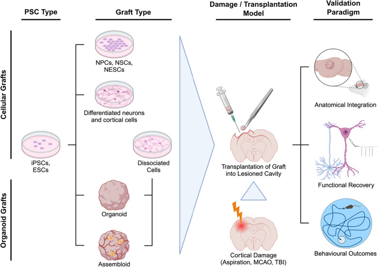
The lack of effective therapeutic options for patients suffering from neurological impairments related to acquired brain damage requires novel translational strategies, among which transplantation of neural tissue is receiving strong attention. One of the most recent developments involves the implantation of brain organoid models, derived from embryonic or induced pluripotent stem cells, into damaged rodent cortices. While this approach is gaining popularity, the extent of graft integration within the host tissue remains poorly understood. This review aims to examine whether xenotransplanting organoids into cortical tissue induces functional recovery and plastic adaptation to the damaged implantation sites. Physiological indications of grafted organoid plasticity and integration into the host included viability, corticogenesis, vascularisation, growth, and the development of area-specific morphological identities. The functional integration into host neural circuitry has been probed by tracing of axonal projection growth according to implantation sites, but also through observations of spontaneous, stimulus evoked, and selectively tuned activity of grafted neurons. Finally, some studies also investigated whether the engraftment procedure facilitated behavioural recovery in tasks requiring motor, memory, or reward-seeking functions. Overall, organoid grafts show signs of progressive anatomical, functional, and behaviourally-relevant integration within the damaged host cortices. Yet, further investigation is necessary before this transplantation approach can be actually translated into a robust method to achieve functional restoration in patients suffering from brain damage.
Keywords: Brain damage; Brain organoids; Functional recovery; Organoid transplantation; Xenotransplantation.
© 2025. The Author(s).
Conflict of interest statement
Declarations. Conflicts of interest: JCS is founder and CEO of OrganoTherapeutics.
Figures
References
-
- Hankey GJ, Jamrozik K, Broadhurst RJ et al (2002) Long-term disability after first-ever stroke and related prognostic factors in the perth community stroke study, 1989–1990. Stroke 33:1034–1040. 10.1161/01.STR.0000012515.66889.24 - PubMed
-
- Thurman DJ, Alverson C, Dunn KA et al (1999) Traumatic brain injury in the United States: a public health perspective. J Head Trauma Rehabil 14:602–615. 10.1097/00001199-199912000-00009 - PubMed
-
- Rahman M, Abbatematteo J, De Leo EK et al (2017) The effects of new or worsened postoperative neurological deficits on survival of patients with glioblastoma. J Neurosurg 127:123–131. 10.3171/2016.7.JNS16396 - PubMed
Publication types
MeSH terms
Grants and funding
LinkOut - more resources
Full Text Sources
