. 2025 Jun 9;15(1):16.
doi: 10.1186/s13395-025-00386-2.

Extensive striated muscle damage in a rat model of Duchenne muscular dystrophy with Dmd exons 10-17 duplication

Affiliations

Extensive striated muscle damage in a rat model of Duchenne muscular dystrophy with Dmd exons 10-17 duplication

Jean-Daniel Masson et al. Skelet Muscle. .

Abstract

Background: Duchenne muscular dystrophy (DMD) mainly affects young boys with out-of-frame mutations in the DMD gene, leading to dystrophin deficiency. This loss disrupts the assembly of the sarcolemmal dystrophin-associated glycoprotein complex, resulting in membrane fragility and damage during muscle contraction-relaxation cycles. Consequently, patients experience progressive muscle weakness, loss of ambulation and cardiorespiratory failure. Gene therapy represents one of the most promising therapeutic approaches, requiring rigorous preclinical validation of candidate strategies. While several preclinical models of dystrophin deficiency mimic point mutations or exon deletions, no existing rat model accurately replicates DMD gene duplications, which account for approximately 10% of DMD cases.

Methods: Using CRISPR/Cas9 genome editing, we generated a ~ 125 kbp duplication encompassing exons 10-17 of the Dmd gene in Sprague Dawley rats. To characterise disease progression in these rats, we assessed biochemical, histological and functional biomarkers at 6 and 10 months of age, comparing them to their healthy littermates.

Results: We established the R-DMDdup10-17 line. The microstructure of limb, diaphragm and cardiac muscles of R-DMDdup10-17 (DMD) rats exhibited dystrophic changes at 6 and 10 months, including loss of myofibres and fibrosis. These alterations led to a significant body mass reduction, muscle weakness (including diaphragm deficiency) and cardiac electrical defects. Premature lethality was observed between 10 and 13 months.

Conclusion: Duplication of the Dmd genomic region encompassing exons 10 to 17 in rats results in dystrophin deficiency, severe striated muscle dystrophy, and premature death. The R-DMDdup10-17 line represents the first reported genetic model of a severe and early lethal duplication variant in the Dmd gene. It provides a critical tool for assessing targeted gene therapies aimed to correct such mutations.

Keywords: Congenital myopathy; ECG; Inter-individual data correction; MYOM3; Myonecrosis; Neuromuscular disorder; Notched T wave; Plethysmography; hs-cTnT.


Conflict of interest statement

Declarations. Ethics approval and consent to participate: The study was conducted in accordance with the 2021/63/EU European Directive on the protection of animals used for scientific purposes. All experimental procedures were reviewed and approved by the local Ethics Committee of the French Ministry of Research and Higher Education (APAFIS#44858-2023091920248268), ensuring compliance with ethical standards for animal research. Competing interests: The authors declare no competing interests.

Figures

Fig. 1
Fig. 1
Early death associated with severe muscle atrophy in dystrophin-deficient R-DMDdup10-17 (DMD) rats. (A) Scheme of CRISPR/Cas9-mediated duplication on the rat chromosome X of a ~ 125 kb genomic region encompassing exons 10 to 17 of the Dmd gene. The wild-type allele (X+) is at the top, with the reading frame indicated by the blue colour of the exons. The allele containing the duplication (XDMD) is at the bottom. Splicing between exon 17 and the duplicated exon 10 induces a premature stop codon in the duplicated exon 10 (white cross on red background) and the absence of translation of subsequent exons (grey). The position and orientation of the primers used for genotyping is indicated by arrows, as is the expected amplicon size for each of the two alleles. (B) Electrophoretic gel of amplicons obtained using a multiplex of the three primers indicated in (A) from genomic DNA of male rats from a litter born from a carrier female and a WT male. A band at 130 bp reveals the WT allele, while a band at 277 bp reveals a male hemizygous for the Dmd allele carrying the duplicated 10–17 region. (C) Body mass of WT and DMD littermates at 6 and 10 months of age. (D) Kaplan-Meier curve for the frequency of WT (black curve) and DMD (orange curve) rat survival. (E) Representative immunofluorescence for laminin (red) and dystrophin (orange) at 6 months on TA, diaphragm and heart sections of WT and DMD littermates, showing complete absence of a dystrophin signal in DMD muscles. Scale bars, 50 μm. (F) Plasma CK levels in 7-month-old WT and DMD littermates. One-tailed unpaired t test. (G) Upper panel: Capillary immunoelectrophoresis plasma protein analysis detecting the two fragments (100 and 130 kDa) of the sarcomeric myomesin-3 (MYOM3) protein in 7-month-old DMD rats, but not in their WT littermates; lower panel: total proteins for each lane. (H) Quantification of plasma MYOM3 levels assessed in (G), corresponding to the sum of both MYOM3-130 and MYOM3-100 signals normalised to total plasma proteins. One-tailed unpaired t test. (I) Plasma hs-cTNT levels in 10-month-old WT and DMD littermates. One-tailed unpaired t test
Fig. 2
Fig. 2
Global muscle atrophy in 10-mo-old R-DMDdup10-17 (DMD) rats. A. Picture of WT and DMD TA at 10 months. B. Mass of WT and DMD TA at 10 months, corrected on body mass (BM). Two-tailed unpaired t test. C. Mass of WT and DMD TA at 10 months, corrected on TL3. Two-tailed unpaired t test. D. Picture of WT and DMD EDL, soleus, and heart at 10 months. E. EDL, soleus, and heart at 10 months, corrected on TL3
Fig. 3
Fig. 3
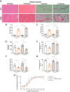
Histological and functional impairment of R-DMDdup10-17 (DMD) skeletal muscles. A. Haematoxylin and eosin (HE, left panels) and Sirius Red (SR, right panels) staining of the tibialis anterior (TA) from 6- and 10-month-old WT and DMD littermates. Scale bars, 50 μm. B. Pathological index calculated on HE-stained transverse sections of TA from 6- and 10-month-old WT and DMD littermates. C. Quantification of fibrotic deposition based on SR-stained area of transverse sections of TA from 6- and 10-month-old WT and DMD littermates. D and E. Corrected maximal force of forelimbs by grip test (D) and corrected force maintenance index (E) in 6- and 10-month-old WT and DMD littermates. F and G. Corrected maximal torque force (F) and force maintenance index (G) of the TA after direct muscle stimulation in 6- and 10-month-old WT and DMD littermates. H. Percentage of force-frequency relationship representative of excitation/contraction coupling ratio in 6- and 10-month-old WT and DMD littermates
Fig. 4
Fig. 4
Reduced spontaneous locomotion in 6-month-old R-DMDdup10-17 (DMD) rats. A. Scheme (upper panel) and picture (lower panel) of the large exercise arena equipped with a mezzanine (Mezza) and a ladder connecting the mezza to the ground. B-E. Total distance moved (B), average of the active ground speed (C), total number of climbing attempts (D), and total number of successful climbing (reaching the mezzanine, E) calculated in video-tracked 6-month-old WT and DMD littermates. Two-tailed unpaired t tests
Fig. 5
Fig. 5
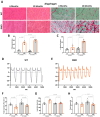
Histological and functional impairment of ventilatory muscles from R-DMDdup10-17 (DMD) rats. A. Haematoxylin and eosin (HE, left panels) and Sirius Red (SR, right panels) staining of diaphragm from 6- and 10-month-old WT and DMD littermates. Scale bars, 50 μm. B. Pathological index calculated on HE-stained transverse sections of diaphragm from 6- and 10-month-old WT and DMD littermates. C. Quantification of fibrotic deposition based on SR-stained area of transverse sections of diaphragm from 6- and 10-month-old WT and DMD littermates. D-E. Representative traces of ventilatory cycles in WT (D) and DMD (E) littermates at 6 months. Negative flow peaks indicate depression in the box, i.e. the inspiration step for the animal, while the positive peaks reflect the expiration. F-H. Quantification of peak inspiratory flow (F), inspiration time (G), and tidal volume (H) measured by whole-body plethysmography in 6-month-old WT and DMD littermates
Fig. 6
Fig. 6
Histological and functional impairment of the heart from R-DMDdup10-17 (DMD) rats. A. Haematoxylin and eosin (HE, left panels) and Sirius Red (SR, right panels) staining of the left ventricle free wall of the heart from 6- and 10-month-old WT and DMD littermates. Scale bars, 50 μm. B. Pathological index calculated on HE-stained transverse sections of the whole heart from 6- and 10-month-old WT and DMD littermates. C. Quantification of fibrotic deposition based on SR-stained area of transverse sections of the whole heart from 6- and 10-month-old WT and DMD littermates. D. Representative averaged ECG trace of 6-month-old WT (grey) and DMD (orange) littermates. Q represents the onset of ventricles depolarization. Tp represents the peak of the T wave (repolarization of the ventricles), and QTp is the time between Q and Tp. E. Quantification of the heart rate-corrected QTp (QTpc) interval in 6- and 10-month-old WT and DMD littermates

Similar articles

References

    1. Burghes AH, Logan C, Hu X, Belfall B, Worton RG, Ray PN. A cDNA clone from the duchenne/becker muscular dystrophy gene. Nature. 1987;328:434–7. - PubMed
    1. Rybakova IN, Patel JR, Ervasti JM. The dystrophin complex forms a mechanically strong link between the sarcolemma and Costameric actin. J Cell Biol. 2000;150:1209–14. - PMC - PubMed
    1. Bladen CL, Salgado D, Monges S, Foncuberta ME, Kekou K, Kosma K, et al. The TREAT-NMD DMD global database: analysis of more than 7,000 Duchenne muscular dystrophy mutations. Hum Mutat. 2015;36:395–402. - PMC - PubMed
    1. Duan D, Goemans N, Takeda S, Mercuri E, Aartsma-Rus A. Duchenne muscular dystrophy. Nat Rev Dis Primers. 2021;7:1–19. - PMC - PubMed
    1. Verhaeg M, Adamzek K, van de Vijver D, Putker K, Engelbeen S, Wijnbergen D, et al. Learning, memory and blood–brain barrier pathology in Duchenne muscular dystrophy mice lacking Dp427, or Dp427 and Dp140. Genes Brain Behav. 2024;23:e12895. - PMC - PubMed

LinkOut - more resources