. 2025 Jun;15(6):e70353.
doi: 10.1002/ctm2.70353.

MLKL‒OPTN axis regulates herpesvirus-induced neurological sequelae

Affiliations

MLKL‒OPTN axis regulates herpesvirus-induced neurological sequelae

Ilina Bhattacharya et al. Clin Transl Med. 2025 Jun.

Abstract

Background: Herpes simplex virus-1 (HSV-1) infections are lifelong and linked to neurological diseases such as multiple sclerosis (MS), yet the underlying mechanisms in the host remain poorly understood.

Methods and results: This study investigates new molecular dynamics following HSV-1 infection, uncovering the pivotal role of the mixed lineage kinase domain-like (MLKL) protein. Beyond its known function in necroptosis, MLKL was found to control HSV-1 transport into the nucleus, tightly regulated by Optineurin (OPTN). We evidenced an essential regulatory interaction between MLKL and OPTN, governing MLKL's activity in both necroptosis-dependent and independent pathways. In vivo, studies using Optn knockout mice demonstrated how this MLKL-OPTN axis contributes to demyelination and neurological symptoms mimicking MS. This axis critically prevents oligodendrocyte death and the associated demyelination during HSV-1 infection. Furthermore, pharmacological interventions with Necrosulfonamide (NSA), an MLKL inhibitor, showed therapeutic potential in preserving myelin integrity and reducing neurological deficits in HSV-1-infected models, suggesting a viable strategy for managing virus-induced neurodegeneration.

Conclusion: Our findings highlight the significant role of MLKL in HSV-1 pathogenesis and suggest that MLKL dysregulation is a key mechanism behind severe neurological damage.

Key points: MLKL plays a significant role in regulating endosomal transport of HSV-1 to nucleus during early stages of infection. Formation of p-MLKL bodies during HSV-1 infection leads to death of oligodendrocyte and subsequent demyelination. OPTN can negatively modulate MLKL levels to restrict infection and consequential oligodendrocyte death during HSV-1 infection.

Keywords: HSV‐1; MLKL; OPTN; cell death; demyelinating disorders.


Conflict of interest statement

The authors declare they have no conflicts of interest.

Figures
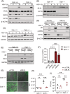
FIGURE 1
FIGURE 1
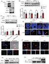
Mixed lineage kinase domain‐like (MLKL) is a cellular factor that promotes herpes simplex virus‐1 (HSV‐1) infection in Optineurin (OPTN)‐deficient conditions. (A) Representative immunoblots of Optn +/+ and Optn ‒/‒ human corneal epithelial (HCE) cells infected with HSV‐1 (.5 MOI) for 6 and 24 h. (B) Representative immunoblots of Optn +/+ and Optn ‒/‒ HCE cells exposed to 1 MOI infection for the 0, 2, 4 and 6 h post‐infection. (C) Representative immunoblots of Optn +/+ and Optn ‒/‒ HeLa cells exposed to 1 MOI infection for the 0, 2, 4 and 6 h post‐infection. (D) siCTRL and siMLKL transfected Optn +/+ and Optn ‒/‒ HCE cells were infected with 1 MOI HSV‐1 for 24 h and the total cell lysates were collected and analysed by immunoblotting against specified antibodies. (E) siCTRL and siMLKL transfected Optn +/+ and Optn ‒/‒ HeLa cells were infected with 1 MOI HSV‐1 for 24 h and the total cell lysates were collected and analysed by immunoblotting against specified antibodies. (F) Glycoprotein B (gB) expression in (E) is quantified relative to GAPDH. (G) siCTRL and siMLKL transfected Optn +/+ and Optn ‒/‒ cells were infected with 1 MOI 17 GFP‒HSV‐1 (green) for 24 h and then imaged using a fluorescent microscope. (H) Quantification of fluorescence intensity of GFP in (G). (I) siCTRL and siMLKL transfected Optn +/+ and Optn ‒/‒ cells were infected with 1 MOI HSV‐1 for 24 h and the total cell lysates were collected to determine the viral titre by a standard viral plaque assay. Data are represented as mean ± standard error of means (SEM) of triplicate samples (F, H and I), and comparable results were obtained from three independent experiments. pfu, plaque‐forming units.
FIGURE 2
FIGURE 2
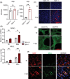
Mixed lineage kinase domain‐like (MLKL) facilitates endosomal trafficking of herpes simplex virus‐1 (HSV‐1) during the early stages of infection. (A) Optn +/+ and Optn ‒/‒ HeLa cells were incubated in serum‐free media for 12 h. These cells were then exposed to epidermal growth factor (EGF) for 30 min on ice. After being washed with PBS, cells were incubated in normal growth media with EGF at 37°C and intracellular uptake of epidermal growth factor receptor (EGFR) was monitored at indicated times by immunofluorescence microscopy. (B) Optn +/+ and Optn ‒/‒ cells were exposed to 10 MOI K26‐RFP HSV‐1 infection for 2 h on ice, followed by incubation at 37°C for 2.5 h. HSV‐1 intracellular transport was monitored by immunofluorescence microscopy. (C) Optn +/+ and Optn ‒/‒ cells were exposed to 1 MOI HSV‐1 infection for 24 h, and HSV‐1 transport to the plasma membrane was examined using transmission electron microscopy. (D) Optn +/+ and Optn ‒/‒ cells were exposed to 10 MOI K26‐RFP HSV‐1 infection for 2 h on ice, followed by incubation with or without Necrosulphonamide (NSA) (MOCK) at 37°C for 2.5 h and immunofluorescence imaging was conducted to track the transport of HSV‐1. (E and F) Optn +/+ and Optn ‒/‒ cells were exposed to 10 MOI K26‐RFP HSV‐1 infection for 2 h on ice, followed by incubation with or without NSA (MOCK) at 37°C for 2.5 and 3 h. Subcellular fractionation was conducted, and (E) nuclear fraction and (F) cytoplasmic fraction were analysed by immunoblotting. All images are representative images and similar results were obtained from three independent experiments.
FIGURE 3
FIGURE 3
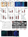
Optineurin (OPTN) utilises the ubiquitin binding domain (UBD) domain to selectively degrade mixed lineage kinase domain‐like (MLKL). (A) Optn +/+ cells were infected with or without 1 MOI herpes simplex virus‐1 (HSV‐1) for 24 h, followed by immunoprecipitation by MLKL and immunoglobulin G (IgG) (control). Total cell lysates were analysed by Western blot with indicated antibodies. (B) Optn +/+ and Optn ‒/‒ cells were subjected to 1 MOI HSV‐1 infection for 12 h. Cycloheximide (CHX) was then added, and cells were collected at the indicated time and analysed by Western blotting with indicated antibodies. (C) Optn +/+ and Optn ‒/‒ cells were subjected to 1 MOI HSV‐1 infection for 24 h with or without bafilomycin (Baf) and cell lysates were analysed by immunoblotting. (D) Optn +/+ cells were exposed to 2 MOI K26‐RFP HSV‐1 infection for 24 h and the colocalisation of Optn and p‐MLKL was observed using immunofluorescence imaging. (E) Optn +/+ and Optn ‒/‒ cells were subjected to 1 MOI HSV‐1 infection for 24 h, and MLKL and LAMP1 (a marker for lysosomal degradation) were examined with immunofluorescence imaging. The white arrows indicate colocalisation of MLKL and LAMP1. (F‒H) Optn ‒/‒ cells were transfected with empty vector control (EV), full‐length OPTN (OPTN), and OPTN 1–424 plasmids and infected with 1 MOI HSV‐1 infection for 24 h, and p‐MLKL expression was examined by (F) immunofluorescence imaging, (G) whole cell lysates were examined by immunoblotting with indicated antibodies and (H) immunoprecipitation was carried out post‐transfection and the cell lysates were immunoblotted with OPTN and MLKL to assess the interaction between the two. Data are represented as mean ± standard error of means (SEM) of triplicate samples (B and C) and similar results were obtained from three independent experiments. NI, non‐infected; WCL, whole cell lysate (WCL).
FIGURE 4
FIGURE 4
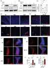
Optineurin (OPTN) restricts the formation of mixed lineage kinase domain‐like (MLKL) bodies during herpes simplex virus‐1 (HSV‐1) infection in mice. Optn +/+ and Optn −/− mice were infected with HSV‐1 (5 × 105 PFU) via corneal scarification. (A) Plaque assays were conducted with corneal swabs collected 2 and 4 days post‐infection (dpi). (B) Receptor‐interacting protein kinase 3 (RIPK3) and MLKL transcripts in the brains of infected mice (harvested 4 dpi) were measured by quantitative polymerase chain reaction (qPCR) analysis. Two‐way analysis of variance (ANOVA) was performed for statistical analysis. (C) p‐MLKL expression was examined in the brainstem of infected mice (harvested 4 dpi) by immunofluorescence imaging. (D) p‐MLKL expression was examined in siCTRL and siOPTN transfected LUHMES cells infected with 2.5 MOI HSV‐1 for 24 h by immunofluorescence imaging. (E) Optn −/− mice were infected with HSV‐1 (5 × 105 PFU) via corneal scarification, and colocalisation of MLKL and myelin basic protein (MBP) (a marker for oligodendrocytes) was examined in the brainstem of infected mice (harvested 8 dpi) by immunofluorescence imaging. NI, non‐infected.
FIGURE 5
FIGURE 5
Optineurin (OPTN) deficiency displays a phenotype similar to demyelinating disorders. Optn +/+ and Optn −/− mice were exposed to no infection and herpes simplex virus‐1 (HSV‐1; 5 × 105 PFU) via corneal scarification. (A) Immunohistochemistry was performed to analyse myelin basic protein (MBP) expression in the cerebellum of non‐infected and infected mice (harvested 8 days post‐infection [dpi]). (B) Immunofluorescence was used to analyse myelin expression in the brainstem of mice indicated by FluoroMyelin (green). (C) Quantification of fluorescence intensity of FluoroMyelin from (B). (D and E) A tape removal test was performed on infected and non‐infected mice, and the time to contact (recognise the tape) and time to remove the tape was noted. (F) Ledge test was performed on infected and non‐infected mice and scores were noted. (G) Rotarod test was performed on infected and non‐infected mice and the time the mice took to stay on the rod without falling was monitored. (H) Wire test was performed on infected and not infected mice and the time it took to hold the wire before falling was noted. (I) MBP concentration in serum was measured by ELISA. (J) Optn transcript was measured in the brains of EAE mice by quantitative polymerase chain reaction (qPCR). 0, 1, 2 and 4 are EAE mice disease progression scores. Refer to the methods section for scoring details. (K) Brain lysates from EAE mice indicate protein levels of MBP, GFAP, mixed lineage kinase domain‐like (MLKL) and OPTN. 0, 1, 2 and 4 are EAE mice disease progression scores. Refer to the methods section for scoring details. (L) p‐MLKL expression was monitored in the spinal cord tissues of EAE mice. S4 is disease progression score 4 for EAE mice. (M) Optn expression levels were examined in the normal brain tissues and multiple sclerosis (MS) brain tissues. The white arrow indicates the MS brain lesion. Data are presented as mean values  ± standard error of means (SEM) (C‒I). Two‐way analysis of variance (ANOVA) was performed for statistical analysis (C‒I). Two‐tailed Student's t‐test was performed for statistical analysis (J). NI, non‐infected.
FIGURE 6
FIGURE 6
Pharmacological inhibition of mixed lineage kinase domain‐like (MLKL) is required to prevent demyelination during Herpes simplex virus‐1 (HSV‐1) infection in Optn‐deficient conditions. (A) Optn ‒/‒ cells were infected with 1 MOI HSV‐1 and treated with DMSO (control), Necrosulphonamide (NSA) and Acyclovir (ACV) for 24 h, and cell lysates were analysed by immunoblotting with indicated antibodies. (B) Optn ‒/‒ cells were infected with 1 MOI HSV‐1 and treated with DMSO (control), Necrostatin 1s (Nec1s) and ACV for 24 h, and cell lysates were analysed by immunoblotting with indicated antibodies. (C) Optn ‒/‒ cells were infected with 1 MOI HSV‐1 and treated with DMSO (control), NSA, Nec1s and ACV for 24 h, and plaque assay was performed. (D, F and H) Optn ‒/‒ mice were infected with HSV‐1 (5 × 105 PFU) via corneal scarification. After 4 days post‐infection (dpi), the mice were treated with two daily doses of DMSO (control), NSA and ACV till 8 dpi. Brainstem slides were probed for (D) p‐MLKL, (E) MLKL and the cerebellum slides were probed for (H) myelin basic protein (MBP) by immunofluorescence imaging. (E, G and I) Optn ‒/‒ mice were infected with HSV‐1 (5 × 105 PFU) via corneal scarification. After 4 dpi, the mice were treated with two daily doses of DMSO (control) and Nec1s till 8 dpi. Brainstem slides were probed for (E) p‐MLKL, (J) MLKL and cerebellum slides were probed for (I) MBP by immunofluorescence imaging. (J and K) Quantification of MBP fluorescence intensity in (H) and (I) respectively. One‐way analysis of variance (ANOVA) was used for statistical analysis (C, J and K).

References

    1. McQuillan G, Kruszon‐Moran D, Flagg EW, Paulose‐Ram R. Prevalence of herpes simplex virus type 1 and type 2 in persons aged 14–49: United States, 2015–2016. NCHS Data Brief. 2018;(304):1‐8. - PubMed
    1. Singh N, Tscharke DC. Herpes simplex virus latency is noisier the closer we look. J Virol. 2020;94(4):e01701‐19. doi: 10.1128/jvi.01701-19 - DOI - PMC - PubMed
    1. van Lint AL, Kleinert L, Clarke SRM, Stock A, Heath WR, Carbone FR. Latent infection with herpes simplex virus is associated with ongoing CD8+ T‐cell stimulation by parenchymal cells within sensory ganglia. J Virol. 2005;79:14843‐14851. - PMC - PubMed
    1. Valerio GS, Lin CC. Ocular manifestations of herpes simplex virus. Curr Opin Ophthalmol. 2019;30(6):525‐531. doi: 10.1097/ICU.0000000000000618 - DOI - PMC - PubMed
    1. Ames J, Yadavalli T, Suryawanshi R, et al. OPTN is a host intrinsic restriction factor against neuroinvasive HSV‐1 infection. Nat Commun. 2021;12(1):5401. doi: 10.1038/s41467-021-25642-z - DOI - PMC - PubMed

LinkOut - more resources