

Volumetric and Diffusion Tensor Imaging Abnormalities Are Associated With Behavioral Changes Post-Concussion in a Youth Pig Model of Mild Traumatic Brain Injury
- PMID: 40491182
- PMCID: PMC12149694
- DOI: 10.1002/nbm.70074
Volumetric and Diffusion Tensor Imaging Abnormalities Are Associated With Behavioral Changes Post-Concussion in a Youth Pig Model of Mild Traumatic Brain Injury
Abstract
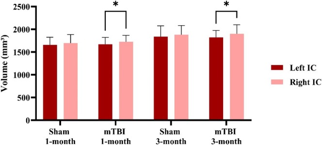
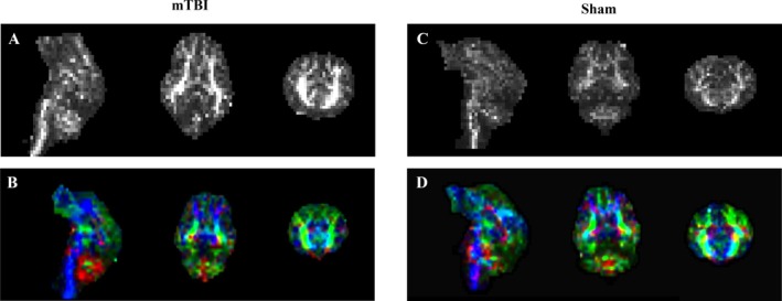
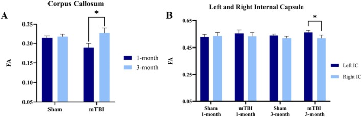
Mild traumatic brain injury (mTBI) caused by sports-related incidents in children and youth often leads to prolonged cognitive impairments but remains difficult to diagnose. In order to identify clinically relevant imaging and behavioral biomarkers associated concussion, a closed-head mTBI was induced in adolescent pigs. Twelve (n = 4 male and n = 8 female), 16-week old Yucatan pigs were tested; n = 6 received mTBI and n = 6 received a sham procedure. T1-weighted imaging was used to assess volumetric alterations in different regions of the brain and diffusion tensor imaging (DTI) to examine microstructural damage in white matter. The pigs were imaged at 1- and 3-month post-injury. Neuropsychological screening for executive function and anxiety were performed before and in the months after the injury. The volumetric analysis showed significant longitudinal changes in pigs with mTBI compared with sham, which may be attributed to swelling and neuroinflammation. Fractional anisotropy (FA) values derived from DTI images demonstrated a 21% increase in corpus callosum from 1 to 3 months in mTBI pigs, which is significantly higher than in sham pigs (4.8%). Additionally, comparisons of the left and right internal capsules revealed a decrease in FA in the right internal capsule for mTBI pigs, which may indicate demyelination. The neuroimaging results suggest that the injury had disrupted the maturation of white and gray matter in the developing brain. Behavioral testing showed that compare to sham pigs, mTBI pigs exhibited 23% increased activity in open field tests, 35% incraesed escape attempts, along with a 65% decrease in interaction with the novel object, suggesting possible memory impairments and cognitive deficits. The correlation analysis showed an associations between volumetric features and behavioral metrics. Furthermore, a machine learning model, which integrated FA, volumetric features and behavioral test metrics, achieved 67% accuracy, indicating its potential to differentiate the two groups. Thus, the imaging biomarkers were indicative of long-term behavioral impairments and could be crucial to the clinical management of concussion in youth.
Keywords: MRI; biomarkers; concussion; pig; recovery; sports injuries; trauma; white matter; youth.
© 2025 The Author(s). NMR in Biomedicine published by John Wiley & Sons Ltd.
Figures


Similar articles
-
Diffusion-Weighted Imaging in Mild Traumatic Brain Injury: A Systematic Review of the Literature.Neuropsychol Rev. 2023 Mar;33(1):42-121. doi: 10.1007/s11065-021-09485-5. Epub 2021 Mar 15. Neuropsychol Rev. 2023. PMID: 33721207
-
A systematic review and data synthesis of longitudinal changes in white matter integrity after mild traumatic brain injury assessed by diffusion tensor imaging in adults.Eur J Radiol. 2022 Feb;147:110117. doi: 10.1016/j.ejrad.2021.110117. Epub 2021 Dec 23. Eur J Radiol. 2022. PMID: 34973540
-
The relationship between blast-related mild traumatic brain injury and executive function is moderated by white matter integrity.Brain Imaging Behav. 2024 Aug;18(4):764-772. doi: 10.1007/s11682-024-00864-z. Epub 2024 Mar 6. Brain Imaging Behav. 2024. PMID: 38448704
-
Diffusion-Tensor Imaging Findings and Cognitive Function Following Hospitalized Mixed-Mechanism Mild Traumatic Brain Injury: A Systematic Review and Meta-Analysis.Arch Phys Med Rehabil. 2017 Nov;98(11):2308-2319. doi: 10.1016/j.apmr.2017.03.019. Epub 2017 Apr 20. Arch Phys Med Rehabil. 2017. PMID: 28433414
-
A Pilot Study Investigating the Use of Serum Glial Fibrillary Acidic Protein to Monitor Changes in Brain White Matter Integrity After Repetitive Head Hits During a Single Collegiate Football Game.J Neurotrauma. 2024 Jul;41(13-14):1597-1608. doi: 10.1089/neu.2023.0307. Epub 2024 Jun 10. J Neurotrauma. 2024. PMID: 38753702 Free PMC article.
References
-
- Ruff R. M., Iverson G. L., Barth J. T., Bush S. S., Broshek D. K., and Committee NPaP , “Recommendations for Diagnosing a Mild Traumatic Brain Injury: A National Academy of Neuropsychology Education Paper,” Archives of Clinical Neuropsychology 24, no. 1 (2009): 3–10, 10.1093/arclin/acp006. - DOI - PubMed
-
- Centers for Disease Control and Prevention , “Nonfatal Traumatic Brain Injuries Related to Sports and Recreation Activities Among Persons Aged ≤19 Years—United States, 2001–2009,” MMWR. Morbidity and Mortality Weekly Report 60, no. 39 (2011): 1337–1342. - PubMed
MeSH terms
Grants and funding
LinkOut - more resources
Full Text Sources
Medical
