Review
. 2025 Jun 9;6(6):e70247.
doi: 10.1002/mco2.70247. eCollection 2025 Jun.

Deep Learning in Digital Breast Tomosynthesis: Current Status, Challenges, and Future Trends

Affiliations
Review

Deep Learning in Digital Breast Tomosynthesis: Current Status, Challenges, and Future Trends

Ruoyun Wang et al. MedComm (2020). .

Abstract

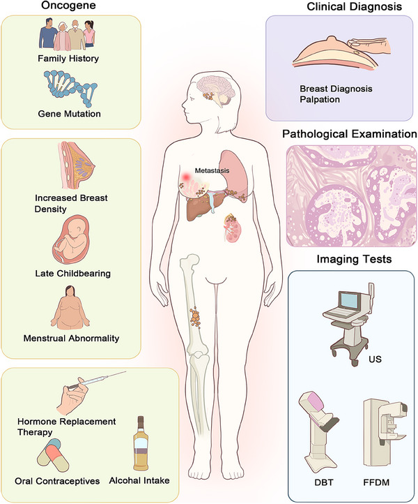
The high-resolution three-dimensional (3D) images generated with digital breast tomosynthesis (DBT) in the screening of breast cancer offer new possibilities for early disease diagnosis. Early detection is especially important as the incidence of breast cancer increases. However, DBT also presents challenges in terms of poorer results for dense breasts, increased false positive rates, slightly higher radiation doses, and increased reading times. Deep learning (DL) has been shown to effectively increase the processing efficiency and diagnostic accuracy of DBT images. This article reviews the application and outlook of DL in DBT-based breast cancer screening. First, the fundamentals and challenges of DBT technology are introduced. The applications of DL in DBT are then grouped into three categories: diagnostic classification of breast diseases, lesion segmentation and detection, and medical image generation. Additionally, the current public databases for mammography are summarized in detail. Finally, this paper analyzes the main challenges in the application of DL techniques in DBT, such as the lack of public datasets and model training issues, and proposes possible directions for future research, including large language models, multisource domain transfer, and data augmentation, to encourage innovative applications of DL in medical imaging.

Keywords: deep learning; digital breast tomosynthesis; early breast cancer screening; medical image analysis; public database.


Conflict of interest statement

The authors declare no conflicts of interest.

Figures

FIGURE 1
FIGURE 1
Breast cancer causes, pathogenesis, and screening methods. The causes of breast cancer include family history, gene mutations, increased breast density, late childbearing, menstrual abnormalities, hormone replacement therapy, oral contraceptives, and alcohol intake. The pathogenesis of breast cancer involves mutations in oncogenes within breast cells, leading to abnormal cell proliferation, tumor formation, and invasion of surrounding tissues. Over time, these cancer cells can break through the basement membrane and metastasize to distant sites via the lymphatic and circulatory systems. Screening methods for breast cancer include clinical diagnosis, pathological examination, and imaging tests such as US, FFDM, and DBT.
FIGURE 2
FIGURE 2
Principles of DM, FFDM, and DBT imaging and the advantages and disadvantages of DBT in clinical applications. DM uses a digital detector to penetrate the breast tissue with X‐rays. The sensor then converts X‐ray absorption into a digital signal that is then processed by a computer to produce a high‐resolution image of the breast. This is achieved through the use of image filtering, feature extraction, and enhancement. FFDM uses a planar detector and a rotating arm to comprehensively scan the breast tissue area, convert the X‐ray signals to digital form, and capture high‐resolution images of the internal structure of the breast via a fully digital detector. The data are then processed and reconstructed by a computer to produce high‐resolution breast images. DBT is based on the imaging principle of obtaining a series of high‐resolution, 2D breast images by taking multiple, low‐dose exposures of an X‐ray tube over a limited angular range. The images are then reconstructed by a computer into high‐resolution tomograms parallel to the detector, resulting in 3D tomograms. DBT has several advantages over DM and FFDM, including enhanced visualization, precise localization, effective differentiation of overlap, improved detection of breast cancer, and improved discrimination between benign and malignant lesions. However, DBT also has some disadvantages, such as being less effective in women with dense breasts, higher radiation dose [396], and longer reading time [87], which can impact efficiency. MGD, mean glandular dose.
FIGURE 3
FIGURE 3
Applying DL models and other AI techniques to DBT data. The traditional AI models applied to DBT include decision trees, random forests, logistic regression, SVM, and multilayer perceptron. Deep learning models for the diagnostic classification of breast diseases on DBT data include CNN, DCNN, AlexNet, DenseNet, RetinaNet, ResNet50, VGG16, VGG19, and Inception V3. The deep learning models used for breast lesion segmentation and detection on DBT data include U‐Net and GCN. Other DL models used for medical image generation from DBT data include Faster R‐CNN, Mask R‐CNN, and YOLO. In addition, a GAN has been used for this purpose. The applications of DL in DBT go beyond early detection and diagnosis of breast cancer. It can also act as a quantitative biomarker, predict molecular subtypes, assess treatment efficacy, and predict patient prognosis.
FIGURE 4
FIGURE 4
Detailed comparison of breast cancer image databases. We make detailed comparisons between databases based on several key attributes, including GRADE, that is, whether information on the severity grading of breast cancer is included; pathological diagnosis, that is, whether benign and malignant information is included; target delineation, that is, whether the delineation of target regions for breast cancer is included; abnormal annotation, that is, whether abnormal annotations are included; and amount, that is, the number of images in the dataset. The yellow triangles indicate that the attribute is present, whereas the blue triangles represent “not applicable,” that is, the data are not provided. The size of the circle indicates the order of magnitude of the images in the dataset. For example, the largest circle represents an order of magnitude of more than 100,000 images, whereas the smallest circle represents an order of magnitude of fewer than 100 images. The representative breast cancer image databases include MIAS, DDSM, LAPIMO, INbreast, BCDR‐DM, CSAW‐CC, OMI‐OB, and VinDr‐Mammo.
FIGURE 5
FIGURE 5
Challenges and future prospects of DL techniques for DBT applications. The challenges are grouped into three categories: data challenges, model challenges, and interpretability challenges. The training of DBT‐DL models depends on the availability of a substantial, high‐quality annotated training set, as there is a paucity of publicly available datasets for DBT. A lack of data can lead to model overfitting. In addition to building public databases, the use of data augmentation, federated learning, encrypted data learning, and other methods is expected to improve model generalizability across different datasets. In addition, the training process of DL models presents numerous challenges. Multisource domain migration, LLM, and knowledge graphs are expected to facilitate the implementation and dissemination of DBT techniques in clinical settings. Since DL models are opaque, resulting in low interpretability. Attention mechanism networks, multiperspective feature fusion, and in‐model interpretation approaches are expected to improve interpretability.

References

    1. Lei S., Zheng R., Zhang S., et al., “Global Patterns of Breast Cancer Incidence and Mortality: A Population‐based Cancer Registry Data Analysis From 2000 to 2020,” Cancer Communications (London) 41, no. 11 (2021): 1183–1194. - PMC - PubMed
    1. Jemal A., Bray F., Center M. M., Ferlay J., Ward E., and Forman D., “Global Cancer Statistics,” CA: A Cancer Journal for Clinicians 61, no. 2 (2011): 69–90. - PubMed
    1. Bray F., Ferlay J., Soerjomataram I., Siegel R. L., Torre L. A., and Jemal A., “Global Cancer Statistics 2018: GLOBOCAN Estimates of Incidence and Mortality Worldwide for 36 Cancers in 185 Countries,” CA: A Cancer Journal for Clinicians 68, no. 6 (2018): 394–424. - PubMed
    1. Huang J., Chan P. S., Lok V., et al., “Global Incidence and Mortality of Breast Cancer: A Trend Analysis,” Aging (Albany NY) 13, no. 4 (2021): 5748–5803. - PMC - PubMed
    1. Bray F., Laversanne M., Sung H., et al., “Global Cancer Statistics 2022: GLOBOCAN Estimates of Incidence and Mortality Worldwide for 36 Cancers in 185 Countries,” CA: A Cancer Journal for Clinicians 74, no. 3 (2024): 229–263. - PubMed

LinkOut - more resources