. 2025 Mar 17;12(6):nwaf095.
doi: 10.1093/nsr/nwaf095. eCollection 2025 Jun.

Ccdc13 is essential for the assembly of ciliary central microtubules

Affiliations

Ccdc13 is essential for the assembly of ciliary central microtubules

Zhimao Wu et al. Natl Sci Rev. .

Abstract

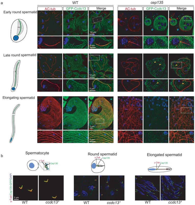
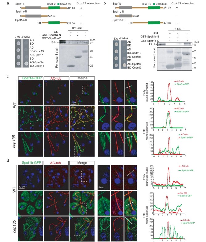
Motile cilia are critical for diverse cellular activities, affecting the survival and development of most eukaryotic organisms. Central microtubules (MTs), which are located in the lumen of ciliary axonemes, are non-centrosomal MTs that are crucial for motile cilia beating. However, the formation mechanism of central MTs remains elusive. Here, by using a Drosophila model, we identify Ccdc13 as a novel regulator for the assembly of central MTs. We show that Ccdc13 localizes along the central MTs and is essential for its formation in sperm flagella, with its deletion consequently affecting the sperm motility and the fertility of male flies. Mechanistically, we demonstrated that Ccdc13 directly interacts with Spef1, acting upstream of Spef1 to regulate central MT elongation. Remarkably, we demonstrated that the role of Ccdc13 in ciliary central MT formation is conserved in mammals. Ccdc13 deficiency in mice leads to the loss of central MTs in motile ependymal cilia, resulting in abnormal cilia motility and hydrocephalus. Our results mark the discovery of Ccdc13 as a novel regulator for ciliary central MT assembly and reveal that the Ccdc13-Spef1 complex is an evolutionarily conserved module that is critical for central MT formation in motile cilia of both flies and mammals.

Keywords: Ccdc13; Drosophila; Spef1; central microtubules; motile cilia.


Figures

Figure 1.
Figure 1.
Ccdc13 is essential for CP MT formation in Drosophila. (a) Generation of Ccdc13 deletion mutant (ccdc131) flies. Schematics display the genomic structure (upper panel) and protein structure (lower panel) of Ccdc13. The gRNA target sites are shown. Ccdc131 has a deletion in its cDNA from nt 516 to 1427, leading to an in-frame deletion of 305 aa (aa 172 to 476) from Ccdc13. Ccdc132 mutant has a deletion in cDNA from nt 511 to 1444, resulting in a reading-frame shift. (b) Genotyping of the ccdc131 mutant and ccdc132 mutant using PCR. The PCR products are 1980 bp long for wild type (WT) and 1065 bp long for the mutant. (c) Western blot analysis of the testes showed the presence of a truncated protein product of Ccdc13 in the Ccdc131 mutant. α-tubulin was used as the loading control. The asterisk indicates the target band. (d) Ccdc131 mutant flies had normal negative geotaxis and hearing. (e) Ccdc131 males were infertile due to the immobility of their sperms. Note that the infertility defect was rescued by expressing GFP–Ccdc13. (f) The sperm flagella in ccdc131 testes showed no defects in elongation compared with those in WT testes. The Nexin–Dynein regulatory complex (N-DRC) protein Gas8 was used to label flagella. (g, h) ccdc131 flagella lacked the CP. In electron microscopy (EM) images of testis (g) cross sections and (h) longitudinal sections, both WT and ccdc131 mutants had 64 spermatids in each cyst. The arrowheads denote representative central MTs in the WT cross sections; the central MT was completely lost in ccdc131 mutants. The quantification data (i) were obtained from >300 flagella in three flies. Scale bars: 200 μm (f), 0.5 μm (g, full-scale images), 100 nm (g, zoomed-in areas). Quantification results are presented as mean ± SD, unless stated otherwise. Student's t-test: ns, no significance; ***P < 0.001.
Figure 2.
Figure 2.
Ccdc13 localizes to the CP in Drosophila. (a) Ccdc13 displayed CP-dependent axonemal localization. In WT testes, Ccdc13 first appeared along the axoneme of the flagella in early-round spermatids and the localization persisted in late-round spermatids and elongating spermatids. In testes of cep135 mutants, whose flagella lacked the CP, Ccdc13 no longer displayed axonemal localization. The flagella were labeled by using AC-tubulin (red). The white asterisk indicates the leaf-blade-shaped mitochondria region in early-round spermatocytes. The arrowhead represents the microtubule-like GFP–Ccdc13 signal in the cytosol of the cep135 mutant. (b) Ccdc13 was not required for the basal body localization of Cep135. The basal body was labeled by using γ-tubulin (red).
Figure 3.
Figure 3.
Ccdc13 interacts with CP protein Spef1 in Drosophila. (a) Ccdc13 exhibited direct interaction with Spef1a in both the Y2H assay (lower left panel) and the GST pull-down assay (lower right). The upper panel displays the schematics of the Spef1a constructs. In the Y2H assay, the interaction of Ccdc13 with Spef1a was evidenced by colony growth on the SD–Ade–Leu–Trp–His plates. In the GST pull-down assay, Ccdc13 directly interacted with the C-terminal of Spef1a whereas no interaction was observed with its N-terminal. L: Leu, W: Trp, H: His, A: Ade. (b) Ccdc13 exhibited direct interaction with Spef1b in both the Y2H assay (lower left panel) and the GST pull-down assay (lower right). The upper panel displays the schematics of the Spef1b constructs. In the Y2H assay, the interaction of Ccdc13 with Spef1b was evidenced by colony growth on the SD–Ade–Leu–Trp–His plates. In the GST pull-down assay, Ccdc13 directly interacted with the C-terminal of Spef1b whereas no interaction was observed with its N-terminal. L: Leu, W: Trp, H: His, A: Ade. (c) Subcellular localizations of Spef1a during spermiogenesis. In early-round spermatids, Spef1a was not observed on the axonemes of the flagella. In late-round spermatids, Spef1a began to appear on the axonemes of the flagella. In cep135 mutants, the Spef1a signal on the axoneme was notably impaired, exhibiting abnormal signal distribution in the cytosol. The right panels depict zoomed-in views of the axonemes (white dotted rectangles) to highlight specific details. Line scans were performed to show correlations between axonemes and Spef1a. (d) Spef1b–GFP localizes to the sperm flagella starting from late-round spermatids and this localization relies on Cep135.
Figure 4.
Figure 4.
Ccdc13 acts upstream of Spef1a in CP MT formation. (a) Generation of Spef1a and Spef1b mutant flies. Schematics display the genomic (upper panel) and protein (lower panel) structures of Spef1a and Spef1b. The gRNA target sites are shown. spef1a1 had a deletion in cDNA from nt 376 to 382, resulting in a frameshift and deletion of the C-terminal half of the protein (aa 126 to the end at 254). spef1b1 had a deletion in cDNA from nt 31 to 136, introducing a stop codon and resulting in the deletion of almost the whole protein (leaving only 10 aa). (b) spef1a was essential for male fertility and sperm motility. Quantification results are presented as mean ± SD. Student's t-test: ns, no significance; ***P < 0.001. (c) Representative EM images of flagellar cross sections from wild type (WT), spef1b1, spef1a1 and spef1a1; spef1b1 fly mutants. The quantification results are presented on the right. (d) Spef1a failed to colocalize with the axoneme in ccdc131 flagella. Arrows indicate typical flagella. Axonemes in dotted rectangles are zoomed-in in the right-hand panels to show details. Line scans were performed to show correlations between axoneme and Spef1a. (e) Axonemal localization of Ccdc13 was detected in only early-round spermatids of spef1a1 flies and spef1a1; spef1b1 double-mutant flies, albeit with a compromised signal compared with that in WT flies. Arrows and asterisks, respectively, indicate typical Ccdc13-positive and Ccdc13-negative flagella in spef1a1 and spef1a1; spef1b1 testes. Axonemes in white dotted rectangles are zoomed-in in the right-hand panels to show details. (f) A model for the role of Ccdc13 and Spef1a in the CP MT formation in sperm flagella of Drosophila. In spermatocytes, Cep135 initiates the formation of a singlet central MT. In early-round spermatids, Ccdc13 emerges on the CP, potentially stabilizes the singlet central MT and/or initiates the formation of the second central MT. Following this, Spef1a is recruited and, together with Ccdc13, promotes the elongation and stabilization of CP MTs.
Figure 5.
Figure 5.
Mammalian Ccdc13 is an important CP protein whose depletion in mice results in hydrocephalus. (a) Ccdc13 specifically localized to motile cilia but not primary cilia in mammalian cells. Cultured mEPCs (top panel) were fixed at Day 15 post serum starvation whereas RPE1 cells (bottom panel) were fixed 24 h post serum starvation. Representative confocal images are presented. (b) Ccdc13 colocalized with Hydin (arrows) in the axoneme lumen of mEPC cilia. Cultured mEPCs were infected with lentivirus to express GPF–Ccdc13 and fixed at Day 7. Hydin served as a CP marker. Representative 3D-SIM images are presented. The framed region is magnified to show details. (c) The strategy for generating Ccdc13 knockout (KO) mice. A Ccdc13 flox allele was generated with two loxP sites flanking Exon 3 (e3). Upon crossing with Cre mice, this was expected to lead to the deletion of Exon 3, causing a frameshift and retaining only 74 amino acids of the N-terminal. (d) Genotyping of Ccdc13−/− mutant mice using PCR. The PCR products were 1532 bp long for WT and 403 bp long for Ccdc13−/− mice. (e) Anti-Ccdc13 staining confirmed the absence of Ccdc13 in Ccdc13−/− multicilia. mEPCs at Day 15 were fixed, followed by immunostaining. (f) Ccdc13−/− mice displayed a dome-shaped skull (arrow). (g) Ccdc13−/− mice exhibited hydrocephalus. Representative HE-stained coronal sections of the brain are presented. Arrows indicate enlarged ventricles. (h) Typical image sequences of ependymal cilia were acquired at 200 frames per second (fps) by using living brain slices of P21 mice of indicated genotypes. Trajectories of cilia are shown in each sample.
Figure 6.
Figure 6.
Mammalian Ccdc13 is critical for CP formation. (a) CP-associated proteins Spef1, Hydin and Spag16 failed to show prominent axonemal localizations in Ccdc13−/− mEPCs. Cultured mEPCs were fixed at Day 15. (b) The localization of Wdr47 was mildly affected in short cilia but almost lost in long cilia in Ccdc13−/−. (c) Ccdc13−/− ependymal multicilia lacked a CP. Representative TEM images from ependymal tissues of P21 mice are presented. Arrowheads indicated central MTs. Quantification results were obtained from three pairs of littermates, with 200 cross sections of cilia scored for each group of mice. (d) Ccdc13 defect impairs the CP formation of respiratory multicilia. Tracheal epithelial tissues of P21 mice were fixed and processed for TEM to observe CP MT (arrowheads) formation. The pie chart summarizes the percentages of the axoneme section with different central MT numbers.
Figure 7.
Figure 7.
Mammalian Ccdc13 interacts with Spef1 and exhibits MT-binding activity. (a) Mouse Ccdc13 directly interacted with mouse Spef1 in GST pull-down assays. Asterisks indicate the position of GST–Ccdc13. (b) Mouse Ccdc13 associated with the C-terminus of mouse Spef1 in vivo. The indicated proteins were transiently expressed in HEK293T cells, followed by co-immunoprecipitation (Co-IP) assays. Asterisks indicate the position of GFP–Spef1 and its truncations. (c) In vivo colocalization of Ccdc13 and the C-terminal of Spef1 in mammalian cells. The colocalization of Spef1-C with Ccdc13 was observed whereas there was no colocalization between Ccdc13 and Spef1-N. (d) Ccdc13 exhibited the ability to bundle MTs, with the N-terminal amino acids 1–300 as the region responsible for this activity. MTs that were polymerized in vitro were incubated with the specified concentrations of the respective proteins and subsequently imaged. (e) Ccdc13 (1–300) effectively co-sedimented with MT pellets in the MT-pelleting assay. P, pellet fraction containing Taxol-stabilized MTs; S, supernatant fraction containing soluble proteins. Results are representative of two independent experiments.

References

    1. Lindemann CB, Lesich KA. Flagellar and ciliary beating: the proven and the possible. J Cell Sci 2010; 123: 519–28. 10.1242/jcs.051326 - DOI - PubMed
    1. Shinohara K, Hamada H. Cilia in left-right symmetry breaking. Cold Spring Harb Perspect Biol 2017; 9: a028282. 10.1101/cshperspect.a028282 - DOI - PMC - PubMed
    1. Sironen A, Shoemark A, Patel M et al. Sperm defects in primary ciliary dyskinesia and related causes of male infertility. Cell Mol Life Sci 2020; 77: 2029–48.10.1007/s00018-019-03389-7 - DOI - PMC - PubMed
    1. Liu H, Zheng J, Zhu L et al. Wdr47, Camsaps, and Katanin cooperate to generate ciliary central microtubules. Nat Commun 2021; 12: 5796. 10.1038/s41467-021-26058-5 - DOI - PMC - PubMed
    1. Yuan S, Wang Z, Peng H et al. Oviductal motile cilia are essential for oocyte pickup but dispensable for sperm and embryo transport. Proc Natl Acad Sci USA 2021; 118: e2102940118. 10.1073/pnas.2102940118 - DOI - PMC - PubMed

LinkOut - more resources