. 2025 Aug;12(32):e06688.
doi: 10.1002/advs.202506688. Epub 2025 Jun 10.

A Novel Class of Multi-substituted Diaryl Scaffold Derivatives Inhibit Glioblastoma Progression by Targeting CD155

Affiliations

A Novel Class of Multi-substituted Diaryl Scaffold Derivatives Inhibit Glioblastoma Progression by Targeting CD155

Yong-Jian Wang et al. Adv Sci (Weinh). 2025 Aug.

Abstract

Glioblastoma (GBM) is the most formidable malignancy in the brain, characterized by a significant resistance to treatment. The immune targeting of glioblastoma stem cells (GSCs) holds great promise. In this study, structural modifications of the lead compound clofoctol is conducted and structure-activity relationship analyses are performed against GBM, yielding a novel blood-brain barrier-permeable compound, B7, featuring a pivotal multi-substituted diaryl scaffold. B7 demonstrates potent anti-GBM effects, significantly inhibiting GSC proliferation, migration, and invasion. Notably, B7 inhibits tumor progression, specifically bolstered natural killer (NK) cell-mediated cytotoxicity, and mitigates the immunosuppressive microenvironment in intracranial xenograft mice implanted with GBM cells and GSCs, as well as in cocultures of GSCs and NK-92 cells. Mechanistically, these anti-GBM effects of B7 are abolished by overexpression of poliovirus receptor cell adhesion molecule (CD155), both in vitro and in vivo. Further exploration reveals that B7 targets CD155 via interaction at five crucial binding sites, namely, L47, L108, L142, M110, and V115 residues. These interactions collectively contribute to the hydrophobic interaction energies within the B7-CD155 complex, modulating the CD155/T cell immunoreceptor with Ig and ITIM domains/CD226 axis to reshape the NK cell-mediated tumor immune microenvironment. In conclusion, this study establishes the therapeutic potential of B7 for glioma, synergistically targeting GSC biology and NK cell immunity for the treatment of GBM.

Keywords: glioblastoma stem cells; multi‐substituted diaryl derivatives; poliovirus receptor cell adhesion molecule (CD155); tumor immune microenvironment.


Conflict of interest statement

The authors declare no conflict of interest.

Figures
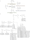
Figure 1
Figure 1
Illustrative representation of the design concept and chemical synthesis route map for the derivatives. A) Strategies employed in derivative design. B) Synthesis methods utilized for target compounds.
Figure 2
Figure 2
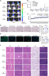
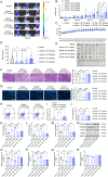
The activity of compound B7 against glioma in vivo. A) Representative bioluminescent images of nude mice, n = 3. B) Quantitative analysis of photon flux of nude mice, n = 6. C) Body weight changes of nude mice, n = 6. D) Representative H&E staining images of tumor tissues in the brain of nude mice, n = 3, bar: 100 µm. E) Representative immunohistochemical staining images for Ki67 in tumor tissues of nude mice, n = 3, bar: 200 µm. F) Representative H&E staining images of heart, liver, kidney, lung, and spleen tissues of nude mice, bar: 400 µm. One‐way ANOVA, followed by Tukey's post hoc test, was used for multiple group comparisons to determine differences between groups. The results were presented as mean ± SD. * P < 0.05, ** P < 0.01, *** P < 0.001 versus Control.
Figure 3
Figure 3
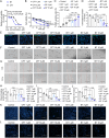
Effects of B7 on the viability and functionality of GSCs. A) Effects of B7 and CFT on the viability of GSCs and IC50 values. B) GSC viability at 12–72 h. C,E) Quantitative analysis (C) and representative images (E) for the evaluation of GSC apoptosis using TUNEL staining, bar: 200 µm. D,F) Quantitative analysis (D) and representative images (F) of the GSC sphere formation assays, bar: 400 µm. G,H) Representative images (G) and quantification (H) for the effects of B7 and CFT on GSC migration in scratch assays, bar: 400 µm. I,K) Quantification (I) and representative images (K) of transwell migration assays, bar: 400 µm. J,L) Quantification (J) and representative images (L) of transwell invasion assays, bar: 400 µm. Comparisons among multiple groups were analyzed using one‐way ANOVA, followed by Tukey's post hoc test. The results were presented as mean ± SD, n = 3. * P < 0.05, ** P < 0.01, *** P < 0.001 versus Control.
Figure 4
Figure 4
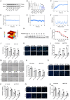
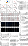
CD155 serves as the target for compound B7 to inhibit GSCs. A,B) Representative images (A) and quantitative analysis (B) of Western blotting analysis for CD155 protein expression. C–G) Molecular dynamics (MD) simulation analysis of B7‐CD155 complex, including the root mean square deviation (RMSD) (C), radius of gyration (Rg) (D), solvent‐accessible surface area (SASA) (E), root mean square fluctuation (RMSF) (F), and free energy landscape (FEL) (G). H,I) Representative images (H) and fitting curve (I) for the evaluation of B7‐CD155 binding efficiency using Cellular Thermal Shift Assay (CETSA). J) The cell viability of GSCs. K,L) Representative images (K) and quantification (L) of TUNEL staining, bar: 200 µm. M,N) Representative images (M) and quantification (N) of the scratch assay, bar: 200 µm. O,P) Representative images (O) and quantification (P) of transwell migration assay, bar: 400 µm. Q,R) Representative images (Q) and quantification (R) of transwell invasion assay, bar: 400 µm. Comparisons across multiple groups were analyzed using one‐way ANOVA and Tukey's post hoc test in panel B. Two‐tailed Student's t‐test was employed for two‐group comparisons in panels J, L, N, P, and R. The results were expressed as mean ± SD, n = 3. * P < 0.05, ** P < 0.01, *** P < 0.001 vs Control or corresponding vehicle.
Figure 5
Figure 5
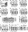
Effects of B7 on the cytotoxicity of NK‐92 cells in a coculture environment. A) Viability of NK‐92 cells cultured alone and within the coculture environment with GSCs. B–F) Levels of lactate dehydrogenase (LDH) (B), interferon‐gamma (IFN‐γ) (C), tumor necrosis factor‐alpha (TNF‐α) (D), granzyme B (GzmB) (E), and perforin (F) released from NK‐92 cells cocultured with GSCs. G–K) Representative images (G) and quantification of TIGIT (H), CD226 (J), and CD96 (K) protein expression using Western blotting analysis. L) Co‐immunoprecipitation (co‐IP) analysis of the interaction of CD155 with TIGIT and CD226. One‐way ANOVA followed by Tukey's post hoc test was employed to analyze group differences. The results were expressed as mean ± SD, n = 3. * P < 0.05, ** P < 0.01, *** P < 0.001 vs Control or corresponding vehicle.
Figure 6
Figure 6
In vivo effects of B7 on tumor progression and regulation of the immune microenvironment associated with CD155. A) Representative bioluminescent images of nude mice, n = 3. B) quantification of photon flux of nude mice intracranially implanted with GSCs‐Luc2 cotransfected with either the empty vehicle or CD155 overexpression plasmid, n = 8. C) Body weight changes of these nude mice, n = 8. D,E) Quantification of tumor volume (D) and tumor images (E) in the nude mouse brains, n = 8. F) H&E staining and quantification of tumor tissues, n = 3, bar: 400 µm. G) Immunohistochemical staining and quantification for Ki67 in tumor tissues of nude mice, n = 3, bar: 200 µm. H,I) Representative flow cytometry images (H) and quantification (I) of CD3/CD49b+ NK cells in different groups of tumor tissues. J–M) The IFN‐γ levels (J), TNF‐α (K), GzmB (L), and perforin (M) levels in tumor tissues of nude mice, n = 3. N–R) Representative images (N) and quantification of CD155 (O), CD226 (P), TIGIT (Q), and CD96 (R) protein expression using Western blotting analysis, n = 3. One‐way ANOVA and Tukey's post hoc test were used to discern intergroup differences. The results were presented as mean ± SD. * P < 0.05, ** P < 0.01, *** P < 0.001 versus corresponding vehicle.
Figure 7
Figure 7
Mutations in CD155 for specific binding to B7. A) Key amino acids in the B7‐CD155 complex using molecular dynamics analysis. B) Schematic diagram of the B7‐CD155 complex and the locations of key amino acids. C–J) Fitting curve of Y121A (C), D117A (D), L47I (E), L108I (F), L142I (G), M110I (H), and V115I (I) and representative Western blotting images (J) demonstrating B7‐CD155 binding efficiency using CETSA. K,L) Representative images (K) and quantification (L) of the effects of mutations on CD155 function and the interaction between B7 and CD155 in scratch assays, bar: 400 µm. M,N) Representative images (M) and quantification (N) of the influence of mutations on migration of B7 in transwell migration assays, bar: 400 µm. O,P) Representative images (O) and quantification (P) of the influence of mutations on invasion of B7 in transwell invasion assays, bar: 400 µm. Q) Viability of NK‐92 cells within the coculture environment with GSCs overexpressing CD155 WT and mutant plasmids. R–V) Levels of LDH (R), IFN‐γ (S), TNF‐α (T), GzmB (U), and perforin (V) released from NK‐92 cells cocultured with GSCs overexpressing CD155 WT and mutant plasmids. W–Z) Representative images (X) and quantification of TIGIT (W), CD226 (Y), and CD96 (Z) protein expression using Western blotting analysis. One‐way ANOVA, followed by Tukey's post hoc test, was used for multiple group comparisons. Two‐tailed Student's t‐test was employed for two‐group comparisons. The results were presented as mean ± SD, n = 3. * P < 0.05, ** P < 0.01, *** P < 0.001 versus corresponding control.

References

    1. Ostrom Q. T., Price M., Neff C., Cioffi G., Waite K. A., Kruchko C., Barnholtz‐Sloan J. S., Neuro. Oncol. 2022, 24, 10.1093/neuonc/noac202. - DOI - PMC - PubMed
    1. Weller M., van den Bent M., Preusser M., Le Rhun E., Tonn J. C., Minniti G., Bendszus M., Balana C., Chinot O., Dirven L., French P., Hegi M. E., Jakola A. S., Platten M., Roth P., Rudà R., Short S., Smits M., Taphoorn M. J. B., von Deimling A., Westphal M., Soffietti R., Reifenberger G., Wick W., Nat. Rev. Clin. Oncol. 2021, 18, 170. - PMC - PubMed
    1. Regmi M., Wang Y., Liu W., Dai Y., Liu S., Ma K., Lin G., Yang J., Liu H., Wu J., Yang C., J. Exp. Clin. Cancer Res. 2024, 43, 47. - PMC - PubMed
    1. Poorva P., Mast J., Cao B., Shah M. V., Pollok K. E., Shen J., Mol. Ther. 2025, 10.1016/j.ymthe.2025.02.043. - DOI - PMC - PubMed
    1. Park J. H., Hothi P., de Lomana A. L. G., Pan M., Calder R., Turkarslan S., Wu W. J., Lee H., Patel A. P., Cobbs C., Huang S., Baliga N. S., Sci. Adv. 2024, 10, 7706. - PMC - PubMed

MeSH terms

LinkOut - more resources