Review
. 2025 Jun 10;22(5):460-480.
doi: 10.20892/j.issn.2095-3941.2024.0628.

The mechanisms and clinical significance of CD8+ T cell exhaustion in anti-tumor immunity

Affiliations
Review

The mechanisms and clinical significance of CD8+ T cell exhaustion in anti-tumor immunity

Tao Zhong et al. Cancer Biol Med. .

Abstract

CD8+ T cell exhaustion, a critical challenge in the immune response to cancer, is characterized by a profound decline in the functionality of effector CD8+ T cells. This state of exhaustion is accompanied by the upregulation of various inhibitory receptors and significant shifts in both transcriptional and epigenetic profiles, thus ultimately leading to inadequate tumor control. Therapeutic strategies aimed at reversing CD8+ T cell exhaustion have the potential to rejuvenate immune responses and enhance treatment efficacy. This review compiles current knowledge regarding the molecular mechanisms underlying CD8+ T cell exhaustion, including the roles of immune checkpoint molecules, the tumor microenvironment, metabolic reprogramming, transcription factors, and epigenetic modifications. Emerging therapeutic approaches designed to combat CD8+ T cell exhaustion are evaluated, with emphasis on the modulation of immune checkpoints; targeting of metabolic and transcriptional changes; and exploration of other innovative strategies, such as epigenetic editing and engineered CAR-T cells. Importantly, we expand the exhaustion concept to immune cells beyond CD8+ T cells, such as CD4+ T cells, natural killer cells, and myeloid populations, thereby highlighting the broader implications of systemic immunosuppression in the cancer context. Finally, we propose avenues for future research aimed at further elucidating the factors and molecular mechanisms associated with CD8+ T cell exhaustion, thereby underscoring the critical need for strategies aimed at reversing this state to improve outcomes in cancer immunotherapy.

Keywords: CD8+ T cell exhaustion; anti-tumor immunity; cancer immunotherapy; immune checkpoint; immune checkpoint inhibitors.


Conflict of interest statement

No potential conflicts of interest are disclosed.

Figures

Figure 1
Figure 1
Functional and molecular hallmarks of CD8+ T cells. (A) Functional effector CD8+ T cells. Functional effector CD8+ T cells express IFN-γ and TNF-α genes under transcriptional and epigenetic regulation. The cells have low inhibitor receptor expression (PD-1, LAG-3, TIGIT, TCR, and CTLA-4) and produce pro-inflammatory cytokines (IFN-γ, TNF-α, and IL-2), which amplify immune responses. Meanwhile, they exhibit efficient functional mitochondrial metabolism through mitochondrial mass and polarized mitochondria. These elements together ensure CD8+ T cells’ high proliferative ability and cytotoxicity for mounting immune responses. (B) Exhausted CD8+ T cells. Exhausted CD8+ T cells are characterized by pronounced expression of inhibitory receptors (PD-1, LAG-3, TIGIT, TCR, and CTLA-4) and diminished production of cytokines (IFN-γ, TNF-α, and IL-2). Exhausted CD8+ T cells exhibit mitochondrial dysfunction, accompanied by diminished mitochondrial mass and polarized mitochondria, and elevated ROS production. The cells show significantly elevated expression of exhaustion-associated genes (PDCD1 and TOX) under transcriptional and epigenetic regulation. Tox is required for exhausted CD8+ T cells’ epigenetic remodeling and survival. Exhausted CD8+ T cells display significantly diminished proliferative ability and cytotoxicity. CTLA-4, cytotoxic T-lymphocyte-associated protein 4; IL-2, interleukin-2; IFN-γ, interferon-gamma; LAG-3, lymphocyte activation gene 3; PD-1, programmed cell death protein 1; PDCD1, programmed cell death 1, also named PD-1; ROS, reactive oxygen species; TCR, T cell receptor; TIGIT, T cell immunoreceptors with Ig and ITIM domains; TNF-α, tumor necrosis factor alpha; TOX, thymocyte selection associated high mobility group box.
Figure 2
Figure 2
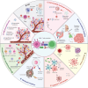
Strategies to revise CD8+ T cell exhaustion in the TME. (A) TME preconditioning. Preconditioning of the TME with radiation or chemotherapy to decrease immunosuppressive cell populations and enhance antigen presentation potentiates the effects of adoptively transferred T cells. (B) TME. TME cells can be targeted to prevent T cell exhaustion. CD8+ T cell activity can be enhanced through strategies targeting Treg-specific pathways such as CTLA-4 or CD25; inhibiting MDSC recruitment and function with agents such as CSF1R inhibitors or chemokine receptor blockers; or reprogramming TAMs from the pro-tumoral M2 phenotype to the anti-tumor M1 state with agents such as CD40 agonists. (C) TME metabolic landscape modulation. Metabolic reprogramming, such as shifting from oxidative phosphorylation to glycolysis in T cells, preventing ATP conversion to adenosine production, or targeting cancer cells through alternative metabolic substrates (e.g., inhibition of lactate dehydrogenase A to decrease lactate production), further enhances CD8+ T cell responses. (D) Gut microbiome targeting. Gut microorganisms such as Bifidobacterium and Akkermansia muciniphila enhance ICI efficacy by promoting T cell priming through microbial metabolites. (E) Oncolytic viruses. Oncolytic viruses designed to selectively infect and lyse tumor cells, thus releasing tumor antigens and inducing local inflammation, provide another innovative approach for reshaping the TME. (F) Adoptive T cell therapies. CAR-T cells with dominant-negative TGF-β receptors and IL-2 or IL-33 secretion ability have shown promise in mitigating TME suppression. (G) Checkpoint inhibitors. Checkpoint inhibitors such as PD-1/PD-L1 blockers primarily neutralize immune checkpoint molecules on the surfaces of T cells, thus preventing them from binding ligands, and subsequently inhibiting T cell function or apoptosis. (H) Targeting hypoxia and VEGF pathways. VEGF inhibitors and inhibitors of hypoxia-inducible factor restore T cell efficacy. Normalizing aberrant tumor vasculature with agents such as VEGF inhibitors can improve oxygenation, subsequently increasing immune cell infiltration and function. ADP, adenosine diphosphate; AMP, adenosine monophosphate; ATP, adenosine triphosphate; CAR-T, chimeric antigen receptor T; CD, cluster of differentiation; CSF1R, colony-stimulating factor 1 receptor; CTLA-4, cytotoxic T-lymphocyte-associated protein 4; HDACs, histone deacetylases; ICIs, immune checkpoint inhibitors; IL-2, interleukin-2; IL-33, interleukin-33; MDSCs, myeloid-derived suppressor cells; MHCI, major histocompatibility complex I; PD-1, cell death protein 1; PD-L1, programmed death-ligand 1; SCFAs, short-chain fatty acids; TAMs, tumor-associated macrophages; TCR, T cell receptor; TGF-β, transforming growth factor beta; TME, tumor microenvironment; Tregs, regulatory T cells; VEGF, vascular endothelial growth factor.
Figure 3
Figure 3
Exhaustion of other populations of immune cells. (A) CD4+ T cell exhaustion. CD4+ T cell exhaustion occurs because of upregulation of inhibitory receptors, prolonged antigen exposure, and the presence of immunosuppressive cytokines. (B) NK cell exhaustion. NK cell exhaustion is triggered by the persistent engagement of the checkpoint NKG2A and killer cell immunoglobulin-like receptors and diminished signaling through the activating receptors (e.g., NKG2D and DNAM-1). (C) B cell exhaustion. B cell exhaustion occurs primarily because of upregulation of inhibitory receptors (such as PD-1 and BTLA) and prolonged antigen exposure. (D) MDSC exhaustion. MDSC exhaustion occurs because of hypoxia, nutrient deprivation, and the presence of immunosuppressive cytokines in the TME. (E) Dendritic cell (DC) exhaustion. DCs are exhausted in the TME because of antigen exposure and the influence of immunosuppressive factors. (F) Monocyte exhaustion. Monocytes are exhausted in the presence of pathogens or various tumor-derived factors. BTLA, B and T lymphocyte attenuator; CTLA-4, cytotoxic T-lymphocyte-associated protein 4; DCs, dendritic cells; DNAM-1, DNAX accessory molecule 1; MDSCs, myeloid-derived suppressor cells; NK, natural killer; NKG2A, natural-killer group 2, member A; NKG2D, natural-killer group 2, member D; PD-1, cell death protein 1; TME, tumor microenvironment.
Figure 4
Figure 4
Determinants of CD8+ T cell exhaustion in cancer. (A) Inhibitory immune checkpoint molecules. The expression of inhibitory immune checkpoint molecules (PD-1, CTLA-4, LAG-3, TCR, and TIGIT) acts as a brake on T cell activity, thus restricting their ability to mount effective anti-tumor immune responses. (B) Tumor microenvironment (TME). The TME is rich in regulatory cells (Tregs, MDSCs, and TAMs), with elevated production of inhibitory cytokines (IL-10 and TGF-β), thereby restricting CD8+ T cell infiltration and function. Tumor cells and associated stromal cells express ectonucleotidases, such as CD39 and CD73, which convert ATP to adenosine. Elevated adenosine binds A2A receptors on T cells and subsequently decreases cytotoxic activity. ROS production further impairs TCR signaling, and upregulates inhibitory receptors and ligands, thus perpetuating the cycle of CD8+ T cell exhaustion. In addition, HIF-1α promotes PD-L1-mediated immune evasion and ultimately CD8+ T cell exhaustion. Moreover, stromal elements such as fibroblasts contribute to a physical and biochemical barrier, thereby modulating immune cell infiltration and activity. (C) Metabolic reprogramming. Glucose dependence and lactate accumulation decrease mitochondrial mass and exacerbate CD8+ T cell exhaustion. Mitochondrial dysfunction decreases FAO and consequently disrupts the balance between NAD+/NADH and ATP production, thus further aggravating CD8+ T cell exhaustion. Insufficiency in key amino acids impairs the nucleotide and polyamine synthesis essential for CD8+ T cell function. Lipid metabolism is altered, and increased lipid droplet accumulation contributes to ROS generation and oxidative stress. (D) Epigenetic regulation. DNA methylation leads to silencing of functional effector genes and downregulation of inhibitory receptors. Histone methylation and acetylation alter transcriptional activity, thereby decreasing effector molecule expression and maintaining the exhausted state. Collectively, these mechanisms underlie the complex crosstalk characterizing the exhaustion phenotype, and profoundly influence the persistence, functionality, and survival of CD8+ T cells within the unfavorable and suppressive tumor milieu. (E) Transcription factors and non-coding RNAs. Transcription factors (such as TCF-1 and LEF-1) interact with β-catenin, thereby activating downstream target genes and maintaining the CD8+ T cell exhaustion state through their stem cell-like characteristics. TOX, NR4A, Blimp-1, and BATF expression orchestrates the phenotypic and functional exhaustion of CD8+ T cells by maintaining an exhausted state. Non-coding RNAs (lncRNAs, circRNAs, and ceRNAs) fine-tune CD8+ T cell exhaustion-associated gene networks. ADP, adenosine diphosphate; AMP, adenosine monophosphate; ATP, adenosine triphosphate; A2AR, A2A receptor; BATF, basic leucine zipper transcription factor; Blimp-1, B-lymphocyte-induced maturation protein 1; CD39, ectonucleoside triphosphate diphosphohydrolase-1; CD73, ecto-5′-nucleotidase; CTLA-4, cytotoxic T-lymphocyte-associated protein 4; ECM, extracellular matrix; FAO, fatty acid oxidation; HIF-1α, hypoxia-inducible factor-1 alpha; IFN-γ, interferon-gamma; IL-10, interleukin-10; LAG-3, lymphocyte activation gene 3; LEF-1, lymphoid enhancer-binding factor 1; MDSCs, myeloid-derived suppressor cells; NAD+, nicotinamide adenine dinucleotide; NADH, nicotinamide adenine dinucleotide, reduced; NR4A, nuclear receptor subfamily 4A; PD-1, cell death protein 1; PD-L1, programmed cell death-ligand 1; ROS, reactive oxygen species; TAMs, tumor-associated macrophages; TCF-1, T cell factor 1; TCR, T cell receptor; TGF-β, transforming growth factor beta; TIGIT, T cell immunoreceptors with Ig and ITIM domains; TME, tumor microenvironment; TNF-α, tumor necrosis factor alpha; TOX, members of the thymocyte selection-associated high-mobility group box; Tregs, regulatory T cells.

References

    1. Hayashi H, Chamoto K, Hatae R, Kurosaki T, Togashi Y, Fukuoka K, et al. Soluble immune checkpoint factors reflect exhaustion of antitumor immunity and response to PD-1 blockade. J Clin Invest. 2024;134:e168318. - PMC - PubMed
    1. Strandgaard T, Nordentoft I, Birkenkamp-Demtroder K, Salminen L, Prip F, Rasmussen J, et al. Field cancerization is associated with tumor development, T-cell exhaustion, and clinical outcomes in bladder cancer. Eur Urol. 2024;85:82–92. - PubMed
    1. Puig-Saus C, Sennino B, Peng S, Wang CL, Pan Z, Yuen B, et al. Neoantigen-targeted CD8+ T cell responses with PD-1 blockade therapy. Nature. 2023;615:697–704. - PMC - PubMed
    1. Kennedy PT, Saulters EL, Duckworth AD, Lim YJ, Woolley JF, Slupsky JR, et al. Soluble CTLA-4 attenuates T cell activation and modulates anti-tumor immunity. Mol Ther. 2024;32:457–68. - PMC - PubMed
    1. Li K, Yuan Z, Lyu J, Ahn E, Davis SJ, Ahmed R, et al. PD-1 suppresses TCR-CD8 cooperativity during T-cell antigen recognition. Nat Commun. 2021;12:2746. - PMC - PubMed

LinkOut - more resources