. 2025 Jul 22;99(7):e0046725.
doi: 10.1128/jvi.00467-25. Epub 2025 Jun 10.

The furin cleavage site is required for pathogenesis, but not transmission, of SARS-CoV-2

Affiliations

The furin cleavage site is required for pathogenesis, but not transmission, of SARS-CoV-2

Angelica L Morgan et al. J Virol. .

Abstract

The SARS-CoV-2 spike, key to viral entry, has two features that differentiate it from other sarbecoviruses: the presence of a furin cleavage site (FCS; PRRAR sequence) and an extended S1/S2 loop characterized by an upstream QTQTN amino acid motif. Our prior works show that shortening the S1/S2 loop by deleting either the FCS (ΔPRRA) or an upstream sequence (ΔQTQTN) ablates spike processing, alters host protease usage, and attenuates infection in vitro and in vivo. With the importance of the loop length established, we evaluated the impact of disrupting the FCS while preserving the S1/S2 loop length. Using reverse genetics, we generated a SARS-CoV-2 mutant that disrupts the FCS (PQQAR) but maintains its extended S1/S2 loop. The SARS-CoV-2 PQQAR mutant has reduced replication, decreased spike processing, and attenuated disease in vivo compared to wild-type SARS-CoV-2. These data, similar to those from the FCS deletion mutant, indicate that loss of the furin cleavage site attenuates SARS-CoV-2 pathogenesis. Importantly, we subsequently found that the PQQAR mutant can be transmitted in the direct contact hamster model despite lacking an intact FCS. However, competition transmission showed that the mutant was attenuated compared to WT SARS-CoV-2. Together, the data suggest that the FCS is required for SARS-CoV-2 pathogenesis but is not strictly required for viral transmission.

Importance: The presence of the furin cleavage site (FCS) within the spike protein of SARS-CoV-2 distinguishes it from other sarbecoviruses found in nature. While prior works have deleted the FCS, these mutant viruses also shortened the S1/S2 loop, which is known to be important for pathogenesis. This study defines the importance of the FCS in the context of the extended SARS-CoV-2 S1/S2 loop. The study finds that the FCS disruption mutant is attenuated in vitro and in vivo. Disruption of the FCS reduces spike processing and changes the usage of the host protease TMPRSS2. Importantly, while not strictly required, the FCS plays a role in SARS-CoV-2 transmission efficiency. Overall, the manuscript demonstrates the importance of the furin cleavage site for SARS-CoV-2 infection, pathogenesis, and transmission.

Keywords: QTQTN; S1/S2 Loop; SARS-CoV-2; entry; furin cleavage site; protease; spike.


Conflict of interest statement

V.D.M. has filed a patent on the reverse genetic system for SARS-CoV-2. All other authors declare no conflicts of interest.

Figures

Fig 1
Fig 1
Generation and in vitro characterization of SARS-CoV-2 PQQAR mutant. (A) Alignment of the S1/S2 cleavage site of SARS-CoV-2 WA1 and series of mutant viruses generated for evaluation including deletion of the furin cleavage site (ΔFCS), truncation of the extended loop (ΔQTQTN), and disruption of the furin cleavage site motif (PQQAR). (B) SARS-CoV-2 spike trimer structure (gray) highlighting the S1/S2 cleavage loop. WT (left) and PQQAR mutant (right) are zoomed with mutated residues (Q682 and Q683) in orange to disrupt the furin cleavage site. (C) Schematic of SARS-CoV-2 spike with PQQAR substitutions identified. (D) Viral titer from Vero E6 infected with WT (black) or PQQAR (orange) SARS-CoV-2 at an MOI of 0.01 (n = 3). (E) Viral titer from Calu-3 2B4 infected with WT or PQQAR SARS-CoV-2 at an MOI of 0.01 (n = 3). Data are mean ± SD. Statistical analysis was conducted using a two-tailed Student’s t test. *P ≤ 0.05; **P ≤ 0.01; and ***P ≤ 0.001.
Fig 2
Fig 2
SARS-CoV-2 PQQAR mutant attenuated in golden Syrian hamsters. (A and B) Schematic of golden Syrian hamster infection with WT (black) or PQQAR mutant (orange) SARS-CoV-2. Three- to four-week-old male hamsters were infected with 105 pfu and monitored for (B) weight loss and disease for 7 days post-infection. (C through E) Viral titers were measured at days 2 and 4 from (C) infected lung, (D) nasal wash, and (E) trachea. Data are representative of mean ± SEM. Statistical analysis was conducted using a two-tailed Student’s t test. *P ≤ 0.05; **P ≤ 0.01; and ***P ≤ 0.001. Experimental schematic was made in BioRender.
Fig 3
Fig 3
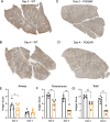
Attenuated antigen staining in PQQAR-infected hamsters. (A–D) Nucleocapsid antigen staining of left lung section from hamsters infected with 105 FFU of either (A and B) WT or (C and D) PQQAR mutant at 2 or 4 dpi. Antigen staining was scored in a blinded manner by location in the (E) airway, (F) parenchyma, and (G) total for WT (black) or PQQAR (orange) infected lungs. Statistical analysis was conducted using a two-tailed Student’s t test. *P ≤ 0.05; **P ≤ 0.01; and ***P ≤ 0.001.
Fig 4
Fig 4
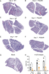
Reduced inflammation and damage in PQQAR-infected lungs. (A through G) Representative H&E staining of the left lung of hamsters infected with 105 pfu of either WT or PQQAR SARS-CoV-2 at (A and B) 2 days, (C and D) 4 days, or (E and F) 7 days post-infection or (G) mock. (H) WT (black), PQQAR (orange), or PBS (gray) lung sections from each day were scored for histopathological analysis, with sections from individual animals averaged and representing a single point. Statistical analysis was conducted using a two-tailed Student’s t test. *P ≤ 0.05; **P ≤ 0.01; ***P ≤ 0.001.
Fig 5
Fig 5
Disruption of FCS alters spike processing and protease usage. (A) Schematic of SARS-CoV-2 virion sucrose cushion purification approach. (B) Lysates from sucrose cushion purified WT, PQQAR, and ΔFCS virions grown in Vero E6 were probed with α-Spike and α-Nucleocapsid (N) antibodies by Western blot. Full-length spike (FL) and S1/S2 cleavage product are indicated. (C) Quantification of densitometry of the proportion between FL (black) and S1/S2 (red) of the total spike shown (lower). (D) Schematic of SARS-CoV-2 entry and protease usage, including knockout of TMPRSS2-mediated entry. (E) Viral titer from Calu-3 TMPRSS2 knock-out cells infected with WT (black) or PQQAR (orange) SARS-CoV-2 at an MOI of 0.01 (n = 3). (F) Viral titer at 48 hpi from Calu3 WT (Fig. 1D) and Calu3 TMPRSS2-/- cells. Statistical analysis was conducted using a two-tailed Student’s t test. *P ≤ 0.05; **P ≤ 0.01; and ***P ≤ 0.001. Entry schematic was made in BioRender.
Fig 6
Fig 6
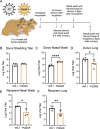
The FCS is not required for SARS-CoV-2 transmission. (A) Schematic of transmission experiment in golden Syrian hamsters. Three- to four-week-old male donor hamsters were intranasally infected with 105 pfu of WT or PQQAR SARS-CoV-2 and individually housed. Donors were subsequently paired 1:1 with recipients 24 hpi and cohoused for 8 h before separating and nasal washing donors. (B through F) Nasal washes and lungs were collected at 2 days post-infection for donors (dpi) (B through D) and post-contact for recipients (E and F). Viral titers were measured using focus-forming assays for donor and recipient samples. Statistical analysis was conducted using a two-tailed Student’s t test. *P ≤ 0.05; **P ≤ 0.01; and ***P ≤ 0.001. Experimental schematic was made in BioRender.
Fig 7
Fig 7
The furin cleavage site impacts SARS-CoV-2 transmission efficiency. (A) Schematic of transmission competition experiment in golden Syrian hamsters. Three- to four-week-old male donor hamsters were intranasally infected with 105 pfu of WT:PQQAR SARS-CoV-2 in a 1:1 ratio and were individually housed. (B through F) After 24 hpi, donors were paired with recipients and cohoused for 12 h before separating and nasal washing on donors. Nasal washes and lungs were collected at 2 and 4 days post-infection for donors (dpi) and post-contact for recipients (dpc). Next-generation sequencing (NGS) was performed on extracted RNA to measure the percentage of WT (gray) and PQQAR (orange) present in the nasal wash and lung of donors (B through D) and recipients (E and F). The expected distribution (B–F, top bar) based on NGS percentage mutant/WT observed in the inoculating dose (two inoculum preparations with RNA sequenced twice from each) if no differences between WT and mutant virus were observed. (G) Level of PQQAR mutation detection in recipient animals (orange) versus background mutations (black). Statistical analysis was conducted using a two-tailed Student’s t test. *P ≤ 0.05; **P ≤ 0.01; and ***P ≤ 0.001. Experimental schematic was made in BioRender.

Update of

Similar articles

References

    1. Gralinski LE, Menachery VD. 2020. Return of the Coronavirus: 2019-nCoV. Viruses 12:135. doi: 10.3390/v12020135 - DOI - PMC - PubMed
    1. Amanat F, Krammer F. 2020. SARS-CoV-2 vaccines: status report. Immunity 52:583–589. doi: 10.1016/j.immuni.2020.03.007 - DOI - PMC - PubMed
    1. Plante JA, Mitchell BM, Plante KS, Debbink K, Weaver SC, Menachery VD. 2021. The variant gambit: COVID-19’s next move. Cell Host Microbe 29:508–515. doi: 10.1016/j.chom.2021.02.020 - DOI - PMC - PubMed
    1. DeGrace MM, Ghedin E, Frieman MB, Krammer F, Grifoni A, Alisoltani A, Alter G, Amara RR, Baric RS, Barouch DH, et al. 2022. Defining the risk of SARS-CoV-2 variants on immune protection. Nature 605:640–652. doi: 10.1038/s41586-022-04690-5 - DOI - PMC - PubMed
    1. Tomalka JA, Suthar MS, Deeks SG, Sekaly RP. 2022. Fighting the SARS-CoV-2 pandemic requires a global approach to understanding the heterogeneity of vaccine responses. Nat Immunol 23:360–370. doi: 10.1038/s41590-022-01130-4 - DOI - PubMed

MeSH terms

LinkOut - more resources