. 2025 Jul 9;16(7):e0081025.
doi: 10.1128/mbio.00810-25. Epub 2025 Jun 10.

Toxoplasma gondii parasites induce a localized myeloid cell immune response surrounding parasites in the brain during acute infection

Affiliations

Toxoplasma gondii parasites induce a localized myeloid cell immune response surrounding parasites in the brain during acute infection

Stephanie B Orchanian et al. mBio. .

Abstract

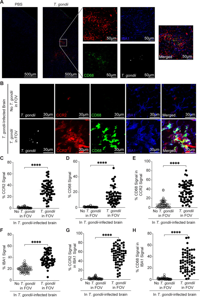
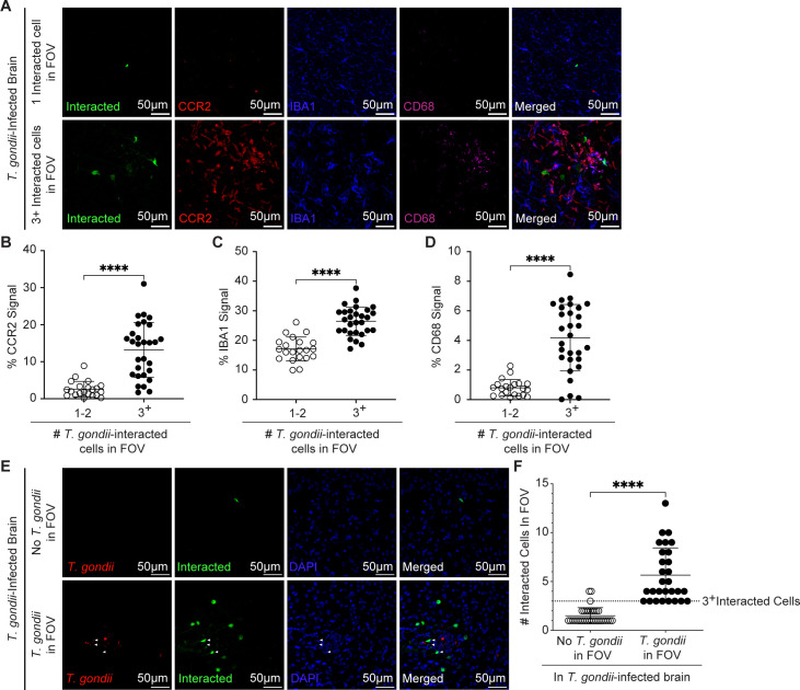
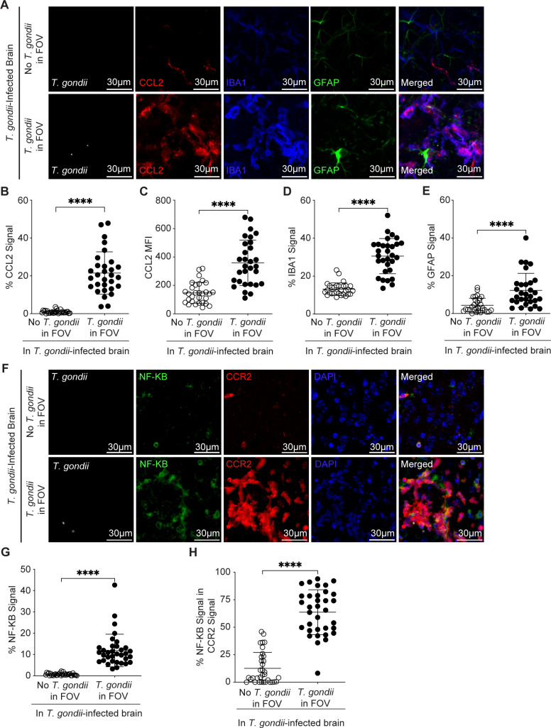
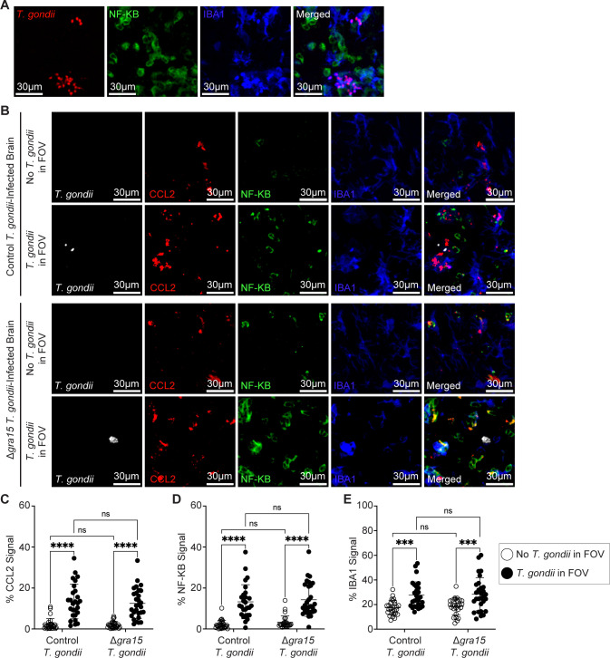
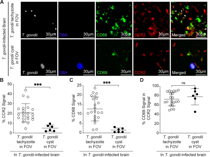
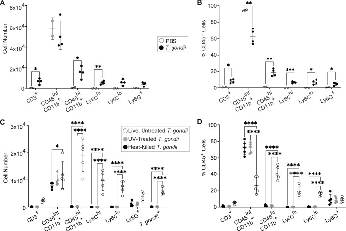
Toxoplasma gondii is a foodborne intracellular parasite that chronically infects the brain. Although T. gondii infection elicits a protective immune response, the nature of the monocyte response in the immediate vicinity of parasites during initial brain infection is not well understood. By infecting mice with T. gondii and comparing areas of the brain containing or not containing parasites, we found an increase in CCR2+ monocytes, IBA1+ myeloid cells, GFAP+ astrocytes, and CD68+ signal near parasites in the brain, indicating immune cell infiltration and phagolysosomal activation in response to the infection. CCR2+CD68+ monocytes were specifically increased near tachyzoites with minimal localization of these cells near cysts in the brain. This monocyte response was also detected near parasite-interacted cells, identified using T. gondii parasites that inject Cre recombinase into "interacted" cells of Ai6 CCR2RFP/+ mice, enabling us to track these events in vivo. The chemokine CCL2 and its transcription factor NF-κB were also upregulated surrounding parasites in the brain; however, the T. gondii effector protein GRA15, which sustains NF-κB activation in infected cells, was not required for CCL2 production, NF-κB activation, or myeloid cell recruitment to parasites in the brain. In contrast, active T. gondii replication played a more significant role, as CCR2+ monocytes were recruited to replication-competent but not replication-deficient T. gondii delivered via intracranial injection into mice. These findings provide novel insights into the drivers of immune cell mobilization and activation in the brain during initial T. gondii central nervous system infection and highlight the importance of parasite replication in this process.

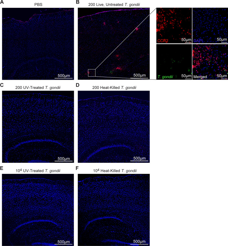
Importance: Toxoplasma gondii is a brain-infecting parasite, and the mobilization of peripheral immune cells to the brain is critical for controlling T. gondii infection. However, the initial events driving these cells to sites of T. gondii infection in the brain are poorly understood. We show that peripheral myeloid immune cell recruitment and activation are specific to areas of the brain containing actively replicating parasites, which are capable of lysing host cells. This local immune response is characterized by focal chemokine production, myeloid cell recruitment, and the activation of phagolysosomal pathways. These highly localized host responses were independent of the parasite effector protein that induces NF-κB activation within infected cells, GRA15. However, the localized monocyte recruitment was dependent on live parasites and active parasite replication. This research highlights the importance of host cell sensing of parasite replication in the brain for immune control of T. gondii infection.

Keywords: Toxoplasma gondii; brain; immune response; monocyte; myeloid cells; neuroinflammation.


Conflict of interest statement

The authors declare no conflict of interest.

Figures

Fig 1
Fig 1
T. gondii infection induces CCR2+ monocyte recruitment and activation surrounding parasites in the brain. CCR2RFP/+ mice were i.p. injected with PBS or type II T. gondii, and brains were harvested and examined at 15 DPI. (A) Representative tile scan of T. gondii (white), CD68+ cells (green), CCR2+ cells (red), and IBA1+ cells (blue) in brains of infected or PBS-injected control mice. Magnified inset shows a FOV with T. gondii parasites. (B) Representative confocal microscopy of T. gondii (white), CD68+ cells (green), CCR2+ cells (red), and IBA1+ cells (blue) in brains of infected mice in FOVs containing T. gondii (bottom row) or >1,500 µm away from T. gondii (top row). Quantification of the percent area per FOV of CCR2+ signal (C), CD68+ signal (D), CD68+ signal within CCR2+ area (E), IBA1+ signal (F), CCR2+ signal within IBA1+ signal (G), or CD68+ signal within IBA1+ signal (H) in FOVs containing parasites or >1,500 µm away from T. gondii in infected brains. In panels C through H, n = 55–62 FOVs within the cerebrum, midbrain, and interbrain from 13 mice per group from three independent experiments. Statistical significance was determined by Student’s t-test in panel F or Mann-Whitney U test in panels C, D, E, G, and H. ****P < 0.0001.
Fig 2
Fig 2
T. gondii-interacted cells are detected near tachyzoites and activated myeloid cells. Ai6 heterozygous CCR2RFP/+ mice were infected with type II toxofilin-Cre T. gondii, and brains were harvested and examined at 15 DPI. (A) Representative confocal microscopy of T. gondii-interacted cells (green), CCR2+ cells (red), IBA1+ cells (blue), and CD68+ cells (magenta) in brains of infected mice in FOVs containing one T. gondii-interacted cell (top row) or three or more (3+) T. gondii-interacted cells (bottom row) in infected brains. Quantification of the percent area per FOV of CCR2+ signal (B), IBA1+ signal (C), or CD68+ signal (D) in FOVs containing parasites or >1,500 µm away from T. gondii in infected brains. (E) Representative confocal microscopy of T. gondii (red), T. gondii-interacted cells (green), and DAPI (blue) in brains of infected mice in FOVs containing parasites (bottom row) or >1,500 µm away from T. gondii (top row) in infected brains. (F) Quantification of total interacted cell number in FOVs containing parasites or >1,500 µm away from T. gondii in infected brains. In panels B through D n = 21–29 FOVs within the cerebrum, midbrain, and interbrain from 9 to 10 mice per group from two to three independent experiments. In panel F n = 29–34 FOVs within the cerebrum, midbrain, and interbrain from 9 mice per group from three independent experiments. Statistical significance was determined by Student’s t-test in panel C or Mann-Whitney U test in panels B, D, and F. ****P < 0.0001.
Fig 3
Fig 3
T. gondii infection induces NF-κB and CCL2 production near parasites. CCL2-RFP mice were infected with type II T. gondii, and brains were harvested and examined at 15 DPI. (A) Representative confocal microscopy of T. gondii (white), GFAP+ cells (green), CCL2+ cells (red), and IBA1+ cells (blue) in brains of infected mice in FOVs containing T. gondii (bottom row) or >1,500 µm away from T. gondii (top row). Quantification of the percent area of CCL2+ signal per FOV (B), MFI of CCL2+ signal (C), percent area of IBA1+ signal per FOV (D), and percent area of GFAP+ signal per FOV (E) in FOVs containing parasites or >1,500 µm away from T. gondii in infected brains. (F) Representative confocal microscopy of T. gondii (red), NF-κB+ cells (green), and DAPI (blue) in brains of infected mice in FOVs containing T. gondii (bottom row) or >1,500 µm away from T. gondii (top row). Quantification of the percent area of NF-κB+ signal per FOV (G) or the percent area of NF-κB+ signal within CCR2+ signal (H) in FOVs containing parasites or >1,500 µm away from T. gondii in infected brains. In panels B through E, n = 30 FOVs within the cerebrum, midbrain, and interbrain from seven mice per group from three independent experiments. In panels G and H, n = 34 FOVs within the cerebrum, midbrain, and interbrain from 11 mice per group from three independent experiments. Statistical significance was determined by Student’s t-test in panel C or Mann-Whitney U test in panels B, D, E, G, and H. ****P < 0.0001.
Fig 4
Fig 4
CCL2, NF-κB, and IBA1 signals are increased near T. gondii in the brain in a GRA15-independent manner. CCL2-RFP mice were infected with parental type II T. gondii or type II GRA15 knockout (∆gra15) T. gondii, and brains were harvested and examined at 15 DPI. (A) Representative confocal microscopy of T. gondii (red), NF-κB+ cells (green), and IBA1+ cells (blue) in brains of mice infected with parental type II T. gondii. (B) Representative confocal microscopy of T. gondii (white), CCL2+ cells (red), NF-κB+ cells (green), and IBA1+ cells (blue) in brains of mice infected with type II T. gondii (top two rows) or ∆gra15 T. gondii (bottom two rows) in FOVs containing T. gondii (bottom row) or >1,500 µm away from T. gondii (top row). Quantification of the percent area per FOV of CCL2+ signal (C), NF-κB+ signal (D), and IBA1+ signal (E) in FOVs containing parasites or >1,500 µm away from T. gondii in infected brains. In panels C through E, n = 29–31 FOVs within the cerebrum, midbrain, and interbrain from six to eight mice per group from three to five independent experiments. Statistical significance was determined by a two-way analysis of variance. ***P < 0.001, ****P < 0.0001.
Fig 5
Fig 5
T. gondii tachyzoites, but not tissue cysts, induce activated CCR2+ monocytes surrounding parasites in the brain. CCR2RFP/+ mice were i.p. injected with type II T. gondii, and brains were harvested and examined at 28 DPI. (A) Representative confocal microscopy of T. gondii (white), Dolichos biflorus agglutinin (DBA; blue), CD68+ cells (green), and CCR2+ cells (red) in brains of infected mice in FOVs containing T. gondii tachyzoites (top row) or a cyst (bottom row). Quantification of the percent area per FOV of CCR2+ signal (C), CD68+ signal (D), or CD68+ signal within CCR2+ area. In panels B through D, n = 6–22 FOVs within the cerebrum, midbrain, and interbrain from 5 mice from three independent experiments. Statistical significance was determined by Student’s t-test. ***P < 0.001.
Fig 6
Fig 6
Intracranial infection with live, replicating T. gondii induces CCR2+ monocyte recruitment to the site of injection. CCR2RFP/+ mice were intracranially injected in the V1 region of the cortex with T. gondii tachyzoites or PBS as a control, and brains were harvested and examined at 7 DPI. Representative confocal microscopy of T. gondii (green), CCR2+ cells (red), DAPI (blue) at the injection site in mice treated with PBS (A) or 200 live, untreated (replication-competent) T. gondii parasites (B), 200 UV-treated T. gondii parasites (C), 200 heat-killed T. gondii parasites (D), 104 UV-treated T. gondii parasites (E), or 104 heat-killed T. gondii parasites (F). Magnified inset shows a FOV from a brain injected with live, untreated T. gondii parasites.
Fig 7
Fig 7
Monocytes and T cells are mobilized to the brain after intracranial infection with live, replicating T. gondii. (A and B) CCR2RFP/+ mice were intracranially injected in the V1 region of the cortex with 200 T. gondii tachyzoites or PBS as a control, and brain immune cell numbers (A) and frequencies of CD45+ cells (B) were quantified by flow cytometry 7 days later. (C and D) WT mice were intracranially injected with 200 heat-killed, UV-treated, or live, untreated T. gondii parasites, and brain immune cell numbers (C) and frequencies of CD45+ cells (D) were quantified by flow cytometry 7 days later. In panels A and B, n = 2–4 mice per group. Statistical significance was determined by Student’s t-test. In panels C and D, n = 5 mice per group. Statistical significance was determined by two-way analysis of variance. *P < 0.05, **P < 0.01, ***P < 0.001, ****P < 0.0001.

Similar articles

References

    1. Kadry H, Noorani B, Cucullo L. 2020. A blood-brain barrier overview on structure, function, impairment, and biomarkers of integrity. Fluids Barriers CNS 17:69. doi: 10.1186/s12987-020-00230-3 - DOI - PMC - PubMed
    1. Kim KS. 2008. Mechanisms of microbial traversal of the blood-brain barrier. Nat Rev Microbiol 6:625–634. doi: 10.1038/nrmicro1952 - DOI - PMC - PubMed
    1. Blanchard N, Dunay IR, Schlüter D. 2015. Persistence of Toxoplasma gondii in the central nervous system: a fine-tuned balance between the parasite, the brain and the immune system. Parasite Immunol 37:150–158. doi: 10.1111/pim.12173 - DOI - PubMed
    1. Webster JP. 2010. Dubey, J.P. toxoplasmosis of animals and humans. Parasites Vectors 3:112. doi: 10.1186/1756-3305-3-112 - DOI
    1. Pappas G, Roussos N, Falagas ME. 2009. Toxoplasmosis snapshots: global status of Toxoplasma gondii seroprevalence and implications for pregnancy and congenital toxoplasmosis. Int J Parasitol 39:1385–1394. doi: 10.1016/j.ijpara.2009.04.003 - DOI - PubMed

LinkOut - more resources