. 2025 Jul 22;99(7):e0053725.
doi: 10.1128/jvi.00537-25. Epub 2025 Jun 10.

Immunogenicity of NSDV GP38 and the role of furin in GP38 proteolytic processing

Affiliations

Immunogenicity of NSDV GP38 and the role of furin in GP38 proteolytic processing

Caroline Bost et al. J Virol. .

Abstract

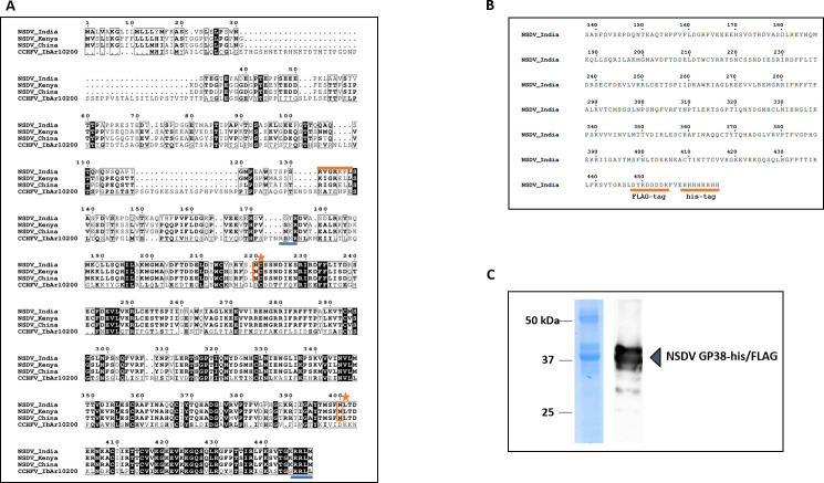
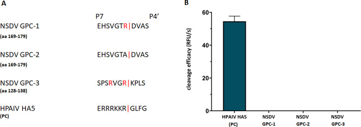
Nairobi sheep disease virus (NSDV) is a tick-borne orthonairovirus, which is genetically related to Crimean-Congo hemorrhagic fever virus (CCHFV), and causes severe hemorrhagic gastroenteritis in infected sheep. CCHFV GP38, a cleavage product of the CCHFV glycoprotein precursor (GPC), has recently attracted attention: not only has GP38 been reported to elicit detectable anti-GP38 antibodies in CCHFV-infected patients, but anti-GP38 antibodies have also been shown to protect mice from lethal CCHFV challenge. While proteolytic cleavage of CCHFV GP38 has been described to involve the proprotein convertases furin and subtilisin/kexin-isozyme-1 (SKI-1), little is known about the processing of NSDV GPC, or the occurrence and immunogenicity of NSDV GP38 in infected sheep. Here, we provide the first evidence for the presence and immunogenicity of NSDV GP38 in infected sheep demonstrating seroconversion by the detection of anti-GP38 antibodies over the course of infection. To further characterize GPC processing in vitro, we investigated the impact of furin overexpression and the effect of a furin inhibitor on NSDV glycoprotein expression, cleavage, and viral infectivity. While virus infectivity remained unaffected, our results suggest that other proteases besides furin may play a role in the proteolytic processing of NSDV GPC at a cleavage site that remains to be explored. Taken together, our findings highlight the immunogenicity of NSDV GP38 in sheep and warrant further research into the similarities and differences in proteolytic cleavage between the glycoproteins of NSDV and other orthonairoviruses, such as CCHFV.

Importance: Nairobi sheep disease virus (NSDV) is a zoonotic orthonairovirus causing severe and often fatal hemorrhagic gastroenteritis in small ruminants. Its genetic relationship to human-pathogenic Crimean-Congo hemorrhagic fever virus (CCHFV) and striking similarities in the clinical picture between CCHFV-infected patients and NSDV-infected ruminants have led to the idea that NSDV could serve as a model organism to study CCHFV pathogenesis. However, knowledge on NSDV-host interactions has been limited. While CCHFV GP38 has recently attracted attention as vaccine candidate and possible virulence factor, the occurrence and role of putative GP38 in other orthonairoviruses has been unclear. This study provides first evidence for the presence and immunogenicity of NSDV GP38 in infected sheep. Furthermore, our data indicate that other proteases besides furin may be involved in the proteolytic cleavage of NSDV GPC. Future studies are needed to determine the proteases involved and to investigate the possible functional role of GP38 in NSDV pathogenesis.

Keywords: CCHFV; GP38; NSDV; furin; orthonairovirus; serology.


Conflict of interest statement

The authors declare no conflict of interest.

Figures

Fig 1
Fig 1
Production and characterization of recombinant NSDV GP38-his/FLAG. (A) Alignment of partial GPC sequences from different NSDV strains (Ganjam virus IG 619 = NSDV_India; NSDV 708 = NSDV_Kenya; NSDV H. longicornis China = NSDV_China) and CCHFV (IbAr10200). Numbering corresponds to NSDV_India GPC. Amino acid sequences are displayed starting from their respective N-terminus. For CCHFV, the furin protease cleavage site (RSKR) and SKI-1/S1P cleavage site (RRLL) are underlined in blue. For NSDV, arginine-containing motifs reported to be conserved across the GPCs of different orthonairoviruses are highlighted in orange. Two potential N-glycosylation sites (Asn220 and Asn400) are highlighted with an orange star. (B) For protein purification, the partial NSDV_India GPC sequence (aa 138–445; NSDV GP38-his/FLAG protein) was fused to a C-terminal 6×His- and FLAG-tag both highlighted with an orange underline. (C) SDS-PAGE of recombinant NSDV GP38-his/FLAG his-tag purified from Sf9 cells followed by Coomassie blue staining and immunoblot analysis using anti-FLAG primary and horseradish peroxidase-conjugated secondary antibodies.
Fig 2
Fig 2
Seroconversion of NSDV- infected sheep against different NSDV antigens. (A) Sera from NSDV-infected sheep (n = 6) collected at different days post infection (dpi) were tested in duplicate in an indirect in-house ELISA based on recombinant NSDV GP38-his/FLAG. Mean of corrected OD values and standard deviations are displayed. (B) Serum samples collected from NSDV-infected sheep (n = 6) at 0 and 28 dpi were tested for reactivity in an indirect ELISA based on NSDV N-his/FLAG, commercially available recombinant Gn and Gc proteins, and GP38-his/FLAG.
Fig 3
Fig 3
Immunoblot analysis of NSDV protein expression in SW13 cells. (A) Immunoblot analysis of NSDV-infected and mock-infected SW13 cells collected at 28 h p.i. For detection, newly generated monoclonal antibody (mAb) 6F6 A3B raised against NSDV GP38-his/FLAG and goat anti-mouse IRDye 800-conjugated antibodies were used. The detection of GAPDH served as a loading control. (B) Cell lysates from multi-cycle infection kinetics were collected at the indicated time post infection (p.i.). For detection, mAb 6F6 A3B and mAb 5H11 C1 (raised against NSDV Gc) and rabbit-derived polyclonal serum (R8253) for nucleoprotein (N) detection were used as primary antibodies followed by incubation with goat anti-mouse or anti-rabbit IRDye 680 or 800CW-conjugated secondary antibodies, respectively. β-Tubulin and GAPDH served as loading controls. Representative blots from three independent experiments performed in duplicates are shown.
Fig 4
Fig 4
NSDV GP38 expression in the supernatant of transfected or NSDV-infected cells. HEK 293T and SW13 cells were transfected with pcDNA NSDV MLD-GP38-Strep (−) or co-transfected with pcDNA NSDV MLD-GP38-Strep and pIR-hfurin encoding for human furin protease (+). Supernatants were harvested at 72 h post-transfection and analyzed by immunoblot using mAb 6F6 A3B. For comparison of NSDV GPC cleavage products, supernatant from NSDV-infected SW13 cells (24 h p.i.) was also analyzed by immunoblot. A representative blot from at least three independent experiments is shown.
Fig 5
Fig 5
Efficiency of cleavage of FRET substrates by furin. (A) FRET substrates spanning the P7-P4′ amino acid sequences of the putative cleavage site motifs containing an N-terminal o-aminobenzoyl fluorophore and a C-terminal Tyr(3 NO2)-NH2 as a quenching residue were synthesized and tested in an enzyme assay with recombinant furin. Three NSDV GPC sequences were tested: GPC-1 covering the monobasic motif at position 175; GPC-2, a negative control for GPC-1 with the arginine of the motif replaced by alanine, and GPC-3 covering another putative dibasic furin motif for cleavage after residue 134. A FRET substrate with a multibasic furin cleavage site motif of hemagglutinin (HA5) of a highly pathogenic avian influenza virus (HPAIV) strain served as positive control (PC). (B) The cleavage of the FRET substrates by recombinant furin was measured in an enzyme kinetic assay. Mean values + SD based on three independent measurements with three independent weights of substrates are displayed.
Fig 6
Fig 6
Impact of furin inhibition on NSDV glycoprotein processing. SW13 cells were infected with NSDV (MOI of 0.1). After removal of inoculum, fresh medium with (+) or without (−) furin inhibitor MI-1148 (30 µM in DMSO) or DMSO alone was added. At 24 h p.i., cell lysates (A) and supernatants (B) were collected and analyzed for N, Gc, and GP38 expression in immunoblot. β-Tubulin and GAPDH served as loading controls. Representative blots from three independent experiments each performed in duplicate are shown. (C) The signal intensities of the GP38 bands were quantified using Li-Cor software Image Studio Lite Ver 5.2. The GP38 signal intensities from inhibitor-treated samples were set in relation to the signal intensities of the untreated samples (=100%). Mean relative signal intensities and standard deviations from three independent experiments each performed in duplicate are shown.
Fig 7
Fig 7
Impact of furin inhibition on NSDV and CCHFV infectivity. SW13 cells were infected with either NSDV or CCHFV at an MOI of 0.1. At 1 h p.i., inoculum was removed and fresh medium with (+) or without (−) MI-1148 was added in a concentration of 30 µM in DMSO for 24 h. DMSO-treated cells served as control. Virus titers were determined by plaque assay. Mean and standard deviation of virus titers are displayed from at least two independent experiments, performed in duplicate. Statistical analysis: unpaired t-test; (*) P ≤ 0.05; ns, not significant (P ≥ 0.05).

Similar articles

References

    1. Kuhn JH, Alkhovsky SV, Avšič-Županc T, Bergeron É, Burt F, Ergünay K, Garrison AR, Marklewitz M, Mirazimi A, Papa A, Pawęska JT, Spengler JR, Palacios G. 2024. ICTV Virus Taxonomy Profile: Nairoviridae 2024. J Gen Virol 105. doi: 10.1099/jgv.0.001974 - DOI - PMC - PubMed
    1. Gargili A, Estrada-Peña A, Spengler JR, Lukashev A, Nuttall PA, Bente DA. 2017. The role of ticks in the maintenance and transmission of Crimean-Congo hemorrhagic fever virus: a review of published field and laboratory studies. Antiviral Res 144:93–119. doi: 10.1016/j.antiviral.2017.05.010 - DOI - PMC - PubMed
    1. Krasteva S, Jara M, Frias-De-Diego A, Machado G. 2020. Nairobi sheep disease virus: a historical and epidemiological perspective. Front Vet Sci 7:419. doi: 10.3389/fvets.2020.00419 - DOI - PMC - PubMed
    1. Kuehnert PA, Stefan CP, Badger CV, Ricks KM. 2021. Crimean-Congo Hemorrhagic Fever Virus (CCHFV): a silent but widespread threat. Curr Trop Med Rep 8:141–147. doi: 10.1007/s40475-021-00235-4 - DOI - PMC - PubMed
    1. Bernard C, Holzmuller P, Bah MT, Bastien M, Combes B, Jori F, Grosbois V, Vial L. 2022. Systematic review on Crimean–Congo hemorrhagic fever enzootic cycle and factors favoring virus transmission: special focus on France, an apparently free-disease area in Europe. Front Vet Sci 9:932304. doi: 10.3389/fvets.2022.932304 - DOI - PMC - PubMed

LinkOut - more resources