. 2025 Jun 10;135(16):e187663.
doi: 10.1172/JCI187663. eCollection 2025 Aug 15.

The macrophage-intrinsic MDA5/IRF5 axis drives HIV-1 intron-containing RNA-induced inflammatory responses

Affiliations

The macrophage-intrinsic MDA5/IRF5 axis drives HIV-1 intron-containing RNA-induced inflammatory responses

Sita Ramaswamy et al. J Clin Invest. .

Abstract

Despite effective antiretroviral therapy, transcriptionally competent HIV-1 reservoirs remain and contribute to persistent immune activation in people living with HIV (PWH). HIV-1-infected macrophages are important mediators of chronic innate immune activation, though the mechanisms remain unclear. We previously reported that nuclear export and cytoplasmic expression of HIV-1 intron-containing RNA (icRNA) activates mitochondrial antiviral signaling (MAVS) protein-mediated type I IFN responses in macrophages. In this study, we demonstrate an essential role of melanoma differentiation-associated protein 5 (MDA5) in sensing HIV-1 icRNA and promoting MAVS-dependent interferon regulatory factor 5 (IRF5) activation in macrophages. Suppression of MDA5 but not retinoic acid-inducible gene I expression nor disruption of the endosomal TLR pathway abrogated HIV-1 icRNA-induced type I IFN responses and IP-10 expression in macrophages. Furthermore, induction of IP-10 in macrophages upon HIV-1 icRNA sensing by MDA5 was dependent on IRF5. Additionally, monocytes and monocyte-derived macrophages (MDMs) from older (>50 years) individuals exhibited constitutively higher levels of IRF5 expression compared with younger (<35 years) individuals, and HIV-1 icRNA-induced IP-10 expression was significantly enhanced in older macrophages, which was attenuated upon ablation of IRF5 expression, suggesting that IRF5 functions as a major mediator of proinflammatory response downstream of MDA5-dependent HIV-1 icRNA sensing, dysregulation of which might contribute to chronic inflammation in older PWH.

Keywords: AIDS/HIV; Aging; Inflammation; Innate immunity; Macrophages.


Conflict of interest statement

Conflict of interest: The authors have declared that no conflict of interest exists.

Figures

Figure 1
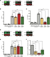
Figure 1. MAVS-mediated sensing of cytoplasmic icRNA triggers an innate immune response.
THP-1/PMA macrophages were infected with LaiΔenv GFP/G (MOI 2) in the presence or absence of (1 μM), raltegravir (RAL; 30 μM), spironolactone (Spiro;100 nM), and KPT335 (KPT; 1 μM). Cells and culture supernatants were harvested at 3 dpi for (A) flow cytometry analysis to measure infection levels (%GFP+) and (B) ELISA to measure IP-10 secretion. (C) Infection levels and (D) IP-10 secretion in THP-1/PMA macrophages infected with either WT (LaiΔenvGFP/G) or HIV-1/M10 were determined at 3 dpi by flow cytometry and ELISA, respectively. (E) MAVS expression in THP1 cells transduced with shCTRL or shMAVS lentivectors was determined by Western blot analysis. (F and G) LaiΔenvGFP/G-infected THP-1/PMA macrophages and cell supernatants were harvested at 3 dpi for (F) flow cytometry analysis and (G) ELISA to measure infection establishment (%GFP+) and IP-10 secretion. Data are displayed as the means ± SEM, with each dot representing an independent experiment. Statistical significance was assessed via 1-way ANOVA with Dunnett’s multiple-comparison test (A and B) or unpaired t test (C, D, F, and G). *P < 0.05, **P < 0.01, ***P < 0.001, and ****P < 0.0001.
Figure 2
Figure 2. MDA5 is required for HIV-1–induced innate immune response in macrophages.
(A) RIG-I, UNC93B1, and MDA5 expression in THP-1 cells transduced with RIG-I, UNC93B1, MDA5, or control shRNA-expressing lentivectors was quantified via RT-qPCR. (B) THP-1/PMA knockdown cell lines were infected with LaiΔenvGFP/G at MOI 2 and harvested at 3 dpi for infection establishment (%GFP+) via flow cytometry. (C) Supernatants from infected THP-1/PMA cells were used to assess IP-10 secretion via ELISA. (D) MDMs were transfected with siRNA targeting RIG-I, UNC93B1, and MDA5 for 2 days, and knockdown of RIG-I, UNC93B1, and MDA5 expression was assessed via RT-qPCR. MDMs were infected with LaiΔenvGFP/G at MOI 1 in the presence of dNs and harvested at 2 dpi for analysis of (E) infection efficiency via flow cytometry and (F) IP-10 mRNA expression via RT-qPCR. Data are displayed as the means ± SEM, with each dot representing an independent experiment (AC) or cells from an independent donor (DF). Statistical significance was assessed via unpaired t test (A and D) or 1-way ANOVA with Dunnett’s multiple comparisons (B, C, E, and F). *P < 0.05, ***P < 0.001, and ****P < 0.0001.
Figure 3
Figure 3. MDA5 recognizes unspliced HIV-1 RNA in the cytoplasm.
HEK293T cells were infected with LaiΔenvGFP/G at MOI 1 and transfected with either MDA5-Flag or RIG-I–Flag at 24 hours after infection. Cytoplasmic fractions were immunoprecipitated with either anti-Flag mAb or IgG-coated beads. (A) Input and (B) IP lysates were run on Western blot to ensure equivalent levels of transfection and immunoprecipitation among conditions. (C) RT-qPCR analysis for HIV-1 usRNA, HIV-1 msRNA, GAPDH, or actin mRNA in transfected and infected HEK293T cells in the presence or absence of EFV. Fold enrichment of immunoprecipitated RNA reported as RNA transcripts detected from each IP condition using anti-Flag mAb or control IgG compared with the input amount. Data are displayed as means ± SEM, with each dot representing a different experiment. Statistical significance was assessed via unpaired t tests (C). **P < 0.01.
Figure 4
Figure 4. IRF5 is necessary for HIV-1 icRNA-induced IP-10 expression in macrophages.
(A) IRF3, IRF5, and IRF7 expression in THP-1 cells transduced with IRF3, IRF5, IRF7, or control shRNA lentivectors was quantified via Western blot analysis and normalized to control shRNA transduced cells. (B and C) THP1/PMA macrophages were infected with LaiΔenvGFP/G at MOI 2, and cells and supernatants were harvested at 3 dpi for analysis via (B) flow cytometry to assess infection levels and (C) ELISA for IP-10 secretion. (D) Expression of IRFs in MDMs transfected with siRNA against IRF3, IRF5, or IRF7 mRNA was assessed via RT-qPCR. (E and F) MDMs were infected with LaiΔenvGFP/G at MOI 1 in the presence of dNs and harvested at 2 dpi for analysis of (E) infection efficiency via flow cytometry and (F) IP-10 expression via RT-qPCR. Data are displayed as the means ± SEM, with each dot representing a different donor. Statistical significance was assessed via 1-way ANOVA with Dunnett’s multiple comparisons (B, C, E, and F). *P < 0.05 and ***P < 0.001.
Figure 5
Figure 5. TRAF6 and IKK-β are required for HIV-1–induced IP-10 expression in macrophages.
(A) TRAF6 or IKK-β expression in THP-1 cells transduced with TRAF6 or IKK-β shRNA lentivectors was quantified via Western blot analysis and normalized to control shRNA transduced cells. THP1/PMA macrophages infected with LaiΔenvGFP/G at MOI 2 and harvested at 3 dpi for analysis via (B) flow cytometry to assess infection levels and (C) ELISA to assess IP-10 secretion. (D) MDMs were transfected with siRNA against TRAF6 or IKK-β mRNA for 2 days, and knockdown of TRAF6 or IKK-β was assessed via Western blot and RT-qPCR. (E and F) MDMs infected with LaiΔenvGFP/G at MOI 1 in the presence of dNs and harvested at 2 dpi for analysis of (E) infection efficiency via flow cytometry and (F) IP-10 expression by RT-qPCR. Data are displayed as the means ± SEM, with each dot representing a separate experiment (AC) with cells from independent donors (DF). Statistical significance was assessed via 1-way ANOVA with Dunnett’s multiple comparisons (B, C, E, and F). **P < 0.01, ***P < 0.001, and ****P < 0.0001.
Figure 6
Figure 6. MDA5, MAVS, TRAF6, and IKK-β are required for HIV-1 icRNA-induced nuclear localization of IRF5 in macrophages.
(A) THP-1/PMA macrophages infected with LaiΔenvGFP/G at MOI 2 and harvested at 3 dpi for analysis via confocal microscopy to assess changes in IRF5 localization. Representative images are shown. Scale bar: 5 mm. (BF) Fluorescence microscopy images were quantified via CellProfiler to assess mean pixel intensity of IRF5 staining that colocalized with DAPI (mean nuclear intensity). Images from 3 independent infection experiments were analyzed and quantified, with each dot representing a field containing approximately 50–150 cells. Statistical significance was assessed via Kruskal-Wallis test with Dunn’s multiple-comparison analysis (BG). ****P < 0.0001.
Figure 7
Figure 7. MDMs and monocytes isolated from older individuals exhibit higher levels of IRF5 expression.
(AF) Constitutive mRNA expression of (A) MDA5, (B) MAVS, (C) TRAF6, (D) IRF3, (E) IRF7, and (F) IRF5 in MDMs and monocytes was assessed via RT-qPCR. (G and H) Western blot analysis for constitutive IRF5 expression in MDMs and monocytes. Data are displayed as mean ± SEM, with each dot representing a donor. Statistical significance was assessed via unpaired t tests (AH). *P < 0.05.
Figure 8
Figure 8. MDMs isolated from older donors exhibit higher levels of HIV-1–induced immune activation.
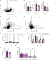
(A) RNA isolated from LaiΔenvGFP/G-infected MDMs (MOI 2) was analyzed via Nanostring nCounter using the Myeloid Innate Immunity V2 panel. Baseline expression of the gene panel was calculated using nSolver and plotted as a ratio of old (HIV) versus young (HIV) with IRFs (red) highlighted. The dashed line represents a P value of 0.05. (B) Raw count values for IRF3, IRF5, and IRF7 were plotted to assess differences. (C and D) Fold changes in innate immune gene expression in HIV-infected MDMs with or without EFV were plotted. Data from MDMs from (C) older (>50 yo) and (D) younger (<35 yo) donors are shown. Differentially expressed ISGs (blue) and IRFs/IRF targets (red) are highlighted. The dashed line represents a P value of 0.05. (E and F) Supernatants were analyzed for (E) p24gag production and (F) IP-10 levels via ELISA. (G) IP-10 levels were normalized to those of p24gag for each donor. (H) MDMs were transfected with either control or IRF5 targeting siRNA prior to infection with LaiΔenvGFP/G. IP-10 and p24gag secretion was measured at 3 dpi. Data are represented as mean ± SEM, with each dot representing an individual donor (B and EG) or with each dot representing a target gene (A, C, and D). Significance was assessed via unpaired 2-tailed t test (A, B, and EG), paired 2-tailed t test (C and D), or 1-way ANOVA with Tukey’s multiple-comparison test (H). *P < 0.05.

References

    1. Paiardini M, Müller-Trutwin M. HIV-associated chronic immune activation. Immunol Rev. 2013;254(1):78–101. doi: 10.1111/imr.12079. - DOI - PMC - PubMed
    1. Babu H, et al. Systemic inflammation and the increased risk of inflamm-aging and age-associated diseases in people living with HIV on long term suppressive antiretroviral therapy. Front Immunol. 2019;10:1965. doi: 10.3389/fimmu.2019.01965. - DOI - PMC - PubMed
    1. North CM, et al. Brief report: systemic inflammation, immune activation, and impaired lung function among people living with HIV in rural Uganda. J Acquir Immune Defic Syndr. 2018;78(5):543–548. doi: 10.1097/QAI.0000000000001711. - DOI - PMC - PubMed
    1. Lv T, et al. HIV-related immune activation and inflammation: current understanding and strategies. J Immunol Res. 2021;2021:7316456. doi: 10.1155/2021/7316456. - DOI - PMC - PubMed
    1. Coghill AE, et al. Advanced stage at diagnosis and elevated mortality among US patients with cancer infected with HIV in the National Cancer Data Base. Cancer. 2019;125(16):2868–2876. doi: 10.1002/cncr.32158. - DOI - PMC - PubMed

MeSH terms