. 2025 Jun 10;135(15):e186599.
doi: 10.1172/JCI186599. eCollection 2025 Aug 1.

Patterns of intra- and intertumor phenotypic heterogeneity in lethal prostate cancer

Affiliations

Patterns of intra- and intertumor phenotypic heterogeneity in lethal prostate cancer

Martine P Roudier et al. J Clin Invest. .

Abstract

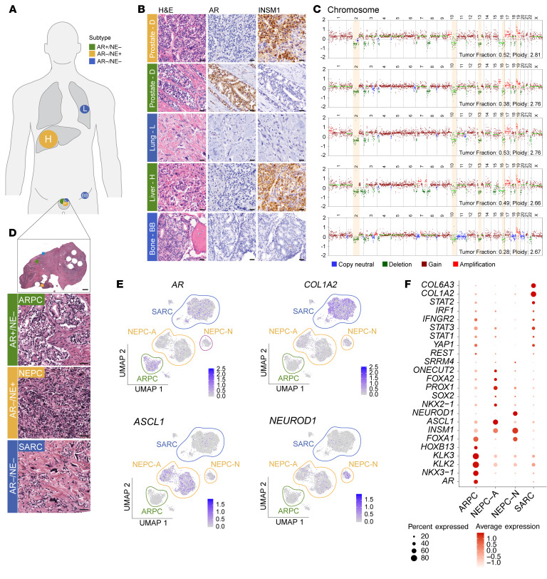
Metastatic prostate cancer (mPC) is a clinically and molecularly heterogeneous disease. While there is increasing recognition of diverse tumor phenotypes across patients, less is known about the molecular and phenotypic heterogeneity present within an individual. In this study, we aimed to define the patterns, extent, and consequences of inter- and intratumoral heterogeneity in lethal prostate cancer. By combining and integrating in situ tissue-based and sequencing approaches, we analyzed over 630 tumor samples from 52 patients with mPC. Our efforts revealed phenotypic heterogeneity at the patient, metastasis, and cellular levels. We observed that intrapatient intertumoral molecular subtype heterogeneity was common in mPC and showed associations with genomic and clinical features. Additionally, cellular proliferation rates varied within a given patient across molecular subtypes and anatomic sites. Single-cell sequencing studies revealed features of morphologically and molecularly divergent tumor cell populations within a single metastatic site. These data provide a deeper insight into the complex patterns of tumoral heterogeneity in mPC with implications for clinical management and the future development of diagnostic and therapeutic approaches.

Keywords: Cell biology; Molecular pathology; Oncology; Prostate cancer; Urology.


Conflict of interest statement

Conflict of interest: PC serves as a consultant for Cure51. SZ is a paid consultant through the network expert companies Guidepoint and GLG. JLZ is currently an employee at AstraZeneca. LDT is a cofounder and has equity in Alpenglow Biosciences. JKL has received research funding from Immunomedics and serves as a scientific advisor for and has equity in PromiCell Therapeutics. CLS serves on the board of directors of Novartis, is a cofounder of ORIC Pharmaceuticals, and is a coinventor of enzalutamide and apalutamide. He is a science advisor to Arsenal, Beigene, Blueprint, Column Group, Foghorn, Housey Pharma, Nextech, KSQ, and PMV Pharmaceuticals. CKCD has received research funding from Bristol Myers Squibb and Intuitive Surgical. HHC has received research funding to her institution from Clovis Oncology, Janssen, Medivation, Promontory Therapeutics, and Sanofi. EYY is a paid consultant and/or received honoraria from Advanced Accelerator Applications/Novartis, Aadi Bioscience, Bayer, Bristol Myers Squibb, Janssen, Lantheus, Loxo, Merck, and Oncternal and has received research funding to his institution from Bayer, Blue Earth, Dendreon, Lantheus, Merck, Oncternal, Seagen, and Tyra. JEH is a paid consultant and/or received honoraria from GSK, ImmunityBio, Daichi-Sankyo, and Seagen. She has received research funding to her institution from Janssen, Macrogenics, Crescendo Biologics, AstraZeneca, Bristol Myers Squibb, and Barinthus. RBM has received research funding to his institution from Janssen, Clovis, Bayer, and INmuneBio. DWL has received research funding to his institution from Veracyte and MDxHealth and is a paid consultant/advisor for Astellas, AstraZeneca, Janssen, and Lantheus. C. Morrissey has received funds from Genentech and Novartis. EC served as a paid consultant to DotQuant. MTS is a paid consultant and/or received honoraria from Sanofi, AstraZeneca, Janssen, Fibrogen, and Pfizer. He has received research funding to his institution from Novartis, Zenith Epigenetics, Eli Lilly, Bristol Myers Squibb, Merck, Immunomedics, Janssen, AstraZeneca, Pfizer, F. Hoffmann-La Roche, Tmunity, SignalOne Bio, Epigenetix, Xencor, Incyte, and Ambrx. PSN served as a paid advisor for Bristol Myers Squibb, Pfizer, Genentech, AstraZeneca, and Janssen. MCH served as a paid consultant/received honoraria from Pfizer and AstraZeneca and has received research funding from Merck, Novartis, Genentech, Promicell, and Bristol Myers Squibb.

Figures

Figure 1
Figure 1. Study cohort and associations between molecular subtype and organ sites.
(A) Summary of the study cohort. A total of 52 patients and 637 samples were analyzed. Ideogram depicts the distribution of samples across anatomic sites. Bar graph shows the percentage and number of patients with tumor involvement of the listed sites. (B) Distribution of organ site involvement across molecular subtypes. (C) Proportion of molecular subtypes across organ sites. (D) Micrographs of H&E and IHC stains representative of the 4 molecular subgroups. Note that a subset of AR–/NE+ tumors demonstrated robust INSM1 but no SYP expression (as shown here). Scale bars: 100 μm.
Figure 2
Figure 2. Patterns of subtype heterogeneity and association with genomic alterations.
(A) Summary of individual sample-level and integrated patient-level assessments of molecular subtypes and subtype heterogeneity, using patient 15-010 as an example. (B) Pie chart shows number and percentages of patients with homogenous and heterogeneous molecular subtype distribution. UpSet plot shows the patterns of molecular subtype co-occurrence across all metastatic sites in 52 patients. (C) HI distributions across different dominant molecular subtypes. P values were derived using the Wilcox-Mann-Whitney test. (D) Comutation plot depicting molecular subtype and genomic features of patients included in the study. Each column represents a patient (case IDs are listed at the bottom). Rows show from top to bottom: IHC-derived dominant molecular subtype (defined as the most commonly observed subtype across all metastases); IHC-derived subtype contribution for each of the four subtypes (AR+/NE–, AR–/NE–, AR+/NE+, and AR–/NE–) (heatmap scaled 0–1) shown as the relative fraction of tumor samples from each subtype in a patient; HI (shown as a heatmap scaled 0–100); whole-exome sequencing–derived key genomic alterations (AR, PTEN, RB1, CHD1, TP53, and BRCA2) shared across all metastases (see legend to the left for alteration type). Asterisks indicate no genomic data are available. CN, copy number. (E) Mosaic plots illustrating the relative distributions of molecular subtypes (along the x axis) and the associated relative distributions of genomic alterations (along the y axis) (see Supplemental Table 4 for details). Dark colors indicate the presence of a given genomic alteration, and light colors show absence. Plots are scaled to the total number of samples in each molecular subtype. (F) HIs for AR and PTEN altered and WT patients. P values were derived using the Wilcox-Mann-Whitney test. (G) Estimated median differences and 95% CIs of HIs between patients with (altered) and without (unaltered) indicated gene alterations (HIaltered – HIunaltered). Numbers in parentheses represent the number of patients, with the first number indicating altered cases.
Figure 3
Figure 3. Diversity of cell proliferation patterns across metastatic sites.
(A) Bar graphs showing the distribution of Ki-67 proliferation indices across different molecular subtypes. P values are derived from 2-sample Mann-Whitney rank-sum tests for Ki-67 levels from individual cores. (B) Comparison of Ki-67 heterogeneity indices (see Methods) for all patients and AR+/NE– tumors only. (C) Scatter plot showing the association between mean Ki-67 indices across all metastases in a given patient. (D) Ki-67 indices across different anatomic sites. Each metastasis is shown as a single dot and color coded according to the IHC-determined molecular subtype. (E) In box plots, horizontal bars indicate the medians and boxes indicate 25th to 75th percentiles and show Ki-67 indices as a function of genomic status of AR and tumor suppressor genes. P values were derived using the Wilcox-Mann-Whitney test.
Figure 4
Figure 4. Clinical features associated with molecular subtypes and intrapatient heterogeneity.
(A) Clinical trajectories for 52 patients included in this study. Bars showing the time from initial diagnosis to death for each patient and are color coded in light blue to indicate the interval from diagnosis to start of androgen deprivation therapy (ADT) and gray for the period after ADT initiation. M indicates the time of first bone metastasis; R indicates first clinical evidence of resistance to ADT. The asterisk indicates that this patient did not receive ADT. Patients are sorted based on dominant molecular subtype (green, AR+/NE–; yellow, AR–/NE+; red, AR+/NE+; and blue, AR–/NE–) and the HI (gray scale heatmap). (B) Summary of prior therapies. Stacked bar graphs showing the number of patients that have (dark color) or have not (light color) received the indicated systemic therapies (abiraterone acetate, enzalutamide, and taxane- and platinum-based chemotherapies) as a function of the dominant molecular subtype. (C) Last recorded PSA serum levels for each patient broken down by dominant molecular subtype. (D) Box plots showing time intervals from first bone metastasis to death for patients with no NE marker positivity compared with patients with any NE marker positivity (INSM1 or SYP H-score ≥ 20). P value was derived using the Wilcox-Mann-Whitney test. (E) Box plot showing time from bone metastasis to death across all 52 patients stratified by dominant molecular subtype. P value was derived using the Kruskal-Wallis test. (F) Time interval from time from bone metastasis to death in patients with a HI of greater than 50% or below. Note that corresponding analyses for other time intervals, diagnosis to death, and ADT to death are shown in Supplemental Figure 5D. In box plots, horizontal bars indicate the medians and boxes indicate 25th to 75th percentiles.
Figure 5
Figure 5. Dissecting molecular subtype pattern at the single-cell level.
UMAP and bar graphs depicting molecular subtype composition based on reanalysis of snRNA-seq data of patients with mPC (40). (A) Example of a homogeneous AR+/NE– tumor (MSK−HP13). (B) A more heterogenous AR+/NE– tumor with admixed AR–/NE– cell populations (MSK−HP03). (C and D) UMAPs from additional mPCs can be found in Supplemental Figure 8. IHC micrographs of AR (AR, NKX3.1) and NE markers (SYP, INSM1, ASCL1, and SOX2) representative of (C) an amphicrine carcinoma (10-056) and (D) a mixed/biphenotypic tumor. Note the absence of NE transcription factor expression in the amphicrine carcinoma. Scale bars: 50 mm. (E) Schematic showing different cell states in AR+/NE+ tumors. Note that despite widespread positivity for AR and NKX3.1, there are distinct cell populations that are negative for these AR markers but positive for SYP, INSM1, ASCL1, and SOX2 (arrows). (F) Coimmunolabeling of mixed/biphasic tumor (07-042) highlights distinct AR–/INSM1+ (arrows) and AR+/INSM1+ (arrowheads) cell populations that show adenocarcinoma and small-cell carcinoma morphology, respectively. Scale bars: 50 μm. (G and H) Integrated snRNA-seq and snATAC-seq UMAPs showing cell clusters with differential expression of AR and NE markers. Note low-level AR expression but high NE marker expression in cluster 3. (I) Pseudo–time analysis using Palantir and (J) cell state densities assessment using Mellon showing cell differentiation trajectories across the 3 clusters. (K) Bubble plot highlighting differential single-gene expression across the 3 clusters.
Figure 6
Figure 6. Inter- and intratumoral heterogeneity at the single-patient level.
(A) Schematic of analyzed metastatic sites and phenotype distribution in patient 13-084. (B) Representative micrographs of 5 lesions demonstrating the spectrum of histomorphological and molecular heterogeneity across different tumor sites. (C) Somatic copy number profiles derived from WGS demonstrating overlapping copy number changes and limited genomic diversity across phenotypically diverse metastases. Shared copy number changes in key genomic regions are highlighted in yellow. (D) Histomorphologic assessment of a prostatic/periprostatic tumor mass shows adjacent AR+/NE– adenocarcinoma (ARPC), AR–/NE+ small-cell carcinoma (NEPC), and AR–/NE– sarcomatoid carcinoma (SARC). (E) UMAP based on snATAC-seq data demonstrating 4 tumor cell clusters based on chromatin accessibility pattern and highlighting 2 distinct NEPC clusters that are characterized by ASCL1 (NEPC-A) and NEUROD1 (NEPC-N) expression (both AR–/NE+). (F) Bubble plots showing cluster-specific gene expression pattern based on snRNA-seq. Scale bars: 50 μm.

Comment in

  • Uncovering phenotypic heterogeneity via research autopsy in lethal prostate cancer

References

    1. Marusyk A, et al. Intra-tumour heterogeneity: a looking glass for cancer? Nat Rev Cancer. 2012;12(5):323–334. doi: 10.1038/nrc3261. - DOI - PubMed
    1. Gerlinger M, et al. Intratumour heterogeneity in urologic cancers: from molecular evidence to clinical implications. Eur Urol. 2015;67(4):729–737. doi: 10.1016/j.eururo.2014.04.014. - DOI - PubMed
    1. Turajlic S, et al. Deterministic evolutionary trajectories influence primary tumor growth: TRACERx renal. Cell. 2018;173(3):595–610. doi: 10.1016/j.cell.2018.03.043. - DOI - PMC - PubMed
    1. Turajlic S, Swanton C. Metastasis as an evolutionary process. Science. 2016;352(6282):169–175. doi: 10.1126/science.aaf2784. - DOI - PubMed
    1. Frankell AM, et al. The evolution of lung cancer and impact of subclonal selection in TRACERx. Nature. 2023;616(7957):525–533. doi: 10.1038/s41586-023-05783-5. - DOI - PMC - PubMed