Review
. 2025 Jun 9;35(11):R545-R562.
doi: 10.1016/j.cub.2025.03.053.

Cellular anatomy of arbuscular mycorrhizal fungi

Affiliations
Review

Cellular anatomy of arbuscular mycorrhizal fungi

Rachael I M Cargill et al. Curr Biol. .

Abstract

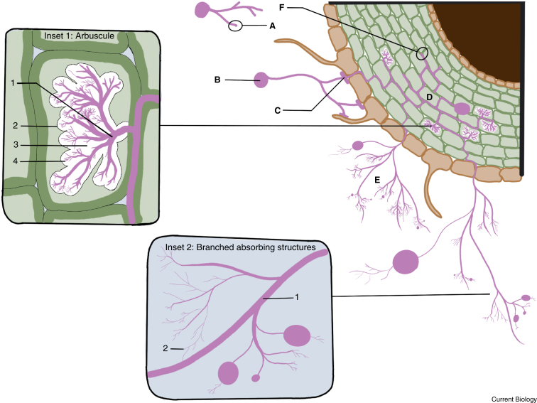
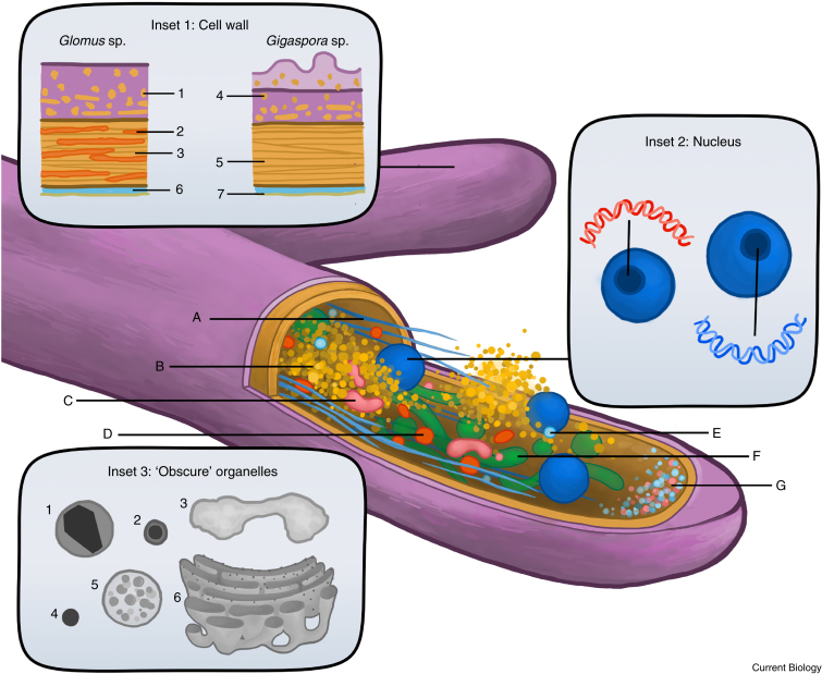
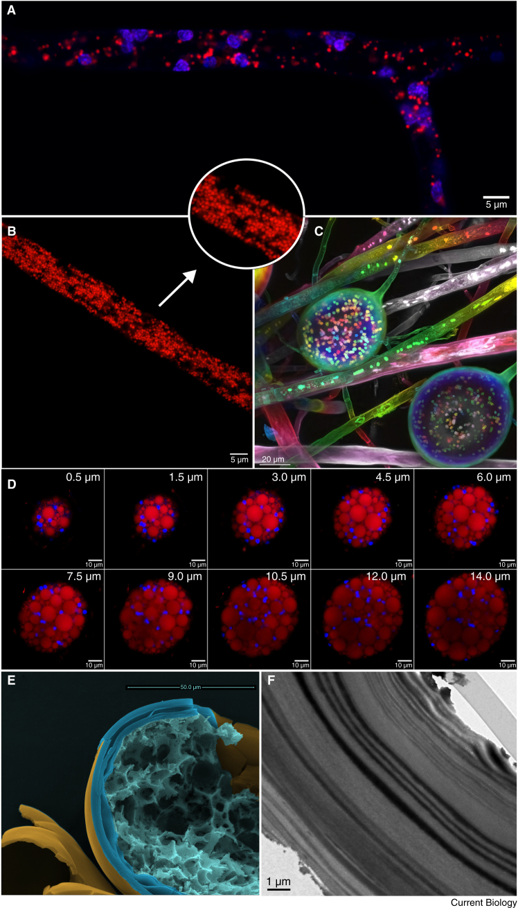
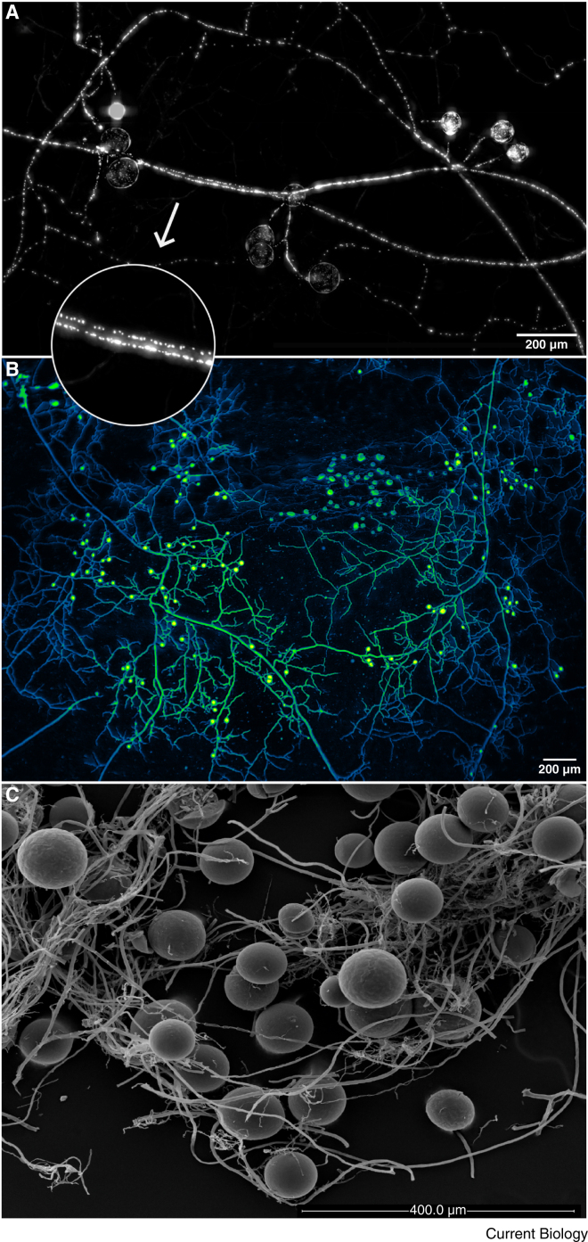
Arbuscular mycorrhizal (AM) fungi are ancient plant mutualists that are ubiquitous across terrestrial ecosystems. These fungi are unique among most eukaryotes because they form multinucleate, open-pipe mycelial networks, where nutrients, organelles, and chemical signals move bidirectionally across a continuous cytoplasm. AM fungi play a crucial role in ecosystem functioning by supporting plant growth, mediating ecosystem diversity, and contributing to carbon cycling. It is estimated that plant communities allocate ∼3.93 Gt CO2e to AM fungi every year, much of which is stored as lipids inside the fungal network. Despite their ecological significance, the cellular biology of AM fungi remains underexplored. Here, we synthesise the current knowledge on AM fungal cellular structure and organisation. We examine AM fungal development at different biological levels - the hypha and its content, hyphal networks and AM fungal spores - and explore key cellular dynamics. This includes cell wall composition, cytoplasmic contents, nuclear and lipid organisation and dynamics, network architecture, and connectivity. We highlight how their unique cellular arrangement enables complex cytoplasmic flow and nutrient exchange processes across their open-pipe mycelial networks. We discuss how both established and novel techniques, including microscopy, culturing, and high-throughput image analysis, are helping to resolve previously unknown aspects of AM fungal biology. By comparing these insights with established knowledge in other, well-studied filamentous fungi, we identify critical knowledge gaps and propose questions for future research to further our understanding of fundamental AM fungal cell biology and its contributions to ecosystem health.


Conflict of interest statement

Declaration of interests The authors declare no competing interests.

Figures

Figure 1
Figure 1
Morphological variation of the arbuscular mycorrhizal (AM) fungal hyphae. Despite being a continuous single cell, AM fungal hyphae demonstrate remarkable morphological and functional diversity. Germ tubes (A) emerge from spores (B) and play a crucial role in initiating fungal growth, soil exploration and host colonisation. Hyphopodia are specialised, swollen structures that develop where fungal hyphae encounter the root epidermis, functioning as adhesion and entry points to plant roots (C), specialised structures formed at the root surface, facilitate host root penetration. Part of the intraradical hyphal network (D), formed within the cortical root cells, arbuscules (Inset 1) develop as highly branched, intracellular structures optimised for nutrient exchange. The arbuscule trunk (Inset 1.1) forms the base of the arbuscule and is enveloped by the periarbuscular membrane (PAM; Inset 1.2), an expansion of the plant plasma membrane. The arbuscular trunk shares protein markers with the plasma membrane of the plant cell. The periarbuscular space (PAS; Inset 1.3) comprises an apoplastic interface between the PAM and the fungal hyphae and facilitates nutrient and signal exchange. The branch domain of the PAM envelops the fine hyphal branches (Inset 1.4) and contains specific phosphate transport proteins. In the soil, branched absorbing structures (BAS; Inset 2) form sub-apically in the extraradical mycelium and are linked to either nutrient absorption, particularly phosphorus, or to spore formation (spore-BAS). Runner hyphae (Inset 2.1), which constitute the skeleton of the extraradical mycelium (ERM) (E), grow out into the soil in search of new hosts and act as conduits for nutrient translocation,. When considering the hyphal growth dynamics, hyphal tips can be found simultaneously intraradically in apical colonisation expansion zones (F) and in thin arbuscule branches (Inset 1.4), and extraradically in the BAS (Inset 2.2) and at the advancing front of the ERM (E). (Image courtesy of Sean C. Maston.)
Figure 2
Figure 2
Illustration of characterised and obscure cellular components in AM fungal hyphae. Schematic illustrations based on published literature showing organelle arrangement within AM fungal hyphae using transmission electron microscopy (TEM) and fluorescence microscopy. Main hypha: (A) microtubules (light blue), (B) lipid droplets (orange), (C) Golgi bodies (pink), (D) mitochondria (red), (E) vesicles (cyan), (F) vacuoles (green) and (G) the apical vesicle crescent (AVC, small light blue and pink spherical structures). Proposed differences in cell wall structure between Glomeraceae and Gigasporaceae species are shown in Inset 1, including differences in wall layering, outer wall structure and glucan content. (Inset 1.1) monomeric chitin, (Inset 1.2) β (1-3) glucans, (Inset 1.3) chitin polymer, (Inset 1.4) monomeric chitin, (Inset 1.5) chitin polymer, (Inset 1.6) extracytoplasmic space, and (Inset 1.7) cell membrane. Inset 2 displays nuclei (blue) and illustrates the homokaryotic and dikaryotic nature of the model AM fungus R. irregularis. In most R. irregularis strains analysed to date, all co-existing nuclei are genetically similar; however, some strains are known to be dikaryotic in which the co-existing nuclei comprise two genetically distinct nuclear populations. The relative abundance of these genomes can be actively adjusted in response to biotic and abiotic factors. Nuclei are not present at the hyphal apex and are observed abundantly in the sub-apical zones of growing hyphae,. Inset 3 illustrates the ‘obscure organelles’ observed mostly in TEM photographs that are yet to be well characterised in AM fungi. (Inset 3.1) membrane-bound crystals, (Inset 3.2) osmophilic granules, (Inset 3.3) glycogen deposits, (Inset 3.4) polyphosphate-like granules, (Inset 3.5) polyvesicular bodies and (Inset 3.6) smooth and rough endoplasmic reticulum (for more details see Box 1). (Image courtesy of Mary-Jane Woodward, Vrije Universiteit, Amsterdam.)
Figure 3
Figure 3
Localisation of nuclei and lipid droplets in hyphae and the multinucleate nature, lipid content, and ultrastructure of spores. (A) Confocal microscopy of a branching hypha with nuclei stained with SYTO13 fluorescent nucleic acid dye (blue) and lipid droplets stained with Nile Red (red). (B) A hyphal section imaged with confocal microscopy showing lipid droplets stained with Nile Red and an insert highlighting accumulations of individual lipid droplets in the centre and near the periphery of the hypha. (C) Multinucleate spores stained with STYO13 and observed with a confocal microscope (ZEISS LSM 800). The image consists of multiple optical slices merged into a single 2D image. The final image was colour coded along the z-axis for depth recognition. (D) The multinucleate nature and lipid content of AM fungal spores. Shown are nuclei (blue) localised to the periphery of the spore and in between the lipid globules (red). Image data consist of multiple optical slices (97 z-stacks with 0.5 μm intervals). Selected slices are shown with the corresponding depth in μm through the spore. Z-stacks were acquired with a silicone immersion 100x lens. (E) Scanning electron microscopy (SEM) of a fractured spore, showing the spore interior and multi-layered wall. (F) TEM photograph of a spore showing a thick wall with laminations. Images in panels A, B, and D were acquired with a Nikon AXR confocal microscope (Amsterdam VUmc, Microscopy and Cytometry Core Facility). All images were acquired from Rhizophagus irregularis specimens.
Figure 4
Figure 4
Techniques for AM fungal network imaging. (A) AM fungal network grown asymbiotically in M-medium with the addition of myristate (a fatty acid that sustains AM fungal growth in the absence of a host158) using a microfluidic chip system showing nuclear distribution in hyphae and spores, with an insert highlighting nuclear spacing in a hyphal section. Following fixation, the network was stained with DAPI (4′,6-diamidino-2-phenylindol) and imaged with a ZEISS Axio Zoom system (10x lens). The image was post-processed to remove uneven background using rolling ball background subtraction (rolling ball radius 50px) (image courtesy of Sander van Otterdijk, Vrije Universiteit, Amsterdam). (B) Image of ERM captured from Glomus aggregatum showing hyphae and spores. The network image was acquired using a 4x lens attached to a bespoke microscope designed to image live ERM without disturbance. False colour was applied in post-processing (image courtesy of Loreto Oyarte Gálvez, Vrije Universiteit and AMOLF, Amsterdam). (C) SEM photograph of intact spores attached to extraradical hyphae.

Similar articles

References

    1. Brundrett M.C., Tedersoo L. Evolutionary history of mycorrhizal symbioses and global host plant diversity. New Phytol. 2018;220:1108–1115. doi: 10.1111/nph.14976. - DOI - PubMed
    1. Hawkins H.-J., Cargill R.I.M., Van Nuland M.E., Hagen S.C., Field K.J., Sheldrake M., Soudzilovskaia N.A., Kiers E.T. Mycorrhizal mycelium as a global carbon pool. Curr. Biol. 2023;33:R560–R573. doi: 10.1016/j.cub.2023.02.027. - DOI - PubMed
    1. Soudzilovskaia N.A., Van Bodegom P.M., Terrer C., Zelfde M.V., McCallum I., McCormack M.L., Fisher J.B., Brundrett M.C., De Sá N.C., Tedersoo L. Global mycorrhizal plant distribution linked to terrestrial carbon stocks. Nat. Commun. 2019;10:5077. doi: 10.1038/s41467-019-13019-2. - DOI - PMC - PubMed
    1. Spatafora J.W., Chang Y., Benny G.L., Lazarus K., Smith M.E., Berbee M.L., Bonito G., Corradi N., Grigoriev I., Gryganskyi A., et al. A phylum-level phylogenetic classification of zygomycete fungi based on genome-scale data. Mycologia. 2016;108:1028–1046. doi: 10.3852/16-042.a. - DOI - PMC - PubMed
    1. Montoliu-Nerin M., Sánchez-García M., Bergin C., Kutschera V.E., Johannesson H., Bever J.D., Rosling A. In-depth phylogenomic analysis of arbuscular mycorrhizal fungi based on a comprehensive set of de novo genome assemblies. Front. Fungal Biol. 2021;2 doi: 10.3389/ffunb.2021.716385. - DOI - PMC - PubMed

LinkOut - more resources