. 2025 Jul 1;64(13):2793-2810.
doi: 10.1021/acs.biochem.5c00168. Epub 2025 Jun 10.

Isoleucine Binding and Regulation of Escherichia coli and Staphylococcus aureus Threonine Dehydratase (IlvA)

Affiliations

Isoleucine Binding and Regulation of Escherichia coli and Staphylococcus aureus Threonine Dehydratase (IlvA)

Mi-Kyung Yun et al. Biochemistry. .

Abstract

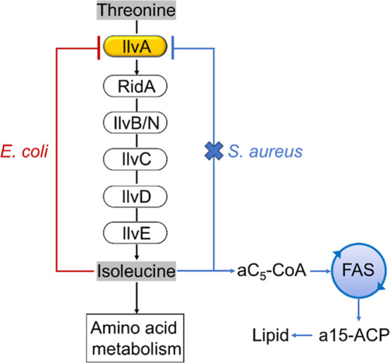
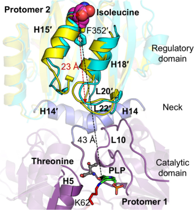
In Staphylococcus aureus, the branched-chain amino acid biosynthetic pathway provides essential intermediates for membrane biosynthesis. Threonine deaminase (IlvA) is the first enzyme in the pathway, and isoleucine feedback regulates the enzyme in Escherichia coli. These studies on E. coli IlvA (EcIlvA) introduced the concept of allosteric regulation. To investigate the regulation of S. aureus IlvA (SaIlvA), we first conducted additional studies on EcIlvA. The previously determined crystal structure of EcIlvA revealed a tetrameric assembly of protomers, each with catalytic and regulatory domains, but the structural basis of isoleucine regulation was not characterized. Here, we present the crystal structure of the EcIlvA regulatory domain bound to isoleucine, which reveals the isoleucine binding site and conformational changes that initiate at Phe352 and propagate 23 Å across the domain. This suggests an allosteric pathway that extends to the active site of the adjacent protomer, mediating regulation across the protomer-protomer interface. The EcIlvA(F352A) mutant binds isoleucine but is feedback-resistant due to the absence of the initiating Phe352. In contrast, SaIlvA is not feedback-regulated by isoleucine and does not bind it. The structure of the SaIlvA regulatory domain reveals a different organization that lacks the isoleucine binding site. Other potential allosteric inhibitors of SaIlvA, including phospholipid intermediates, do not affect enzyme activity. We propose that the absence of feedback inhibition in SaIlvA is due to its role in membrane biosynthesis. These findings enhance our understanding of IlvA's allosteric regulation and offer opportunities for engineering feedback-resistant IlvA variants for biotechnological use.


Figures

1
1
Role of threonine deaminase (IlvA) in the initiation of isoleucine biosynthesis. Threonine dehydratase/deaminase (IlvA) initiates the isoleucine arm of the branched-chain amino acid biosynthesis. IlvA converts threonine to 2-iminobutyrate, and RidA accelerates the spontaneous conversion of 2-iminobutyrate to 2-ketobuytrate, which is condensed with pyruvate by acetolactate synthase (IlvB/N) followed by ketoacid isomeroreductase (IlvC), dihydroxyacid dehydrase (IlvD) and branched-chain amino acid transaminase (IlvE) to result in isoleucine. Isoleucine is a potent feedback regulator of EcIlvA that maintains the intracellular isoleucine homeostasis by tuning the isoleucine biosynthetic rate to its utilization for protein synthesis. SaIlvA is not feedback regulated by isoleucine but is important for both amino acid metabolism and lipid metabolism in .
2
2
Crystal structures of IlvA. (A) Structure of the EcIlvA monomer at 1.87 Å resolution (PDB ID: 9D2Q). It consists of an amino terminal catalytic domain (purple) connected to a carboxy terminal regulatory domain (yellow) by helix H14 (the neck, blue). This new higher resolution EcIlvA structure clarifies the conformation of 3 regions in the previously reported structure (PDB ID: 1TDJ) (dashed red circles, 1 to 3). The PLP cofactor (green balls) is covalently attached to Lys62 in the catalytic domain and there is a glycerol (salmon balls) bound in the regulatory domain. (B) The aligned electron density map occupied by EcIlvA in solution was determined by small-angle X-ray scattering (SAXS) and matches the crystal structure of the tetramer. (C) The EcIlvA regulatory domain in the new structure (PDB ID; 9D2Q) (yellow) overlaid with the regulatory domain in (PDB ID: 1TDJ) (gray) illustrating how the conformation of loops L14, L16 and L21 are altered by glycerol binding. Inset, a close-up view of the area where the conformational change occurs upon binding of isoleucine. (D) The regulatory domain conformational change that occurs within the pocket in the presence of bound glycerol. Glycerol is depicted as salmon balls-sticks, hydrogen bond interactions are shown with dotted red lines, and water molecules are red spheres. (E) The glycerol binding pocket in the ligand-free EcIlvA regulatory domain (PDB ID: 1TDJ). A water molecule is shown with red sphere. (F) The dose response curve shows the effect of glycerol on the activity of EcIlvA. The initial velocity was measured in the presence of 7 mM threonine. Isoleucine was used as the inhibitor control compound.
3
3
Crystal structure of the EcIlvA_R·Ile complex. (A) Overview of the EcIlvA_R·Ile complex protomer with one isoleucine molecule (magenta balls) bound (PDB ID: 9D2R). The rotated view (below) illustrates the positions of the two helices (H17 and H20) that are added to the core structure consisting of two ACT domains inked by loop L19. Five glycerol molecules are depicted as gray sticks. (B) The structure of dimeric EcIlvA_R·Ile complex with the protomers shown in cyan and salmon. The rotated view (below) shows how helices H17 and H20 mediate the formation of the dimer. (C) The EcIlvA_R·Ile dimer fits well into the aligned electron density experimentally determined by SAXS in solution. (D) Superimposition of EcIlvA_R·Ile (cyan) with the glycerol (salmon balls-sticks) bound regulatory domain of EcIlvA (yellow) (PDB ID: 9D2Q). Isoleucine (magenta balls-sticks) is bound between loops L14 and L21 triggering structural changes in the two adjacent helices H15 and H18 and loops L15, L20, and L22. A glycerol molecule, depicted as gray sticks, is bound around Loop L15 of EcIlvA_R·Ile. (E) Close-up views of the isoleucine binding site. Isoleucine is depicted as magenta balls-sticks, water molecules (Wat) are red balls and hydrogen bonds are dashed red lines. (F) An overlay of the EcIlvA_R·Ile complex (cyan) with the EcIlvA regulatory domain (yellow; PDB ID: 9D2Q). The change in the side chain rotamer of Phe352 must occur to accommodate isoleucine and leads to a cascade of structural changes along helices H15 and H18 that result in the movement of loops L15 and L20.
4
4
Isoleucine effect on the activity and stability of EcIlvA. (A) Effect of isoleucine on the steady-state kinetics of EcIlvA. (B) Effect of isoleucine on the steady-state kinetics of EcIlvA­(F352A). The theoretical curve was analyzed by the allosteric sigmoidal equation (Y = V max*h/(K half^h + h)) using GraphPad Prism 9.4.1. for both EcIlvA and EcIlvA­(F352A). K half is the concentration of substrate that produces a half-maximal enzyme velocity, V max is the maximum enzyme velocity, and h is the Hill slope. (C,D) Dose–response curves showing the inhibition of EcIlvA (C) and EcIlvA­(F352A) (D) by isoleucine. Enzyme activity was measured in triplicate with 7 mM threonine. The resulting IC50 values were determined by nonlinear regression using the [log­(inhibitor)] vs normalized response – variable slope model in GraphPad Prism and are presented as mean ± standard error (SE). (E) The thermal stability of EcIlvA and EcIlvA­(F352A) in the absence and presence of 5 mM isoleucine. (F) The thermal stability of EcIlvA_R and EcIlvA_R­(F352A) in the absence and presence of 5 mM isoleucine. Data represented as mean ± standard deviation (error bars) from triplicate sets.
5
5
Crystal structure of EcIlvA_R­(F352A). (A) Crystal structure of the EcIlvA_R­(F352A)·Ile complex (PDB ID: 9D2S). EcIlvA_R­(F352A)·Ile is superimposed on EcIlvA (top, PDB ID: 9D2Q) and EcIlvA_R (bottom, PDB ID: 9D2R). (B) Close-up view of the isoleucine binding site of EcIlvA_R­(F352A)·Ile (brown) and EcIlvA (yellow). (C) Close-up view of the isoleucine binding site in EcIlvA_R­(F352A)·Ile (brown) and EcIlvA_R·Ile (cyan).
6
6
Isoleucine effect on SaIlvA. (A) Effect of isoleucine on the steady-state kinetics of SaIlvA. The theoretical curve was analyzed by the Michaelis–Menten equation using GraphPad Prism 10.1.2. The k cat value was calculated to be 64.4 ± 1.3 s–1. K m and k cat values are presented as mean values ± standard error (SE). (B) Effect of isoleucine on the activity of SaIlvA. Inset, plot at the high concentration of isoleucine (0–5 mM). The initial velocity was measured in the presence of 30 mM threonine. (C) The thermal stability of SaIlvA in the absence and presence of 5 mM isoleucine. Data represented as mean ± standard deviation (error bars) from triplicate sets.
7
7
Crystal structure of SaIlvA_R. (A) Tetramer of the SaIlvA_R crystal structure (PDB ID: 9D2T). Each monomer of the tetramer is related by an approximate 2-fold axis (read symbol and arrows). Two tetramers are in the asymmetric unit. (B) Dimeric structure of SaIlvA_R. Protomers A and B are shown in green and orange, respectively. (C) Monomeric structure of EcIlvA_R·Ile (PDB ID: 9D2R). The two ACT domains are shown in cyan ribbon with highlighted yellow and blue. The bound isoleucine is shown with magenta balls-sticks. (D) The overlaid structures of dimeric SaIlvA_R (green and orange) and monomeric EcIlvA_R·Ile (cyan). The protomer A of SaIlvA_R is superimposed on the ACT1 domain of EcIlvA_R·Ile. The conformational difference of helix H15 is shown as a black arrow.
8
8
Modeled quaternary structure of SaIlvA. (A) Tetramer of SaIlvA_R is shown in green (protomer A), orange (protomer B), blue (protomer C), and yellow (protomer D). The N-terminus of each protomer is shown as a sphere. The N-termini of protomers A and C align in the same direction, while those of the protomers B and D face the opposite way. (B) SaIlvA tetramer model was generated using AlphaFold model of SaIlvA dimer and SaIlvA_R tetramer crystal structure. The four C-terminal regulatory domains form the core of the tetramer. (C) The tetramer model fitted into the electron density determined by small-angle X-ray scattering (SAXS) in solution. (D) The SaIlvA octamer model, which consists of two tetramers, fitted into the electron density determined by small-angle X-ray scattering (SAXS) in solution. A tetramer model is shown in green, orange, blue, and yellow, and the other tetramer is shown in cyan. (E) The hexamer model generated using AlphaFold. (F) The hexamer model fitted into the electron density determined by small-angle X-ray scattering (SAXS) in solution. (G) Protomer A makes a catalytic interaction dimer with protomer C and makes a regulatory interaction dimer with protomer B. The regulatory domains (green and orange) in the hexamer structure resemble the dimeric structure of the SaIlvA_R crystal structure (gray).
9
9
Complementation of ΔIlvA. (A) Growth curve of ΔIlvA containing empty vector (pCN38) or pSaIlvA or pEcIlvA grown in complete defined medium (CDM) or CDM minus isoleucine. (B) Fatty acid analysis by gas chromatography of ΔIlvA containing empty vector (pCN38) or pSaIlvA or pEcIlvA grown in CDM. (C) Fatty acid analysis by gas chromatography of ΔIlvA containing empty vector (pCN38) or pSaIlvA or pEcIlvA grown in CDM minus isoleucine. (D) Growth curve of ΔIlvA containing pEcIlvA (F352A) mutant grown in complete defined medium (CDM) or CDM minus isoleucine. (E) Fatty acid analysis by gas chromatography of ΔIlvA pEcIlvA (F352A) mutant grown in CDM. (F) Fatty acid analysis by gas chromatography of ΔIlvA pEcIlvA (F352A) mutant grown in CDM minus isoleucine. The data in A and D are representative of experiments done twice. The data in B, C, E, and F are from triplicate sets (mean ± SE). The value for the t-test is given in red.
10
10
Allosteric regulation of isoleucine on EcIlvA. Loop interactions between the catalytic and regulatory domain in the active site of EcIlvA. The catalytic domain of protomer 1, regulatory domain of protomer 2 and necks of both protomers of EcIlvA are shown in purple, yellow, and blue, respectively. The EcIlvA_R·Ile structure (cyan cartoon) is superimposed on the EcIlvA structure. The features of protomer 2 are indicated with a prime. The docked threonine molecule is represented by ball-and-sticks with gray carbon atoms, blue nitrogen atom, and red oxygen atoms. PLP and the active-site lysine (K62) are shown as sticks. The red arrow indicates the distance from isoleucine to L20 loop, while the black arrow indicates the distance from isoleucine to PLP of the partner protomer.

Update of

Similar articles

References

    1. Amorim Franco T. M., Blanchard J. S.. Bacterial Branched-Chain Amino Acid Biosynthesis: Structures, Mechanisms, and Drugability. Biochemistry. 2017;56:5849–5865. doi: 10.1021/acs.biochem.7b00849. - DOI - PMC - PubMed
    1. Liang Y. F., Long Z. X., Zhang Y. J., Luo C. Y., Yan L. T., Gao W. Y., Li H.. The chemical mechanisms of the enzymes in the branched-chain amino acids biosynthetic pathway and their applications. Biochimie. 2021;184:72–87. doi: 10.1016/j.biochi.2021.02.008. - DOI - PubMed
    1. Irons J. L., Hodge-Hanson K., Downs D. M.. RidA Proteins Protect against Metabolic Damage by Reactive Intermediates. Microbiol. Mol. Biol. Rev. 2020;84:e00024. doi: 10.1128/mmbr.00024-20. - DOI - PMC - PubMed
    1. Shen W., Borchert A. J., Downs D. M.. 2-Aminoacrylate stress damages diverse PLP-dependent enzymes in vivo. J. Biol. Chem. 2022;298:101970. doi: 10.1016/j.jbc.2022.101970. - DOI - PMC - PubMed
    1. Umbarger H. E.. Evidence for a negative-feedback mechanism in the biosynthesis of isoleucine. Science. 1956;123:848. doi: 10.1126/science.123.3202.848-a. - DOI - PubMed

MeSH terms

LinkOut - more resources