. 2025 Jun 10;16(1):443.
doi: 10.1038/s41419-025-07758-y.

Loss of RNF41 promotes bladder cancer metastasis through increasing NUDC stability to enhance tubulin polymerization

Affiliations

Loss of RNF41 promotes bladder cancer metastasis through increasing NUDC stability to enhance tubulin polymerization

Fuchun Zheng et al. Cell Death Dis. .

Abstract

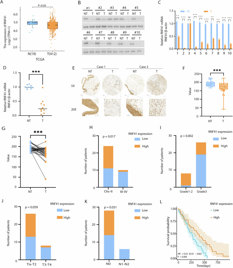
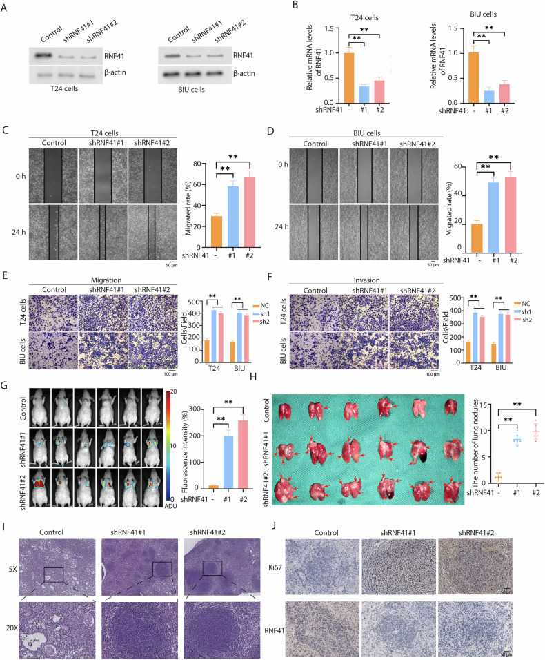
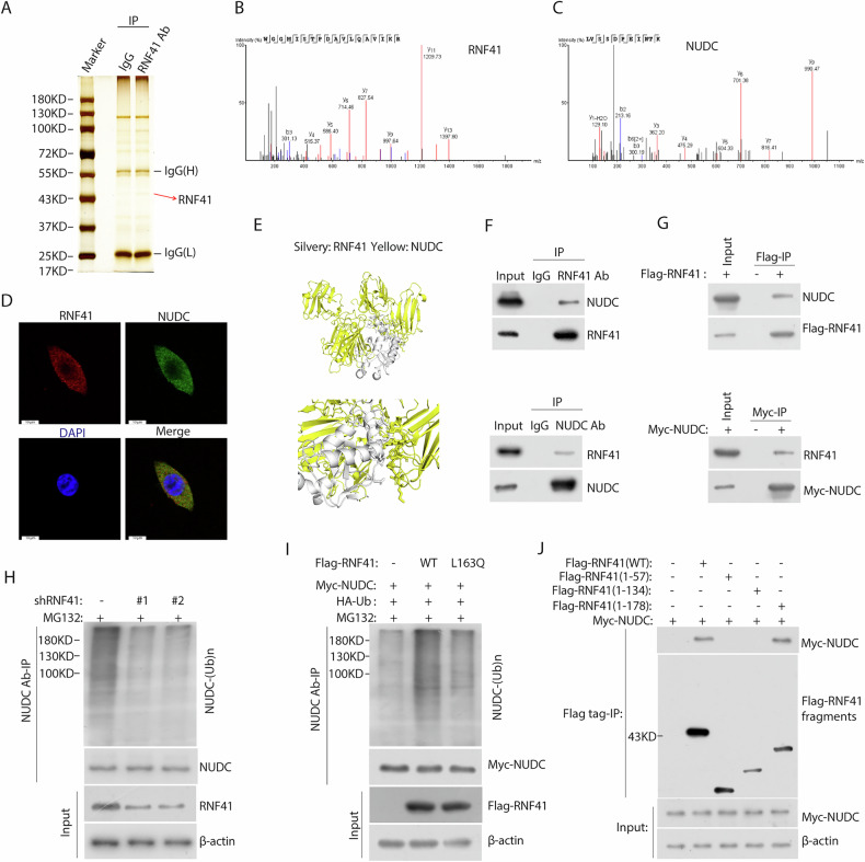
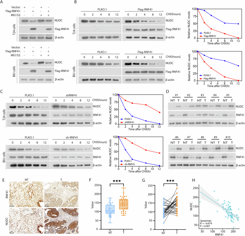
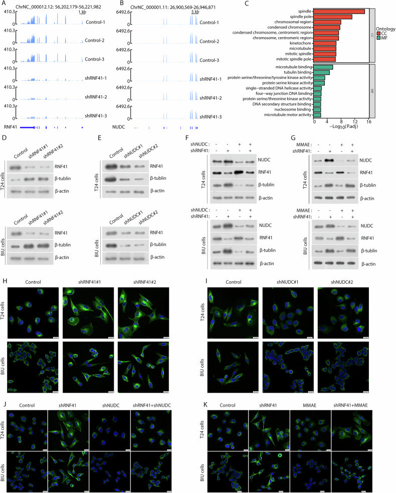
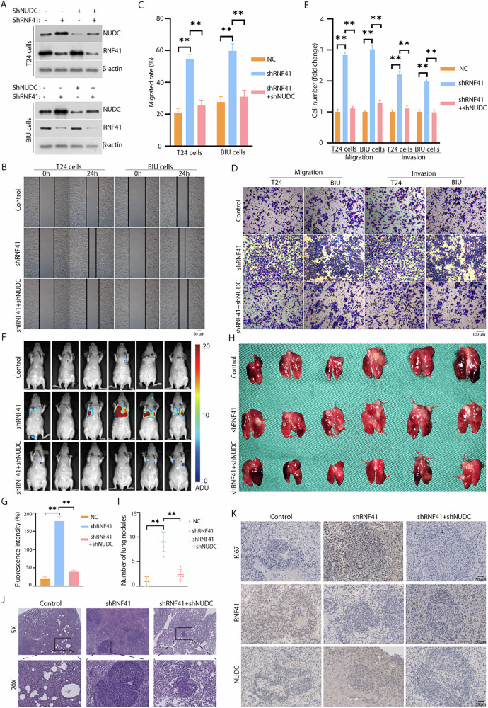
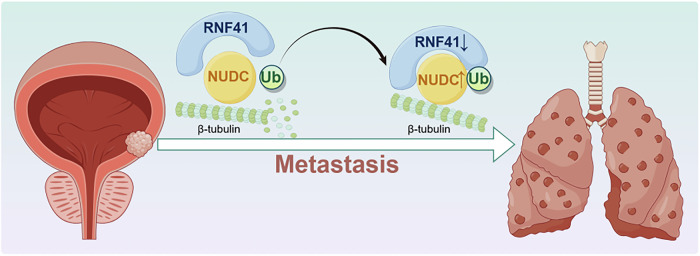
Bladder cancer (BCa) is a representative of urological cancer with a high recurrence and metastasis is the leading cause of death from BCa. The underlying mechanism of BCa metastasis remains poorly defined. Here, we found RNF41 was significantly downregulated in BCa tissue and low level of RNF41 is associated with BCa progression and poor prognosis. RNF41 knockdown promoted cell migration and invasion in both in vitro and in tail vein lung metastasis model, while ectopic RNF41 expression exhibited the opposite effects. Mechanically, we revealed that RNF41 directly interacted with NUDC and ubiquitinates NUDC to promote its degradation. Clinically, RNF41 was significantly downregulated in metastatic BCa tissues and negatively associated with NUDC expression. Furthermore, we identified that RNF41 promoted BCa lung metastasis through NUDC regulated β-tubulin depolymerization. In summary, these findings support that RNF41 was a tumor suppressor in BCa metastasis and highlights that targeting RNF41-NUDC-β-tubulin axis could be a valuable strategy to ameliorate BCa progression and metastasis.


Conflict of interest statement

Competing interests: The authors declare no competing interests. Ethics approval: The animal experiments performed here have been approved by the Animal Protection and Use Committee of The First Affiliated Hospital of Nanchang University.

Figures

Fig. 1
Fig. 1. Downregulation of RNF41 in bladder cancer (BCa).
A RNF41 expression in normal tissues (n = 19) vs. BCa tissues (n = 412) from TCGA database. B Western blot showing RNF41 protein levels in 10 fresh BCa tissues (T) and matched paracancerous tissues (NT). C, D Real-time PCR analysis of RNF41 mRNA expression in 10 fresh BCa tissues and matched paracancerous tissues. E Representative immunohistochemistry (IHC) images showing RNF41 protein expression in BCa and adjacent tissues using tissue chips. Scale bar, 50 μm. F, G RNF41 protein downregulation in adjacent non-tumor tissues and BCa tissues (F) and in paired adjacent and matched BCa tissues (G). HK Correlation of low RNF41 expression with higher stage (p = 0.017), grade (p = 0.002), clinical T3/T4 (p = 0.059), and N1 (p = 0.031). L Overall survival analysis in 129 BCa patients, showing significantly shorter survival in those with low RNF41 expression (p = 0.005). Data provided by The First Affiliated Hospital of Nanchang University.
Fig. 2
Fig. 2. Inhibition of RNF41 promotes migration of BCa cells.
A, B Knockdown efficiency of RNF41 by shRNA, measured via western blot and quantitative RT-PCR (**p < 0.01). C, D Migration abilities of T24 and BIU cells after RNF41 knockdown assessed using a wound healing assay (**p < 0.01). Scale bar, 50 μm. E, F Representative images and quantification of migrated or invading cells in a transwell assay, both without and with matrix penetration (**p < 0.01). Scale bar, 100 μm. G Fluorescence imaging of experimental nude mice injected with T24-Luc cells, comparing fluorescence intensity (**p < 0.01). H Lung imaging and quantification of pulmonary nodules (**p < 0.01). I Hematoxylin and eosin (HE) staining of lungs with metastatic tumors. J Ki67 and RNF41 immunohistochemistry (IHC) staining of lungs with metastatic tumors. Statistical analyses were performed using ANOVA. *p < 0.05; **p < 0.01. Scale bar, 100 μm.
Fig. 3
Fig. 3. RNF41 directly interacts with and ubiquitinates NUDC.
A Proteins interacting with RNF41 were detected using co-immunoprecipitation (co-IP) and silver staining in T24 cells. B, C Peptide fragment mass spectrum for RNF41 and NUDC protein. D Fluorescence microscopy revealed co-localization of RNF41 (red) and NUDC (green) in T24 cells, with nuclear staining by 4’, 6-diamidino-2-phenylindole (DAPI; blue). Scale bar, 10 μm. E Protein docking map illustrating the predicted interaction between RNF41 and NUDC. F Endogenous co-immunoprecipitation results showing the interaction of RNF41 and NUDC in T24 cells. G Co-immunoprecipitation of FLAG-RNF41 and MYC-NUDC in T24 cells, transfected with FLAG-RNF41 and MYC-NUDC plasmids for 36 h. H HEK293T cells co-transfected with the indicated shRNA underwent immunoprecipitation (IP) with anti-NUDC antibody, followed by immunoblotting (IB) with antibodies against NUDC, RNF41, and β-actin. Cells were treated with 20 μM MG132 for 6 h prior to harvesting. I HEK293T cells co-transfected with Myc-NUDC, HA-ubiquitin (HA-Ub), and Flag-tagged RNF41 WT or RNF41 L163Q, with lysates subjected to IP using anti-NUDC antibody followed by IB with antibodies against Myc-tagged, Flag-tagged proteins, and β-actin. Cells were treated with 20 μM MG132 for 6 h before collection. J HEK293T cells were co-transfected with Myc-NUDC and Flag-tagged full-length RNF41 or its deletion mutants, followed by immunoprecipitation with Flag beads and immunoblotting with antibodies against Myc and Flag.
Fig. 4
Fig. 4. RNF41 regulates the stability of NUDC.
A T24 and BIU cells transfected with the specified constructs were treated with or without MG132 for 6 h before collection. RNF41 and NUDC proteins were analyzed using the indicated antibodies. B Left: FLAG-RNF41 plasmid was introduced into T24 and BIU cells, followed by the addition of 50 μg/ml cycloheximide (CHX) at 0, 2, 4, 8, and 12 h. Cells were then harvested, and western blot analysis with NUDC antibodies determined the half-life of NUDC protein. Right: Quantification of NUDC protein levels. C Left: RNF41 shRNA was transfected into T24 and BIU cells for 36 h, followed by treatment with 50 μg/ml CHX at specified intervals (0, 2, 4, 8, 12 h) before collection. The half-life of NUDC protein was assessed via western blot. Right: Quantification of NUDC protein levels. D Expression analysis of RNF41 and NUDC in 10 freshly paired metastatic BCa tissue samples (T) and matched adjacent non-tumor tissues (NT). β-actin served as the loading control. E Representative staining images of RNF41 and NUDC in BCa tissues. Scale bar, 50 μm. F, G Compared to adjacent tissues, NUDC protein levels were elevated in BCa tissues (F), and in paired samples, NUDC was also found to be upregulated in BCa tissues (G). H Correlation analysis between RNF41 and NUDC expression levels in BCa tissues, with statistical analyses performed using the χ² test (P < 0.001). R indicates Pearson’s correlation coefficient.
Fig. 5
Fig. 5. RNF41 regulates NUDC-induced tubulin depolymerization.
Sequencing peaks of RNF41 (B) and NUDC (A) after RNF41 knockdown. C Gene Ontology (GO) analysis of differentially expressed genes following RNF41 knockdown in T24 cells. CC represents cellular component; MF denotes molecular function. D Western blot analysis was performed to assess protein levels in T24 and BIU cells stably expressing RNF41 shRNA. E Western blot was conducted on T24 and BIU cells stably expressing NUDC shRNA to evaluate the indicated protein levels. F T24 and BIU cells stably expressing either NUDC or RNF41 shRNA plasmids, with or without NUDC shRNA, underwent western blot analysis to measure specific protein levels. G T24 and BIU cells with stable RNF41 shRNA expression were treated with or without Monomethyl auristatin E (MMAE) (5 μM) and subsequently analyzed by western blot for the specified proteins. H Confocal microscopy images of T24 and BIU cells stably expressing Control or RNF41 shRNA. I Confocal microscopy images of T24 and BIU cells stably expressing Control or NUDC shRNA. J Confocal images of T24 and BIU cells stably expressing Control, RNF41 shRNA, and/or NUDC shRNA. K Confocal images of T24 and BIU cells stably expressing Control, RNF41 shRNA, and/or MMAE. Tubulin (green) was visualized using β-tubulin antibodies, while DNA (blue) was stained with DAPI. Scale bar, 20 μm.
Fig. 6
Fig. 6. NUDC knockdown rescue the enhanced migratory and invasive abilities caused by RNF41 knockdown, both in vitro and in vivo.
A The effectiveness of RNF41 and NUDC knockdown was assessed using western blot analysis. B, C Wound healing assays were conducted to evaluate the migratory potential of BCa cells, with migration rates calculated (**p < 0.01). Scale bar, 50 μm. D, E Representative images and quantification of migrated or invading cells from a transwell assay, conducted both with and without matrix penetration (**p < 0.01). Scale bar, 100 μm. F, G Fluorescence imaging of experimental nude mice injected with T24-Luc cells, comparing fluorescence intensity (**p < 0.01). H, I Lung imaging and quantification of pulmonary nodules (**p < 0.01). J Hematoxylin and eosin (HE) staining of lung tissue containing metastatic tumors. K Immunohistochemical staining for Ki67, RNF41, and NUDC in lung tissues with metastatic tumors. Scale bar, 50 μm.
Fig. 7
Fig. 7. RNF41 induces canonical polyubiquitination of NUDC to enhance its stability and induce β-tubulin remodeling, which promotes BCa metastasis.
Created with figdraw.com.

Similar articles

References

    1. Siegel RL, Miller KD, Fuchs HE, Jemal A. Cancer statistics, 2022. CA Cancer J Clin. 2022;72:7–33. - PubMed
    1. Catto JWF, Gordon K, Collinson M, Poad H, Twiddy M, Johnson M, et al. Radical cystectomy against intravesical BCG for high-risk high-grade nonmuscle invasive bladder cancer: results from the randomized controlled BRAVO-feasibility study. J Clin Oncol J Am Soc Clin Oncol. 2021;39:202–14. - PMC - PubMed
    1. Patel VG, Oh WK, Galsky MD. Treatment of muscle-invasive and advanced bladder cancer in 2020. CA Cancer J Clin. 2020;70:404–23. - PubMed
    1. Shinagare AB, Ramaiya NH, Jagannathan JP, Fennessy FM, Taplin ME, Van den Abbeele AD. Metastatic pattern of bladder cancer: correlation with the characteristics of the primary tumor. Am J Roentgenol. 2011;196:117–22. - PubMed
    1. Bianchi M, Roghmann F, Becker A, Sukumar S, Briganti A, Menon M, et al. Age-stratified distribution of metastatic sites in bladder cancer: A population-based analysis. Can Urol Assoc J. 2014;8:E148–58. - PMC - PubMed

MeSH terms

LinkOut - more resources