. 2025 Jun 10;16(1):5136.
doi: 10.1038/s41467-025-59596-3.

Iterative transcription factor screening enables rapid generation of microglia-like cells from human iPSC

Affiliations

Iterative transcription factor screening enables rapid generation of microglia-like cells from human iPSC

Songlei Liu et al. Nat Commun. .

Abstract

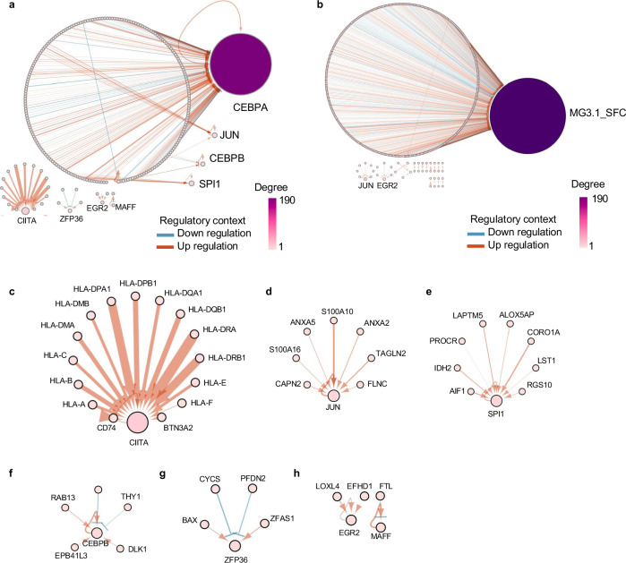
Differentiation of induced pluripotent stem cells (iPSCs) into specialized cell types is essential for uncovering cell-type specific molecular mechanisms and interrogating cellular function. Transcription factor screens have enabled efficient production of a few cell types; however, engineering cell types that require complex transcription factor combinations remains challenging. Here, we report an iterative, high-throughput single-cell transcription factor screening method that enables the identification of transcription factor combinations for specialized cell differentiation, which we validated by differentiating human microglia-like cells. We found that the expression of six transcription factors, SPI1, CEBPA, FLI1, MEF2C, CEBPB, and IRF8, is sufficient to differentiate human iPSC into cells with transcriptional and functional similarity to primary human microglia within 4 days. Through this screening method, we also describe a novel computational method allowing the exploration of single-cell RNA sequencing data derived from transcription factor perturbation assays to construct causal gene regulatory networks for future cell fate engineering.


Conflict of interest statement

Competing interests: S.L. and G.M.C. are listed as inventors of a patent related to work on this article. G.M.C., P.K., and A.H.M.N. are co-founders/employees/advisors at, and have equity in GC Therapeutics, Inc, and are inventors on patents filed by the Presidents and Fellows of Harvard College. Full disclosure for GMC is available at arep.med.harvard.edu/gmc/tech.html . The remaining authors declare no competing interests.

Figures

Fig. 1
Fig. 1. First round of pooled screening identified an initial set of TFs for inducing microglia gene expression.
a Workflow of the first pooled TF screen. Two independent transductions were performed for the first pooled TF screen. This panel is generated using BioRender under an academic license. b Flow cytometry analysis of stem cell (TRA-1-60) and microglia (P2RY12, CD11b, CX3CR1) proteins in the PGP1 + 40 TF pool before and after Dox induction from two independent pooled TF transductions. c Representative image of cells with low TRA-1-60 expression in the Dox+ group to be sorted for scRNA-seq. d UMAP clustering of two independently transfected and differentiated PGP1 iPSC pools. Colors represent clusters identified by Seurat at 0.3 resolution. e Expression of microglia (ITGAM, CX3CR1, TMEM119, P2RY12, TREM2) and spiked-in stem cell (POU5F1) genes in scRNA-seq. f Primer designs for co-amplification of TF and cell barcodes in 10x Genomics 3’ workflow. This panel is generated using BioRender under an academic license. g Number of TFs per cell counted from normalized and binarized TF expression matrix. h Ranking of the 40 TFs after one-sided Wilcoxon rank sum test with the two tested groups being with or without microglia gene expression. Blue highlights top-ranking TFs. i Flow cytometry validation of a single transduction of top-ranking TFs for inducing microglia protein expression. C = CEBPA, F = FLI1, S = SPI1. “Pool” refers to simultaneous transfection of multiple single-TF plasmids, no polycistronic cassette used. All source data are provided as a Source Data file. Raw and processed data are provided in GEO accession GSE287847.
Fig. 2
Fig. 2. Second iteration of pooled TF screen using MG3.1-SFC as baseline identified additional TFs for improved microglia differentiation.
a Workflow of the second pooled TF screen performed in duplicate. This panel is generated using BioRender under an academic license. b Polycistronic cassette design for performing dual-drug selection to achieve 3 + X TF screen. c Normalized mRNA expression from the polycistronic cassette (SPI1, FLI1, CEBPA) and stem cells (POU5F1) from two independent TF transductions. d TF barcode counting enabled the identification of stem cells (“No TF BC”), MG3.1-SFC, and cells with additional TFs (“SFC + X”). e Example histograms of TF barcode raw counts in single cells. f Number of TFs per cell counted from normalized and binarized TF expression matrix. g Ranking of the 42 TFs after one-sided Wilcoxon rank sum test with the two tested groups being with or without microglia gene expression. Blue highlights top-ranking TFs. Grey highlights the SFC polycistronic cassette. h Flow cytometry validation of a single transduction of the top-ranking TFs for improving microglia protein expression. i Polycistronic cassette designs for varying TF orders. This panel is generated using BioRender under an academic license. j Flow cytometry analysis of a single transduction of different arrangements of the six-TF recipe in comparison with MG3.1-SFC. All source data are provided as a Source Data file. Raw and processed data are provided in GEO accession GSE287850.
Fig. 3
Fig. 3. Time-resolved bulk RNA-seq analysis of TFiMGLs reveals differentiation kinetics and shared molecular signatures with primary microglia.
a Expression of the six induced TFs over time measured by bulk RNA-seq (n = 2 for each day). b Expression of stem cell (POU5F1) and microglia (ITGAM, CX3CR1, TMEM119, P2RY12, TREM2) genes over time (n = 2 for each day) measured by bulk RNA-seq. c PCA plot for the bulk transcriptome of TFiMGLs (MG6.4) over time (n = 5 for day 0, n = 2 for days 1–6). d PCA of bulk RNA-seq data from multiple sources containing primary microglia. MG Microglia, DC dendritic cell, HPC hematopoietic progenitor, iMGL growth factor-induced microglia-like cell, Mono monocyte. e GSEA of TFiMGLs versus iPS using two microglia marker gene sets from MSigDB: M40168 and M39077. Raw and processed data are provided in GEO accession GSE287851.
Fig. 4
Fig. 4. Single cell transcriptome analysis shows microglia like identity 2 days after SPI1, FLI1, CEBPA, CEBPB, IRF8, and MEF2C induction.
a UMAP clustering of a single TFiMGL induction at 2-, 4-, and 6-days post induction switching to either stem cell media or microglia media from day 2. Colors represent clusters identified by day and media condition. D2: day 2, D4 + MT: day 4 stem cell media, D4 + MG: day 4 microglia media, D6 + MT: day 6 stem cell media, D6 + MG: day 6 microglia media. Top 5 cell-type enrichments per TFiMGL condition using (b) Descartes single cell atlas or (c) Cell Marker Augmented single cell atlas as reference. Enrichment p-value was computed using Fisher’s exact test or the hypergeometric test and adjusted p-value was computed using the Benjamini-Hochberg method for correction for multiple hypotheses. d Proportion of cells per TFiMGL condition expressing microglia marker gene P2RY12. e Normalized mRNA expression of P2RY12 on overall TFiMGL population. f Top 5 cell-type enrichments of P2RY12+ cell subset using Descartes single cell atlas as reference. g Proportion of cells per TFiMGL condition expressing microglia marker gene CX3XR1. h Normalized mRNA expression of CX3CR1 on overall TFiMGL population. i Top 5 cell-type enrichments of CX3CR1+ cell subset using Descartes single cell atlas as reference. j Mean expression levels of known pan-microglial and mature human microglia genes from different TFiMGL condition (rows). Microglial marker genes were obtained from previously published studies. Genes directly overexpressed are outlined in red. km Mean expression levels of (k) proliferation-associated microglia, (l) neural-associated microglia, and (m) immune-associated microglia transcriptional signatures found in various regions of the developing human brain. All source data are provided as a Source Data file. Raw and processed data are provided in GEO accession GSE287852.
Fig. 5
Fig. 5. TFiMGLs differentiate quickly, are phagocytic and responsive to ADP stimulation.
a Representative immunofluorescence images of stem cell (OCT4), Dox-induced (PU.1), and microglia (CD11b, P2RY12, CX3CR1) proteins on day 4. Scale bar: 20 µm. b Flow cytometry quantification of microglia protein expression on day 4 (n = 3). c PCA of TFiMGLs transcriptome after 24 h treatment with IFNγ, fAβ, or TDP43. (n = 3 per treatment). d, e Pathway analysis of significantly differentially expressed genes after treatment with IFNγ or TDP43. One-sided Fisher’s exact test was used to identify enriched categories with FDR control using Benjamini–Hochberg procedure. f Representative graph of flow cytometry analysis of the uptake of pHrodo-labeled S. aureus Bioparticles over time (n = 3). g Microscopy analysis of particle uptake combined with microglia surface protein staining. Graphs shown are representative of three independent experiments with similar results. h Calcium imaging with Fluo-4 AM after stimulation with 150 µM ADP and peak quantification. Images are taken once every 3 s. ADP was added at t0 Graphs shown are representative of two independent experiments with similar results. i Quantification of fluorescent signals from all cells in the field of view in panel h over a period of 10 min. j Peak dynamics analysis shows a fast rise and slow decay pattern of the intracellular calcium concentration. All source data are provided as a Source Data file. Raw and processed data are provided in GEO accession GSE287853.
Fig. 6
Fig. 6. TF-gene regression analysis for studying causal gene-regulatory networks.
The width of edges is correlated with coefficient values, the larger the value the wider the edge. A red edge means upregulation while a blue edge means downregulation. Edges were selected with these criteria: Abs(coefficient) >0.1 and −log10(p-value) >20. F-test was used to determine if any of the independent variables were significant in the regression model. a Global network for the first pooled screen. b Global network for the second pooled screen. Sub-network for (c) CIITA, (d) JUN, (e) SPI1, (f) CEBP3, (g) ZFP36, (h) EGR2. Source data are provided in Supplementary Data file.

References

    1. Regev, A., Teichmann, S., Lander, E., Amit, I. & Benoist, C. Science forum: the human cell atlas. elife6, e27041 (2017). - PMC - PubMed
    1. Ng, A. H. M. et al. A comprehensive library of human transcription factors for cell fate engineering. Nat. Biotechnol.39, 510–519 (2021). - PMC - PubMed
    1. Gosselin, D. et al. An environment-dependent transcriptional network specifies human microglia identity. Science356, eaal3222 (2017). - PMC - PubMed
    1. Smith, A. M. & Dragunow, M. The human side of microglia. Trends Neurosci.37, 125–135 (2014). - PubMed
    1. Watkins, L. R. & Hutchinson, M. R. A concern on comparing ‘apples’ and ‘oranges’ when differences between microglia used in human and rodent studies go far, far beyond simply species: comment on Smith and Dragunow. Trends Neurosci.37, 189–190 (2014). - PubMed

MeSH terms

Substances

LinkOut - more resources