. 2025 Jun 10;20(1):83.
doi: 10.1186/s13020-025-01121-1.

The gut microbiota-mediated ferroptosis pathway: a key mechanism of ginsenoside Rd against metabolism-associated fatty liver disease

Affiliations

The gut microbiota-mediated ferroptosis pathway: a key mechanism of ginsenoside Rd against metabolism-associated fatty liver disease

Wenjing Liu et al. Chin Med. .

Abstract

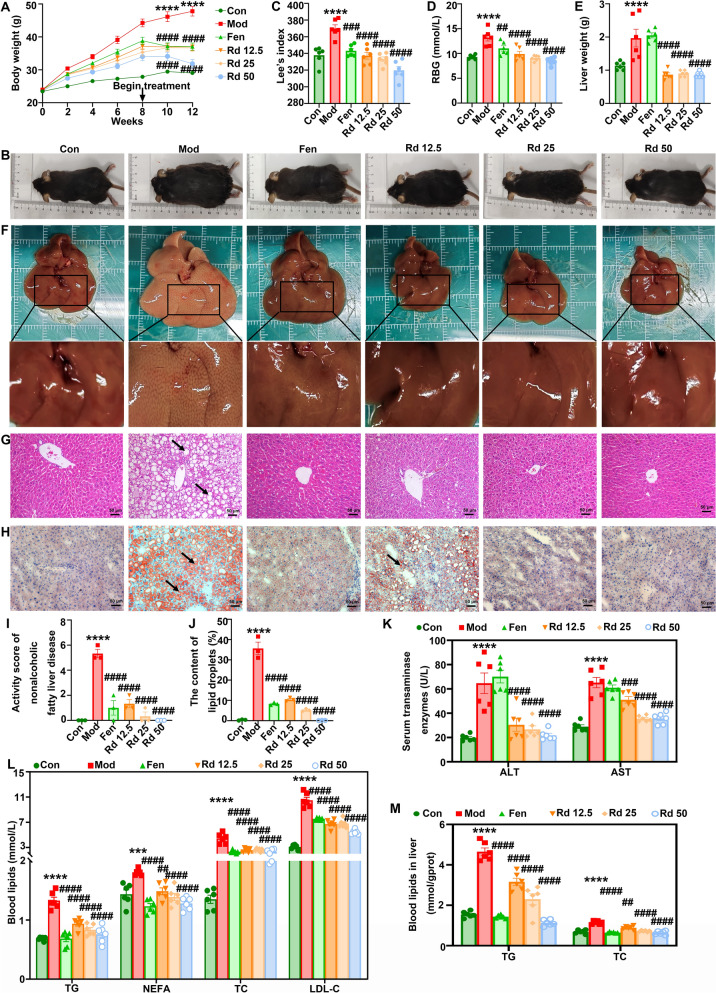
Background: Ginsenoside Rd (G-Rd), found in Panax species, has shown therapeutic potential against metabolism-associated fatty liver disease (MAFLD), but its mechanism has not been well elucidated. This study investigated the key mechanisms of G-Rd in modulating the gut microbiome and lipid peroxidation-mediated ferroptosis pathway in MAFLD.

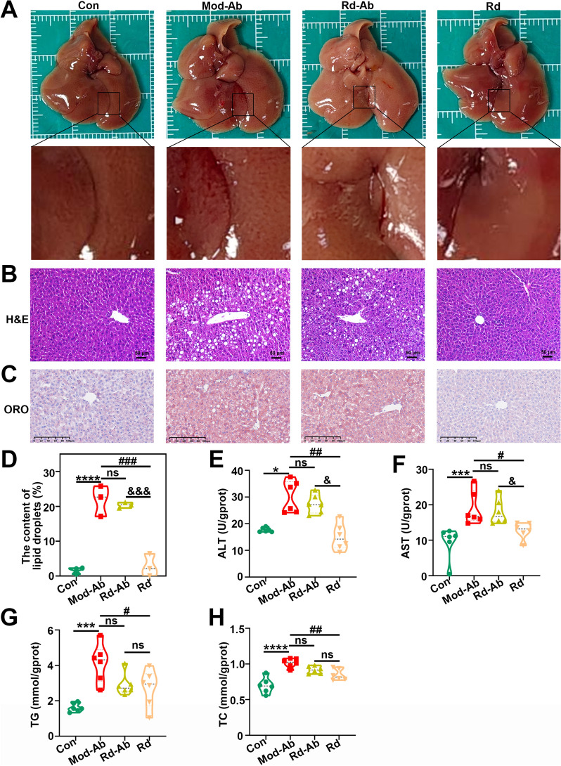
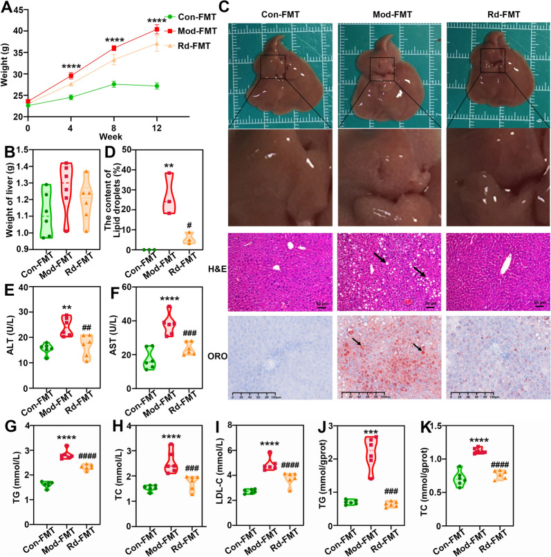
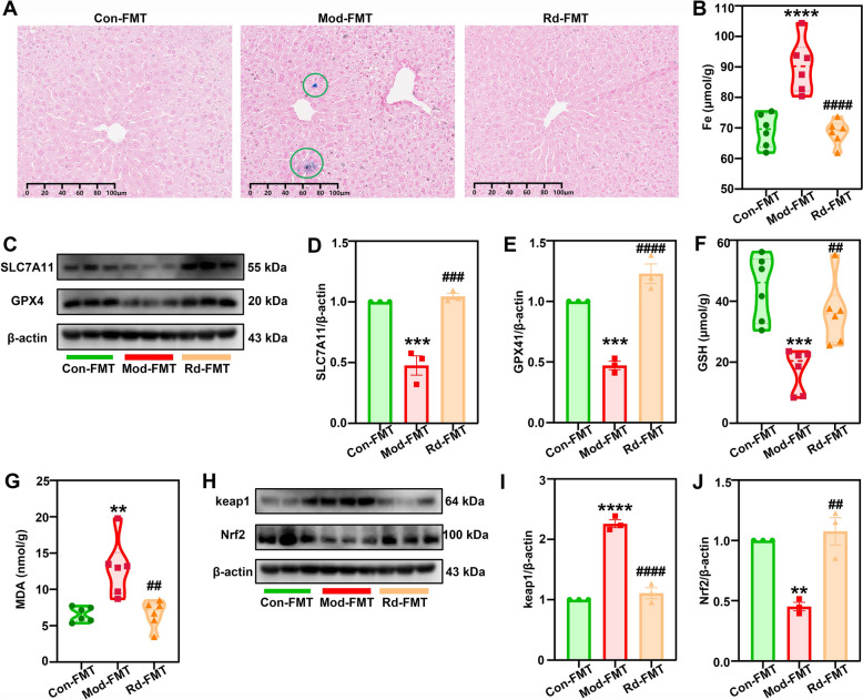
Methods: A high-fat diet-induced MAFLD model was established. Ultrastructural changes in liver tissue were observed using transmission electron microscopy. Metagenomics were employed to detect alterations in gut microbiota and their metabolites. Biochemical analysis and immunohistochemistry were used to examine liver injury, blood lipids, lipid peroxidation-related indicators, and tissue iron content.

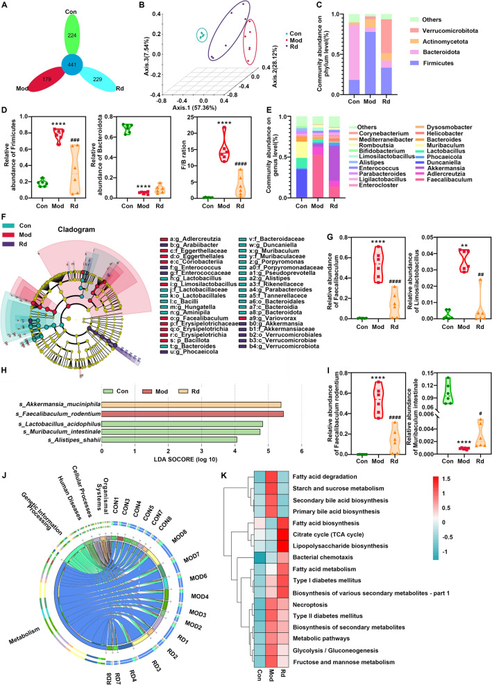
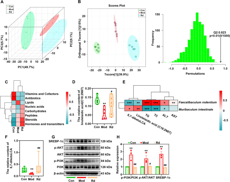
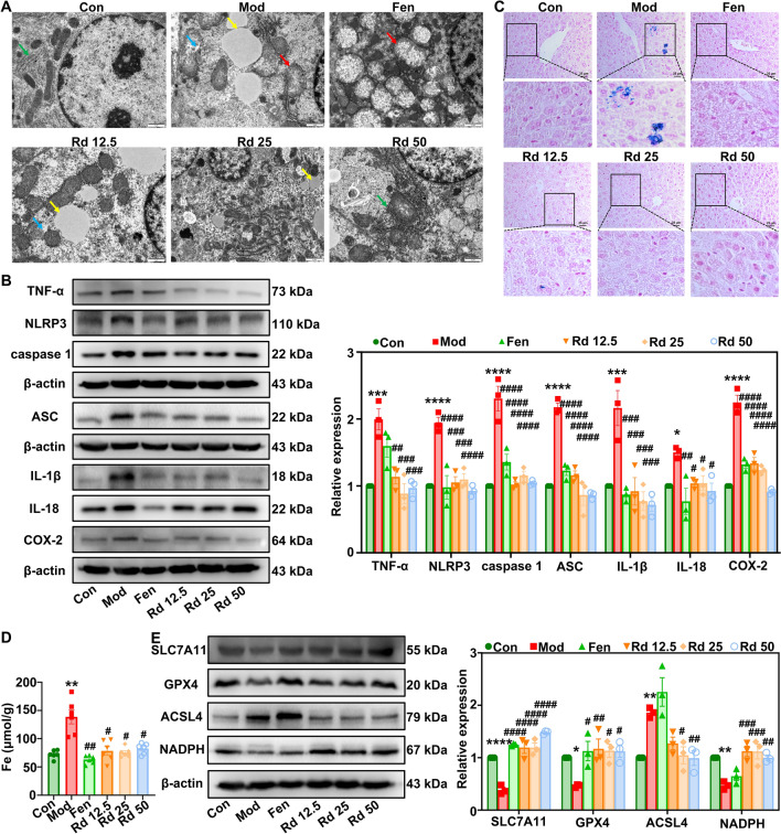
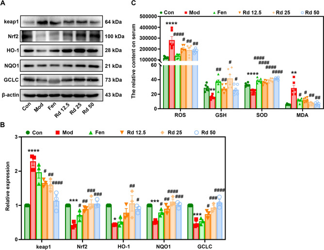
Results: G-Rd significantly reduced liver injury and steatosis in MAFLD mice and downregulated the elevated relative abundance of Firmicutes and the Firmicutes/Bacteroidetes ratio. It also significantly reduced the abundances of Faecalibaculum rodentium while increasing Muribaculum intestinale, with its functional role being relevant to lipid metabolism regulation. Moreover, G-Rd ameliorated mitochondrial damage and inhibited the ferroptosis pathway in the liver, which was associated with antioxidant-related factors mediated by Nrf2 signaling. The liver protective effect of G-Rd was driven by the regulation of gut microbiota, as demonstrated by antibiotic cocktail treatment and fecal microbiota transplantation.

Conclusions: G-Rd attenuated HFD-induced MAFLD by alleviating liver oxidative stress, lipid peroxidation, and ferroptosis through modulation of the gut microbiota. The antioxidant and anti-ferroptotic actions of G-Rd, mediated via the Nrf2 pathway, were found to contribute to the amelioration of liver injury and hepatic steatosis in MAFLD.

Keywords: Ferroptosis; Ginsenoside Rd; Gut microbiota; Lipid peroxidation; MAFLD; Nrf2.


Conflict of interest statement

Declarations. Ethics approval and consent to participate: All experimental procedures received approval from the Animal Experimentation Ethics Committee of Fujian University of Traditional Chinese Medicine (Approval No.: FJTCM IACUC20221151). Competing interests: The authors declare that they have no known competing financial interests or personal relationships that could have appeared to influence the work reported in this paper.

Figures

Fig. 1
Fig. 1
G-Rd attenuated liver injury and hepatic lipid accumulation in MAFLD mice (n = 6 per group, ≥ 3 individual experiments each sample). G-Rd (12.5, 25 and 50 mg/kg) significantly reduced (A) body weight (g), (B) body size, (C) Lee’s index (%), (D) RBG levels (mmol/L), and (E) liver weight (g). Representative (F) images of hepatic morphology, (G) H&E staining (scale bar = 50 μm), and (H) Oil Red O staining (scale bar = 100 μm). (I) The activity score of MAFLD: total score ≥ 5 = NASH, < 3 = non-NASH, 3–4 = limit to NASH. J Positive rate of Oil Red O staining (%), representing the lipid droplet content. Serum levels of (K) ALT, AST (U/L), (L) TG, NEFA, TC and LDL-C and (M) levels of TG, TC in liver. TC ***p < 0.001, ****p < 0.0001 vs. Con; ##p < 0.01, ###p < 0.001, vs. Mod group. Con: Control group, Mod: Model group, Fen: Fenofibrate group, Rd 12.5: 12.5 mg/kg ginsenoside Rd group, Rd 25: 25 mg/kg ginsenoside Rd group, Rd 50: 50 mg/kg ginsenoside Rd group
Fig. 2
Fig. 2
G-Rd restored the impaired diversity and modulated abundances of the gut microbiome in MAFLD mice (n = 6 in each group). A OTUs analyse for the diversity of gut microbiome in each group. B The β-diversity analysed by PCoA. C The relative abundance of the four top-ranked phyla among the three groups. D The relative abundance of Firmicutes and Bacteroidetes and the F/T ratio. E The relative abundance of the twenty top-ranked genera among the three groups. F Feature genera in the three groups at the LDA4 level analysed by LDA effect size. G The relative abundance of Faecalibaculum and Limosilactobacillus. H Feature phyla in the CON, MOD, and G-Rd groups at the LDA4 level analysed by LEfSe. I The relative abundance of Faecalibaculum rodentium and Muribaculum intestinale. J Altered KEGG pathways (level 1) among the three groups as shown in the Circos plots. K Heat map of the altered KEGG pathways. ****p < 0.0001 vs. Con; #p < 0.05, ##p < 0.01, ###p < 0.001 vs. Mod group. Con: Control group, Mod: Model group, Rd: 25 mg/kg ginsenoside Rd group
Fig. 3
Fig. 3
G-Rd restored lipid metabolism in MAFLD mice (n = 6 in each group). A The β-diversity analysed by PCoA. B The orthogonal partial least squares discriminant analysis (OPLS-DA) among the three group. C Heatmap analysis of metabolites. D The relative content of linolelaidic acid (C18:2N6T). E The correlation analysis of gut microbiota, metabolites and pharmacodynamics indexes of MAFLD. F The relative content of 6,7-diketoLCA. G-H Immunoblotting analysis of relative protein expressions of PI3K, p-PI3K, AKT, p-AKT, SREBP-1c to β-actin.
Fig. 4
Fig. 4
G-Rd exhibited anti-ferroptosis effects in MAFLD mice (n = 6 per group, ≥ 3 individual experiments each sample). A Representative images of hepatic ultrastructure, Scale bar = 1 μm. B Protein expressions and statistical analysis of TNF-α, NLRP3, cleaved caspase-1, ASC, IL-1β, IL-18, COX-2 normalized to β-actin. C Perl’s iron staining (scale bar = 25 μm. D Iron content. E Protein expressions and statistical analysis of SLC7A11, GPX4, ACSL4, NADPH to β-actin. *p < 0.05, **p < 0.01, ***p < 0.001, ****p < 0.0001 vs. Con; #p < 0.05, ##p < 0.01, ###p < 0.001, ####p < 0.0001 vs. Mod group. Con: Control group, Mod: Model group, Fen: Fenofibrate group, Rd 12.5: 12.5 mg/kg ginsenoside Rd group, Rd 25: 25 mg/kg ginsenoside Rd group, Rd 50: 50 mg/kg ginsenoside Rd group
Fig. 5
Fig. 5
G-Rd exhibited antioxidant effects to inhibit lipid peroxidation via the Nrf2 pathway (n = 6 per group, ≥ 3 individual experiments per sample). A Immunoblotting analysis and B statistical analysis of relative protein expressions of keap1, Nrf2, HO-1, NQO1, GCLC normalized to β-actin. C Expressions of ROS, GSH, SOD and MDA. *p < 0.05, **p < 0.01, ***p < 0.001, ****p < 0.0001 vs. Con; #p < 0.05, ##p < 0.01, ###p < 0.001, ####p < 0.0001 vs. Mod group. Con: Control group, Mod: Model group, Fen: Fenofibrate group, Rd 12.5: 12.5 mg/kg ginsenoside Rd group, Rd 25: 25 mg/kg ginsenoside Rd group, Rd 50: 50 mg/kg ginsenoside Rd group
Fig. 6
Fig. 6
Role of antibiotics in gut microbiome disruption and the effect of Rd on MAFLD development. (n = 6 per group, ≥ 3 individual experiments per sample). A Liver appearance observed under a light microscope, B H&E staining (scale bar = 50 μm), C Oil Red O staining (scale bar = 100 μm). D Positive rate of Oil Red O staining (%), representing the lipid droplet content. Liver levels of ALT (E), AST (U/gropt) (F) and TG (G) and TC (H). *p < 0.05, ***p < 0.001, ****p < 0.0001 vs. Con; #p < 0.05, ##p < 0.01, ###p < 0.001 vs. Mod group; &p < 0.05, &&&p < 0.001 vs. Rd. Con: Control group, Mod-Ab: antibiotic + HFD group, Rd-Ab: antibiotic + HFD + Rd group, Rd: HFD + Rd 25 mg/kg group
Fig. 7
Fig. 7
G-Rd alleviated liver injury and hepatic steatosis in a gut-dependent manner (n = 6 per group, ≥ 3 individual experiments per sample). A Body weight (g), B Liver weight (g). Representative C images of hepatic morphology, H&E staining (scale bar = 50 μm), and Oil Red O staining (scale bar = 100 μm). D Positive rate of Oil Red O staining (%), representing the lipid droplet content. Serum levels of ALT (E), AST (U/L) (F) and TG (G), TC (H) and LDL-C (I). Levels of TG (J) and TC (K). **p < 0.01, ****p < 0.0001 vs. Con; #p < 0.05, ##p < 0.01, ###p < 0.001, ####p < 0.0001 vs. Mod group. Con: Control-FMT group, Mod: Model-FMT group, Rd: 25 mg/kg ginsenoside Rd-FMT group
Fig. 8
Fig. 8
G-Rd exhibited anti-ferroptosis effects in a gut-dependent manner (n = 6 per group, ≥ 3 individual experiments each sample). A Perl’s iron staining (scale bar = 100 μm). B Iron content. C Immunoblotting analysis and (D, E) statistical analysis of relative protein expressions of SLC7A11, GPX4 normalised to β-actin. Expressions of GSH (F), and MDA (G). H Immunoblotting analysis and statistical analysis (I, J) of relative protein expressions of = Nrf2 and keap1 normalized to β-actin. **p < 0.01, ***p < 0.001, ****p < 0.0001 vs. Con; ##p < 0.01, ###p < 0.001, ####p < 0.0001 vs. Mod group. Con: Control-FMT group, Mod: Model-FMT group, Rd: 25 mg/kg ginsenoside Rd-FMT group

References

    1. Riazi K, Azhari H, Charette JH, Underwood FE, King JA, Afshar EE, Swain MG, Congly SE, Kaplan GG, Shaheen AA. The prevalence and incidence of NAFLD worldwide: a systematic review and meta-analysis. Lancet Gastroenterol Hepatol. 2022;79:851–61. 10.1016/s2468-1253(22)00165-0. - PubMed
    1. Estes C, Anstee QM, Arias-Loste MT, Bantel H, Bellentani S, Caballeria J, Colombo M, Craxi A, Crespo J, Day CP, et al. Modeling NAFLD disease burden in China, France, Germany, Italy, Japan, Spain, United Kingdom, and United States for the period 2016–2030. J Hepatol. 2018;694:896–904. 10.1016/j.jhep.2018.05.036. - PubMed
    1. Teng ML, Ng CH, Huang DQ, Chan KE, Tan DJ, Lim WH, Yang JD, Tan E, Muthiah MD. Global incidence and prevalence of nonalcoholic fatty liver disease. Clin Mol Hepatol. 2023. 10.3350/cmh.2022.0365. - PMC - PubMed
    1. Powell EE, Wong VW, Rinella M. Non-alcoholic fatty liver disease. Lancet. 2021. 10.1016/s0140-6736(20)32511-3. - PubMed
    1. Polyzos SA, Kountouras J, Mantzoros CS. Obesity and nonalcoholic fatty liver disease: from pathophysiology to therapeutics. Metabolism. 2019;92:82–97. 10.1016/j.metabol.2018.11.014. - PubMed

LinkOut - more resources