

Diesel exhaust particles disrupt blood-retina barrier integrity via TLR2 and TLR4 activation
- PMID: 40495488
- PMCID: PMC12313399
- DOI: 10.5483/BMBRep.2025-0013
Diesel exhaust particles disrupt blood-retina barrier integrity via TLR2 and TLR4 activation
Abstract
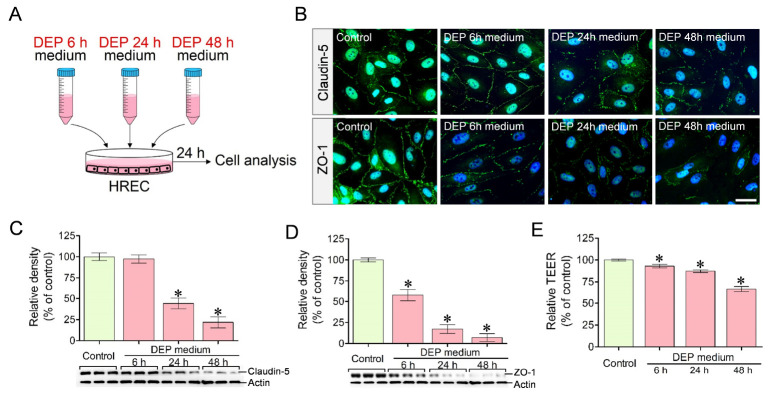
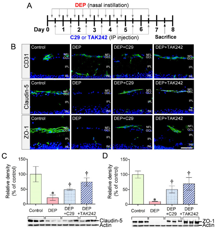
Diesel exhaust particles (DEPs), a major component of air pollution, are well-known to induce inflammation and vascular dysfunction. However, the molecular mechanisms linking DEP exposure to the disruption of the blood–retina barrier (BRB) remain poorly understood. Toll-like receptors (TLRs), particularly TLR2 and TLR4, play critical roles in inflammatory signaling and may contribute to DEP-induced retinal endothelial dysfunction. This study investigates the involvement of TLR2 and TLR4 in mediating DEP-induced disruption of the BRB and evaluates the protective effects of TLR inhibition using both in vitro and in vivo experiments. U937 human macrophages were exposed to DEPs of ultrafine size (<0.2 μm), and the mRNA expression of TNF-α and IL-1β was quantified. Conditioned media from DEP-exposed U937 cultures were then used to treat human retinal endothelial cells (HRECs). DEP exposure significantly increased TNF-α and IL-1β mRNA expression in U937 macrophages. Conditioned media from DEP-exposed U937 macrophages reduced claudin-5 and ZO-1 expression in HRECs, resulting in increased BRB permeability. Inhibition of TLR2 and TLR4 using C29 and TAK242, respectively, significantly attenuated TNF-α and IL-1β mRNA expression in DEP-exposed U937 macrophages and preserved BRB integrity by maintaining claudin-5 and ZO-1 expression in HRECs. In the mouse model, DEP exposure caused a marked reduction in claudin-5 and ZO-1 levels in retinal vessels, whereas treatment with C29 and TAK242 mitigated the loss of these tight junction proteins. This study demonstrates that DEPs induce inflammation and BRB dysfunction through TLR2 and TLR4 activation, leading to increased vascular permeability and potential retinal damage. Furthermore, TLR2 and TLR4 inhibition may be a promising therapeutic strategy to protect retinal health from air pollution–induced damage. [BMB Reports 2025; 58(7): 300-306].
Conflict of interest statement
The authors have no conflicting interests.
Figures


Similar articles
-
Probiotics Function as Immunomodulators in the Intestine in C57Bl/6 Male Mice Exposed to Inhaled Diesel Exhaust Particles on a High-Fat Diet.Cells. 2022 Apr 25;11(9):1445. doi: 10.3390/cells11091445. Cells. 2022. PMID: 35563751 Free PMC article.
-
The interventional role and mechanism of total flavonoids in lychee seeds on rats with liver fibrosis.Sci Rep. 2025 Jul 7;15(1):24320. doi: 10.1038/s41598-025-10007-z. Sci Rep. 2025. PMID: 40624122 Free PMC article.
-
The foreign body response to biomaterial implants is reduced by co-inhibition of TLR2 and TLR4.Acta Biomater. 2025 Jul 1;201:320-335. doi: 10.1016/j.actbio.2025.06.020. Epub 2025 Jun 17. Acta Biomater. 2025. PMID: 40516838
-
Toll like receptor expression induced by exercise in obesity and metabolic syndrome: A systematic review.Exerc Immunol Rev. 2018;24:60-71. Exerc Immunol Rev. 2018. PMID: 29461969
-
Role of Toll-like receptor 2 during infection of Leptospira spp: A systematic review.PLoS One. 2024 Dec 27;19(12):e0312466. doi: 10.1371/journal.pone.0312466. eCollection 2024. PLoS One. 2024. PMID: 39729468 Free PMC article.
References
-
- Andrysík Z, Vondráček J, Marvanová S, et al. Activation of the aryl hydrocarbon receptor is the major toxic mode of action of an organic extract of a reference urban dust particulate matter mixture: the role of polycyclic aromatic hydrocarbons. Mutat Res. 2011;714:53–62. doi: 10.1016/j.mrfmmm.2011.06.011. - DOI - PubMed
MeSH terms
Substances
LinkOut - more resources
Full Text Sources
