

Unilateral pubic hair growth: a paracrine finding indicative of underlying leydig cell tumor in a prepubertal boy
- PMID: 40496178
- PMCID: PMC12148672
- DOI: 10.1016/j.eucr.2025.103071
Unilateral pubic hair growth: a paracrine finding indicative of underlying leydig cell tumor in a prepubertal boy
Abstract
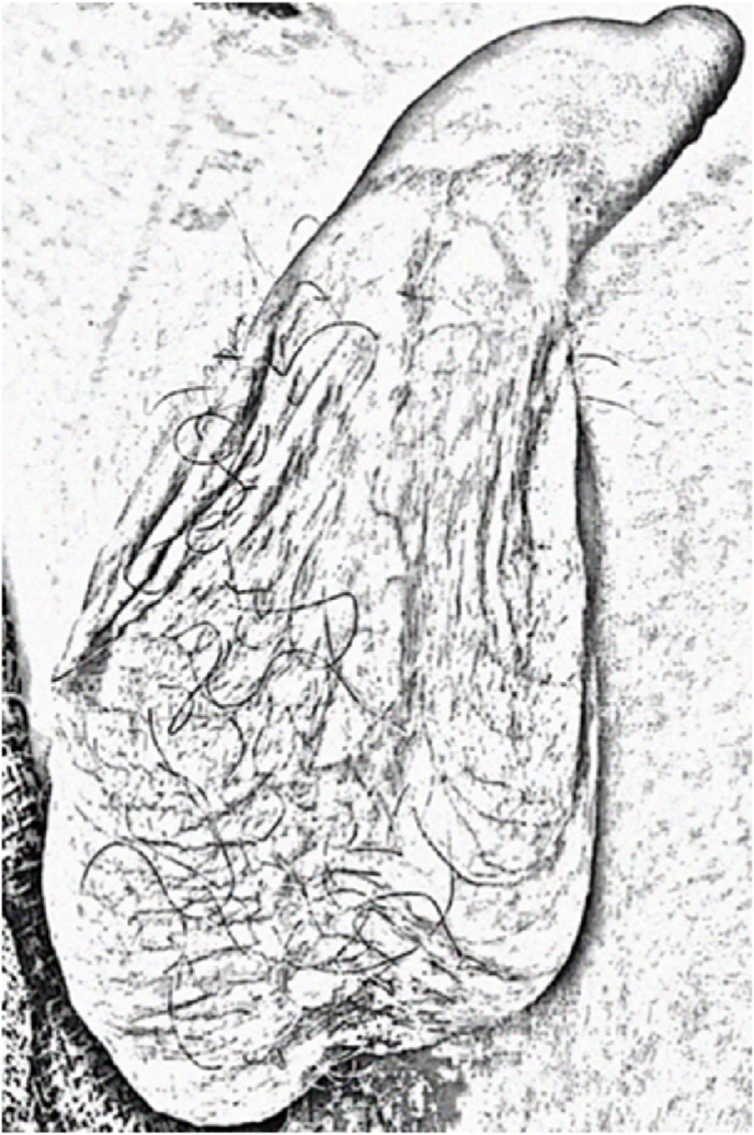
Leydig cell tumors (LCTs) are rare in children and typically present with testicular masses and signs of early puberty. We report a case of a 6-year-old boy presenting solely with precocious pubic hair growth. Ultrasound revealed a small right testicular mass and elevated serum testosterone. Intra-operatively, hair growth was more prominent on the affected hemi-scrotum. Testis-sparing surgery confirmed a benign LCT with adjacent increased spermatogenesis. This is the first known report of ipsilateral pubic hair as a presenting sign of LCT. Localized androgen excess may be an early clinical indicator to guide timely diagnosis and treatment.
Keywords: Leydig cell tumor; Paracrine effects; Precocious puberty; Pubic hair; Sex cord-gonadal stromal tumor; Testis mass; Testis neoplasm; Testis-sparing surgery.
© 2025 The Authors.
Figures


Similar articles
-
Precocious Puberty Associated with Testicular Hormone-secreting Leydig Cell Tumor.Cureus. 2019 Dec 22;11(12):e6441. doi: 10.7759/cureus.6441. Cureus. 2019. PMID: 31893190 Free PMC article.
-
Precocious Pseudo-Puberty with Testicular Enlargement: Two Cases of Leydig Cell Tumor with Different Histopathological Results.Res Rep Urol. 2020 Nov 23;12:577-582. doi: 10.2147/RRU.S277216. eCollection 2020. Res Rep Urol. 2020. PMID: 33262958 Free PMC article.
-
Spermatogenesis in pre-pubertal boys with Leydig cell neoplasms suggests paracrine stimulation by testosterone.J Pediatr Urol. 2021 Feb;17(1):48.e1-48.e6. doi: 10.1016/j.jpurol.2020.10.015. Epub 2020 Oct 15. J Pediatr Urol. 2021. PMID: 33129671
-
Leydig cell hyperplasia in children: Case series and review.J Pediatr Urol. 2017 Apr;13(2):158-163. doi: 10.1016/j.jpurol.2016.12.028. Epub 2017 Feb 10. J Pediatr Urol. 2017. PMID: 28238607 Review.
-
Benign scrotal masses in children - some new lessons learned.J Pediatr Surg. 2016 Oct;51(10):1737-42. doi: 10.1016/j.jpedsurg.2016.07.016. Epub 2016 Aug 5. J Pediatr Surg. 2016. PMID: 27558482 Review.
References
-
- Pohl H.G., Shukla A.R., Metcalf P.D., et al. Prepubertal testis tumors: actual prevalence rate of histological types. J Urol. 2004;172(6 Part 1):2370–2372. - PubMed
-
- Ahmed H.U., Arya M., Muneer A., Mushtaq I., Sebire N.J. Testicular and paratesticular tumours in the prepubertal population. Lancet Oncol. 2010;11(5):476–483. - PubMed
-
- Woodward P.J., Schwab C.M., Sesterhenn I.A. From the archives of the AFIP: extratesticular scrotal masses: radiologic-pathologic correlation. Radiographics. 2003;23(1):215–240. - PubMed
-
- Moul J.W. Timely diagnosis of testicular cancer. Urol Clin. 2007;34(2):109–117. - PubMed
Publication types
LinkOut - more resources
Full Text Sources
