

Pancreas cystic lesion with surprise findings on confocal laser endomicroscopy
- PMID: 40496486
- PMCID: PMC12146039
- DOI: 10.1016/j.vgie.2025.02.005
Pancreas cystic lesion with surprise findings on confocal laser endomicroscopy
Abstract
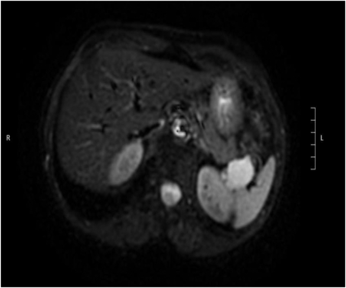
Background and aims: The incidental diagnosis of pancreatic cystic lesions has been increasing, ranging from 2% to 45%, as determined by computed tomography or magnetic resonance imaging. This report describes the case of a 74-year-old female patient referred for a finding on magnetic resonance imaging of a unilocular cystic lesion in the pancreas tail.
Methods: Based on the cyst's size and its unclear nature, the patient was subjected to a repeat EUS at our institution, which showed an anechoic 35 × 20-mm finely septated lesion in the pancreatic tail. To help determine the nature of the cyst, EUS-guided needle-based confocal laser endomicroscopy (EUS-nCLE) was used because of its ability to visualize the cyst wall mucosal layer to a micrometer resolution.
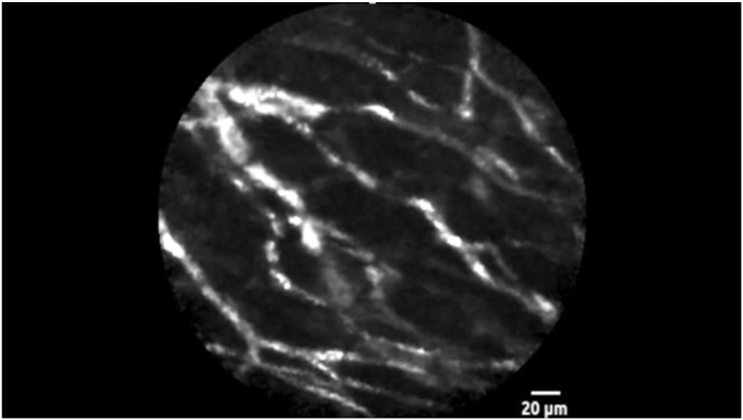
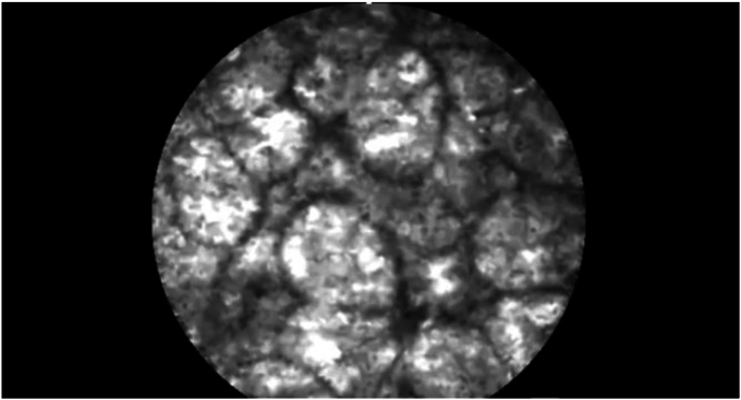
Results: EUS-nCLE of the cyst wall showed an intersecting network of vessels, with background arrangement of gray oval structures and at times background arrangement of dark lobular structures. The findings were not consistent with a mucinous pancreas cyst, serous cystadenoma, or cystic neuroendocrine tumor. As for the NGS, the cyst fluid was positive for both KRAS and PIK3CA pathogenic mutations. After the patient's distal pancreatectomy, histologic examination of the lesion entirely revealed a vascular malformation. The lesion did not have a distinct wall and was surrounded by fat and pancreas tissue. This vascular malformation is a form of lymphangioma.
Conclusion: Lymphangiomas of the pancreas are rare, accounting for 0.2% of all pancreatic lesions. Targeted NGS performed at our institution on the surgical specimen showed absence of KRAS and PIK3CA mutations, suggesting an erroneous or false-positive initial analysis of the cyst fluid. The gray oval structures observed during EUS-nCLE correspond to adipocytes marking part of the cyst border. The dark lobular structures (coffee beans) observed during EUS-nCLE correspond to pancreatic acini marking another part of the cyst border. When these EUS-nCLE patterns are observed in a pancreas cystic lesion in the absence of any epithelial pattern, close follow-up with cross-sectional imaging should be considered instead, especially if the lesion is in a pancreas location that entails major surgery.
© 2025 American Society for Gastrointestinal Endoscopy. Published by Elsevier Inc.
Conflict of interest statement
The following authors disclosed financial relationships: S. El-Dika: Consultant for Boston Scientific. All other authors disclosed no financial relationships.
Figures
References
-
- Krishna S.G., Brugge W.R., Dewitt J.M., et al. Needle-based confocal laser endomicroscopy for the diagnosis of pancreatic cystic lesions: an international external interobserver and intraobserver study (with videos) [published correction appears in. Gastrointest Endosc. 2018;87:1599. - PubMed
LinkOut - more resources
Full Text Sources
Research Materials
Miscellaneous
