Defining the end of puberty in boys: INSL3 and the acute determinants of adult Leydig-cell functional capacity
- PMID: 40496564
- PMCID: PMC12148858
- DOI: 10.3389/fendo.2025.1574760
Defining the end of puberty in boys: INSL3 and the acute determinants of adult Leydig-cell functional capacity
Abstract
Introduction: Testicular Leydig cells are responsible for producing almost all the testosterone required by men throughout the lifespan, with reduced testosterone (hypogonadism) correlating with age-linked morbidity and mortality. Leydig cells derive from stem cells within the testes after birth. These undergo proliferation and differentiation during puberty to achieve their final adult status in young adulthood, after which there appears to be no further cell division and only very limited attrition into old age. Leydig-cell functional capacity reflects the total number and differentiation status of the Leydig-cell population within an individual and can be assessed by measuring in blood the constitutive Leydig-cell hormone insulin-like peptide 3 (INSL3). In adult men, this varies by more than 10-fold between individuals and correlates with later morbidity. Such INSL3 variance appears to have its origin already in young men, though what determines this is largely unknown.
Methods: Here, we have used the ALSPAC (Avon Longitudinal Study of Parents and Children) cohort of boys and young men to estimate when the adult-type Leydig-cell population becomes established, that is, when puberty ends, and the contemporary anthropometric and lifestyle parameters that influence this.
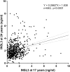
Results and discussion: At 17 years, mean INSL3 is not yet maximal, with high variance due to both longitudinal (timing of pubertal trajectory) and cross-sectional influences, whereas at 24 years, circulating INSL3 concentration has stabilized to its final adult status, even showing a small decreasing trend with age. Maximal INSL3 (i.e., peak puberty) was calculated to be at approximately 22 years in this cohort. Both contemporary body mass index and smoking status, though not inflammatory parameters, were contributory factors to INSL3 concentration. However, the major source of INSL3 variance in young men was shown to be already established at 17 years, with causative influences evidently occurring prior to this age, and showing that early life parameters are important for determining later adult health in men.
Keywords: ALSPAC; INSL3; Leydig cell; hypogonadism; puberty.
Copyright © 2025 Tulumcu, Ivell, Alhujaili and Anand-Ivell.
Conflict of interest statement
The authors declare that the research was conducted in the absence of any commercial or financial relationships that could be construed as a potential conflict of interest. The author(s), RI and RA-I, declared that they were an editorial board member of Frontiers, at the time of submission. This had no impact on the peer review process and the final decision.
Figures


Similar articles
-
The Leydig cell biomarker INSL3 as a predictor of age-related morbidity: Findings from the EMAS cohort.Front Endocrinol (Lausanne). 2022 Nov 8;13:1016107. doi: 10.3389/fendo.2022.1016107. eCollection 2022. Front Endocrinol (Lausanne). 2022. PMID: 36425465 Free PMC article.
-
Male seminal parameters are not associated with Leydig cell functional capacity in men.Andrology. 2021 Jul;9(4):1126-1136. doi: 10.1111/andr.13001. Epub 2021 Mar 24. Andrology. 2021. PMID: 33715296
-
Association of age, hormonal, and lifestyle factors with the Leydig cell biomarker INSL3 in aging men from the European Male Aging Study cohort.Andrology. 2022 Oct;10(7):1328-1338. doi: 10.1111/andr.13220. Epub 2022 Jul 11. Andrology. 2022. PMID: 35770372 Free PMC article.
-
INSL3 as a biomarker of Leydig cell functionality.Biol Reprod. 2013 Jun 13;88(6):147. doi: 10.1095/biolreprod.113.108969. Print 2013 Jun. Biol Reprod. 2013. PMID: 23595905 Review.
-
INSL3: A Marker of Leydig Cell Function and Testis-Bone-Skeletal Muscle Network.Protein Pept Lett. 2020;27(12):1246-1252. doi: 10.2174/0929866527666200925105739. Protein Pept Lett. 2020. PMID: 32981493 Review.
References
MeSH terms
Substances
Grants and funding
LinkOut - more resources
Full Text Sources
Medical

