

From virtual to reality: application of a novel 3D printing hollow model for early-stage lung cancer in the clinical teaching of thoracoscopic sublobar resection
- PMID: 40496613
- PMCID: PMC12149187
- DOI: 10.3389/fonc.2025.1526592
From virtual to reality: application of a novel 3D printing hollow model for early-stage lung cancer in the clinical teaching of thoracoscopic sublobar resection
Abstract
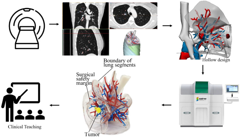
Background: The integration of medical-engineering interdisciplinary technology has transformed clinical skills and anatomical knowledge teaching. Three-dimensional printing (3DP), an innovative tool, shows promise in enhancing surgical training and anatomical understanding. This study evaluates the educational efficacy of a 3DP lung cancer model optimized for surgery in teaching thoracoscopic sublobar resection.
Methods: A total of 62 clinical interns were randomly assigned into two groups: a 3D visualization (3DV) model group and a 3DP model group. Pre- and post-teaching test scores were compared to assess the effectiveness of both models in enhancing anatomical knowledge and surgical skills. Additionally, feedback was collected from the interns regarding the advantages of each model.
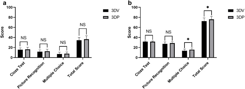
Results: There was no significant difference in the pre-teaching test scores between the two groups (P > 0.05). However, post-teaching scores in the 3DP group were significantly higher than those in the 3DV group (P < 0.05). Survey feedback revealed that the 3DV group excelled in convenience (P < 0.001), while the 3DP group demonstrated superiority in the ease of knowledge acquisition and understanding of vascular spatial relationships (P < 0.001). No significant differences were found between the two groups regarding model intuitiveness and identification of the lung segment range influenced by the safety margin (P > 0.05).
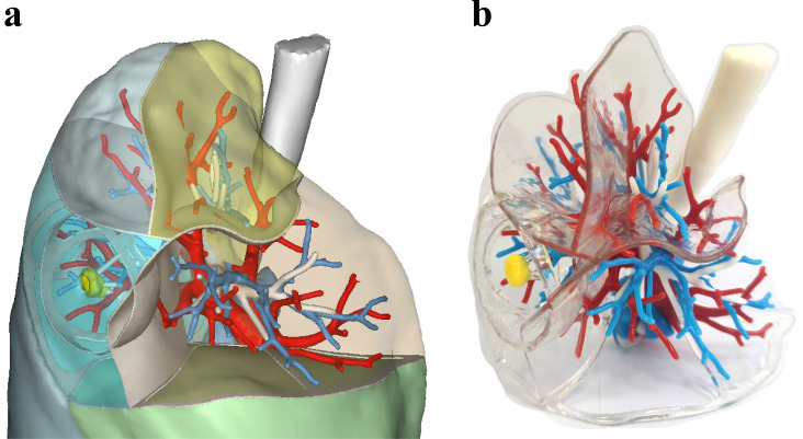
Conclusion: The 3DP model, featuring a transparent hollow sublobar boundary, significantly improved comprehension of complex anatomical relationships and enhanced teaching outcomes in surgical skills. It offers an innovative and effective tool for teaching thoracoscopic sublobar resection, with potential applications in surgical navigation.
Keywords: 3D printing; 3D visualization; clinical teaching; medical education; sublobar resection.
Copyright © 2025 Yang, Miao, Li, Yu, Guo, Li, Zhang, Cheng, Li and Zhang.
Conflict of interest statement
The authors declare that the research was conducted in the absence of any commercial or financial relationships that could be construed as a potential conflict of interest.
Figures
Similar articles
-
Do Three-dimensional Visualization and Three-dimensional Printing Improve Hepatic Segment Anatomy Teaching? A Randomized Controlled Study.J Surg Educ. 2016 Mar-Apr;73(2):264-9. doi: 10.1016/j.jsurg.2015.10.002. J Surg Educ. 2016. PMID: 26868314 Clinical Trial.
-
Integration of case-based learning and three-dimensional printing for tetralogy of fallot instruction in clinical medical undergraduates: a randomized controlled trial.BMC Med Educ. 2024 May 24;24(1):571. doi: 10.1186/s12909-024-05583-z. BMC Med Educ. 2024. PMID: 38789956 Free PMC article. Clinical Trial.
-
Full-sized realistic 3D printed models of liver and tumour anatomy: a useful tool for the clinical medicine education of beginning trainees.BMC Med Educ. 2023 Aug 15;23(1):574. doi: 10.1186/s12909-023-04535-3. BMC Med Educ. 2023. PMID: 37582729 Free PMC article.
-
Role of 3D printing technology in paediatric teaching and training: a systematic review.BMJ Paediatr Open. 2021 Dec;5(1):e001050. doi: 10.1136/bmjpo-2021-001050. BMJ Paediatr Open. 2021. PMID: 35290958 Free PMC article.
-
Three-dimensional printing in anatomy teaching: current evidence.Surg Radiol Anat. 2020 Jul;42(7):835-841. doi: 10.1007/s00276-020-02470-2. Epub 2020 Apr 28. Surg Radiol Anat. 2020. PMID: 32346753 Review.
References
-
- Ngo TD, Kashani A, Imbalzano G, Nguyen KTQ, Hui D. Additive manufacturing (3D printing): A review of materials, methods, applications and challenges. Composites Part B: Engineering. (2018) 143:172–96. doi: 10.1016/j.compositesb.2018.02.012 - DOI
-
- Zhao J, Gong X, Ding J, Xiong K, Zhuang K, Huang R, et al. Integration of case-based learning and three-dimensional printing for tetralogy of fallot instruction in clinical medical undergraduates: a randomized controlled trial. BMC Med Educ. (2024) 24:571. doi: 10.1186/s12909-024-05583-z - DOI - PMC - PubMed
LinkOut - more resources
Full Text Sources