. 2025 May 21:51:431-449.
doi: 10.1016/j.bioactmat.2025.05.012. eCollection 2025 Sep.

Smart biomimetic "nano-med-fireman" blocking inflammation and lactate metabolism crosstalk for normalized spatiotemporal photo-immunotherapy

Affiliations

Smart biomimetic "nano-med-fireman" blocking inflammation and lactate metabolism crosstalk for normalized spatiotemporal photo-immunotherapy

Honglin Huang et al. Bioact Mater. .

Abstract

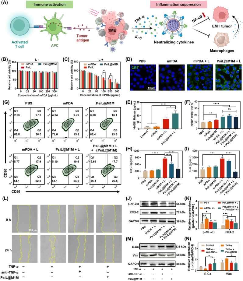
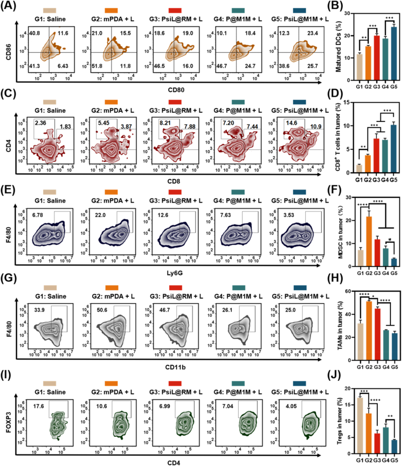
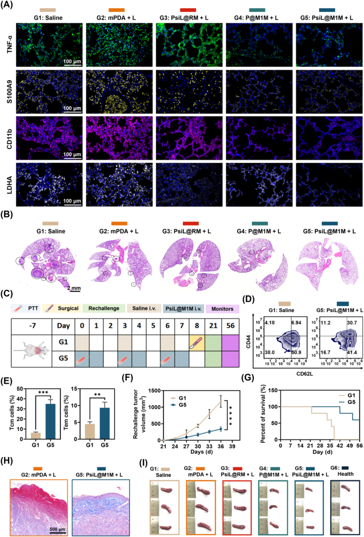
The immense complexity and interconnectedness of inflammation and metabolism in the primary tumor ecosystem and pre-metastatic niches (PMN) present an enormous challenge for developing advanced nano-medicines for cancer immunotherapy. Herein, an intelligent tumor- and PMN-tropic bioactive "nano-med-fireman" (PsiL@M1M) was developed to not only arouse an appropriate photothermal immune cascade but also to "extinguish" the accompanying excessive inflammation. Ultimately, it aims to revitalize the immunosuppressive tumor microenvironment (TME) to spatiotemporally and effectively inhibit tumor metastasis and recurrence. PsiL@M1M was devised by incorporating the siRNA of lactate dehydrogenase A (siLDHA) on the functionalized photothermal mesoporous polydopamine (mPDA) and coupled with an M1-type macrophage membrane (M1M) to enable the capacity of inflammation targeting and modulation. PsiL@M1M actively accumulated and destroyed the primary tumor via photothermal therapy and subsequently mitigated the photothermal therapy-induced inflammatory cascade (e.g., epithelial mesenchymal transition) via cytokine neutralization, eliminating the supply of tumor-derived secretory factors as "nutrients" for PMN. Concurrently, siLDHA interfered with lactate (LA) production, inhibited inflammation-LA communication and relieved immune checkpoint, thus profoundly reversing the immunosuppressive microenvironment of both the primary tumor and PMN. Through cooperation, PsiL@M1M initiated normalized hypo-inflammatory photo-immunotherapy against both local tumor growth and spontaneous metastasis/recurrence. Our study provides a paradigm for a new generation of photothermal nano-medicines for the whole process of tumor treatment.

Keywords: Cytokine neutralization; Inflammation modulation; Lactate metabolism regulation; Photothermal immunotherapy; Pre-metastatic niche reprogramming.


Conflict of interest statement

The authors declare that they have no conflicts of interest.

Figures

Image 1
Graphical abstract
Scheme 1
Scheme 1
Schematic representation of (A) the construction of “nano-med-fireman” (PsiL@M1M) and (B) the elicited spatiotemporal and hypo-inflammatory photo-immunotherapy against primary tumor and lung metastasis by inflammation neutralization and LA metabolic modulation.
Fig. 1
Fig. 1
Characterization of “nano-med-fireman” PsiL@M1M. (A) TEM images of mPDA and PsiL@M1M. (B) Size and surface zeta potential of mPDA, mPDAD, PsiL, and PsiL@M1M. (C) Agarose gel analysis with different weight ratios of siLDHA/mPDAD (from 1:1 to 1:6). The expression of pro-inflammatory cytokine receptors, including (D) TNFR and (E) IL-6R, in macrophages with or without LPS stimulation was evaluated by flow cytometry. Characterized proteins of M1Φ, M1M, and PsiL@M1M were compared by (F) Western blot and (G) SDS‒PAGE. (H) Changes in the hydrodynamic size and PDI of PsiL@M1M in saline over 7 days. (I) Infrared thermal images of mPDA and PsiL@M1M (equivalent to mPDA = 200 μg/mL). (J) Cycled heating profile of PsiL@M1M. (K) Cumulative release of siLDHA during the laser on/off treatments at different pH values (7.4/5.5). (L) Workflow of the in vitro inflammatory cytokine neutralization assay. (M) PsiL@M1M (200 μg/mL) neutralizes TNF-α and IL-6 capacity at different time points. (N) TNF-α or (O) IL-6 removal with PsiL@M1M, compared with mPDA, PsiL@RM, and PsiL@M0M. Quantification of the neutralization rate of (P) TNF-α or (Q) IL-6 with a fixed amount of TNF-α or IL-6 (500 pg/mL) while varying the amount of added PsiL@M1M (0–400 μg/mL). The Kd and Bmax of PsiL@M1M for (R) TNF-α and (S) IL-6. (T) TNF-α and IL-6 release ratio after neutralization with PsiL@M1M (200 μg/mL).
Fig. 2
Fig. 2
Hypo-inflammatory photo-immunotherapy mediated by PsiL@M1Min vitro. (A) Schematic representation of PsiL@M1M-mediated immune activation and inflammation suppression. Viability of 4T1 cancer cells treated with PsiL@M1M and relevant controls (B) in the dark (C) or with laser irradiation. (D) Surface-exposed CRT in 4T1 cells and (E) HMGB1 release after treatment under different conditions. (F) Percentages of CD80 and CD86-positive DCs under different stimulation conditions and (G) representative flow cytometry plots. Concentrations of (H) TNF-α and (I) IL-6 in the supernatants of DC2.4 cells cocultured with 4T1 cells treated with different nanoformulations. (J) Western blot and (K) corresponding quantification of inflammation-associated proteins. (L) Cell migration was evaluated by a scratch wound healing assay. (M) Western blot and (N) corresponding quantification of EMT-associated proteins.
Fig. 3
Fig. 3
Targeted regulation of LA metabolism and inhibition of immune checkpoints by PsiL@M1M. (A) Schematic representation of the effect of intracellular gene silencing of PsiL@M1M on PD-L1 and macrophage. Flow cytometry analysis of the cellular uptake of free siLDHA, PsiL, and PsiL@M1M in (B) 4T1 or (C) M2 Φ. (D) Confocal fluorescence images of 4T1 cells incubated with PsiL@M1M for 2 h or 6 h and (EF) the red/green fluorescence overlapping curves. (G) LDHA mRNA levels in 4T1 cells subjected to different treatments measured via RT‒qPCR, and (H) LDHA protein expression measured via Western blot. (I) Concentrations of LA and (J) pH values in the supernatants of 4T1 cells. Immunofluorescence of PD-L1 (K) and its protein expression (L) in 4T1 cells. (M) Representative images and (N) quantification of the relative migration of macrophages from the transwell experiments.
Fig. 4
Fig. 4
Targeted capacity of PsiL@M1M to primary tumor and lung PMN in vivo. (A) In vivo fluorescence imaging of the whole body (yellow circles indicate tumors) in orthotropic 4T1 tumor-bearing mice. (B) Images of the organs and tumors of the mice in each group at 48 h and (C) semiquantitative analysis of fluorescence intensity. (D) HE images and immunohistochemical staining of TNF-α, IL-6, S100A9, and CD11b (brown) in the lung tissues of different groups of mice (the black arrow indicates aberrant aggregation of inflammatory cells). (E) Plasma TNF-α and IL-6 levels were measured by ELISA in each group. (F) In vivo fluorescence imaging of lungs from orthotropic 4T1 tumor-bearing mice.
Fig. 5
Fig. 5
In vivo antitumor efficacy of PsiL@M1M in tumor-bearing mice. (A) Workflow of PsiL@M1M in tumor-bearing mice. (B) Changes in the relative tumor volume of the mice treated with different formulations for 21 days. (C) Sacrificed tumor tissue images and (D) the mean tumor weight on day 21. (E) HE and immunofluorescence staining of CRT, (F) TNF-α, and IL-6 in tumor sections. (G) Plasma TNF-α and IL-6 levels measured by ELISA in each group. (H) Immunofluorescence staining of LDHA and PD-L1 in tumor sections. (I) Concentrations of LA in the tumor homogenates.
Fig. 6
Fig. 6
TME reprogramming by disrupting the crosstalk of inflammation and LA metabolism. (A) Representative flow cytometry plots and (B) corresponding percentages of mature DCs in the lymph nodes. (C) Representative flow cytometry plots and (D) corresponding percentages of antitumor immune cells (CD3+ CD8+) in the primary tumor after treatment. Representative flow cytometry panel and corresponding percentages of MDSCs (E, F), TAMs (G, H), and Tregs (I, J) in the primary tumor after treatment.
Fig. 7
Fig. 7
Transcriptomic analysis. (A) Venn diagram of the genes detected in the saline, mPDA + L, and PsiL@M1M + L groups. (B) Volcano plot of DEGs in the saline vs. mPDA + L group and (C) saline vs. PsiL@M1M + L group. (D) Heatmap of the 60 DEGs related to immune suppression, positive immune regulation, EMT processes, and LA metabolism-related genes in the saline, mPDA + L, and PsiL@M1M + L groups. (E) GO enrichment analyses of DEGs in the saline vs. mPDA + L groups, (F) and saline vs. PsiL@M1M + L.
Fig. 8
Fig. 8
PMN microenvironment remodeling and spontaneous tumor metastasis and recurrence inhibition by PsiL@M1M. (A) Immunofluorescence staining of TNF-α, S100A9, CD11b, and LDHA in lung tissues after various treatments. (B) HE analysis of lung sections. The black circles denote the metastatic foci. (C) Workflow of the immune memory and tumor rechallenge study. (D) Representative flow cytometry plots and (E) corresponding percentages of Tcm and Tem cells in the spleen on day 21. (F) Growth curve of rechallenged tumors in 4T1 tumor-bearing mice with various treatments. (G) Survival rate of the mice after tumor rechallenge. (H) Masson staining of tumor and skin tissues after various treatments. (I) Morphological changes ofthe spleen after various treatments for 21 days.

Similar articles

References

    1. Hanahan D. Hallmarks of cancer: new dimensions. Cancer Discov. 2022;12(1):31–46. - PubMed
    1. Propper D.J., Balkwill F.R. Harnessing cytokines and chemokines for cancer therapy. Nat. Rev. Clin. Oncol. 2022;19(4):237–253. - PubMed
    1. de Visser K.E., Joyce J.A. The evolving tumor microenvironment: from cancer initiation to metastatic outgrowth. Cancer Cell. 2023;41(3):374–403. - PubMed
    1. Diakos C.I., Charles K.A., McMillan D.C., Clarke S.J. Cancer-related inflammation and treatment effectiveness. Lancet Oncol. 2014;15(11):e493–503. - PubMed
    1. Massagué J., Obenauf A.C. Metastatic colonization by circulating tumour cells. Nature. 2016;529(7586):298–306. - PMC - PubMed

LinkOut - more resources