

Distinct changes in the morphology of cortical and subcortical grey matter associated with age-related hearing loss and tinnitus in the UK Biobank participants
- PMID: 40496672
- PMCID: PMC12149739
- DOI: 10.1093/braincomms/fcaf203
Distinct changes in the morphology of cortical and subcortical grey matter associated with age-related hearing loss and tinnitus in the UK Biobank participants
Abstract
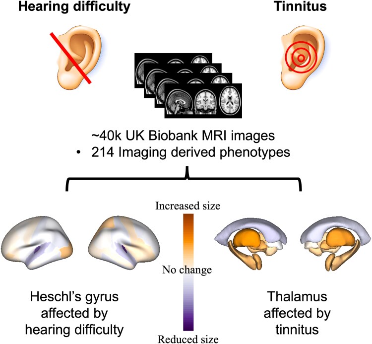
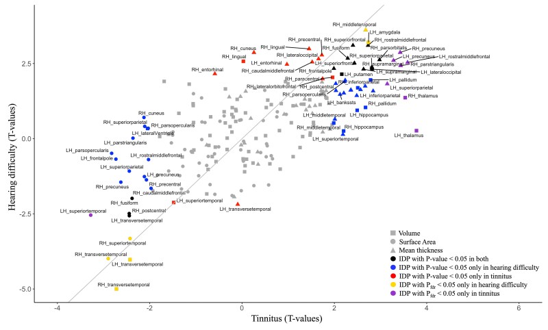
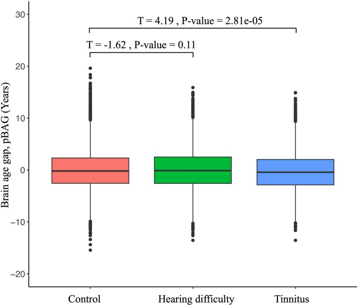
Prevalence of both hearing loss and tinnitus increases with age. However, neuroimaging studies of both conditions report inconsistent changes in brain morphology likely due to small sample size and variable methodology. Structural and functional neuroimaging studies in hearing loss and tinnitus have revealed distinct neural correlates, and further replication is needed to confirm these findings. This study aims to investigate the effects of hearing loss and tinnitus on the brain morphology in a well-powered sample. We utilized self-reported hearing difficulty and tinnitus in participants with magnetic resonance imaging (MRI) in the UK Biobank cohort. Control participants without hearing difficulty and tinnitus were age and sex matched leading to total sample sizes of 13 074 and 6242 for self-reported hearing difficulty and tinnitus, respectively. We utilized the rich UK Biobank dataset (i) to reveal these brain changes in a well-powered large study of hearing loss and tinnitus, (ii) to document the effect of confounding factors on these associations, (iii) to discriminate the effects of tinnitus versus hearing difficulty on the brain and (iv) to estimate the brain-age gap in hearing difficulty and tinnitus subjects compared with controls. Hearing difficulty is significantly associated with smaller grey matter volumes exclusively in the bilateral transverse temporal regions, whereas tinnitus is associated with larger volumes of bilateral hippocampi and thalami when compared with the control group. Furthermore, correcting for confounders (i.e. diabetes, cardiovascular disease, age, sex, smoking, alcohol consumption and Townsend deprivation index) during statistical analysis helped to better delineate the impact of hearing status on brain structural changes. The brain-age gap analysis showed that participants with tinnitus appeared to have significantly younger brains than controls, whereas participants with hearing difficulty did not differ significantly from the control group. Altogether, our results confirmed previous findings and suggest the enlargement of bilateral thalami as the main effect in people with tinnitus. We also established that there are independent and distinct brain pathologies between hearing difficulty and tinnitus. Therefore, the self-reported measure is a reasonable approach to assess the hearing loss and tinnitus pathologies.
Keywords: Heschl’s gyrus; age-related hearing loss; neuroimaging; thalamus; tinnitus.
© The Author(s) 2025. Published by Oxford University Press on behalf of the Guarantors of Brain.
Conflict of interest statement
The authors report no competing interests.
Figures
Similar articles
-
Neuroanatomical Alterations in Tinnitus Assessed with Magnetic Resonance Imaging.Front Aging Neurosci. 2016 Sep 21;8:221. doi: 10.3389/fnagi.2016.00221. eCollection 2016. Front Aging Neurosci. 2016. PMID: 27708577 Free PMC article.
-
Reduced volume of Heschl's gyrus in tinnitus.Neuroimage. 2009 Apr 15;45(3):927-39. doi: 10.1016/j.neuroimage.2008.12.045. Epub 2009 Jan 6. Neuroimage. 2009. PMID: 19168138
-
Altered Amplitude of Low-Frequency Fluctuations and Degree Centrality in Patients with Acute Subjective Tinnitus: A Resting-State Functional Magnetic Resonance Imaging Study.J Integr Neurosci. 2022 Jun 21;21(4):116. doi: 10.31083/j.jin2104116. J Integr Neurosci. 2022. PMID: 35864767
-
The thalamus and tinnitus: Bridging the gap between animal data and findings in humans.Hear Res. 2021 Aug;407:108280. doi: 10.1016/j.heares.2021.108280. Epub 2021 Jun 13. Hear Res. 2021. PMID: 34175683 Review.
-
A Pilot Study to Evaluate a Residual Inhibition Technique in Hearing Aids for Suppression of Tinnitus.Semin Hear. 2023 Jun 28;45(1):123-140. doi: 10.1055/s-0043-1770153. eCollection 2024 Feb. Semin Hear. 2023. PMID: 38370522 Free PMC article. Review.
References
-
- World Health Organization. World report on hearing. World Health Organization; 2021. ISBN: 9789240020481. Accessed 3 March 2021. https://iris.who.int/handle/10665/339913
-
- Gates GA, Mills JH. Presbycusis. The Lancet. 2005;366(9491):1111–1120. - PubMed
LinkOut - more resources
Full Text Sources
Miscellaneous