. 2025 May 20:32:101891.
doi: 10.1016/j.mtbio.2025.101891. eCollection 2025 Jun.

A 'two-missile' nanoplatform for targeting triple-negative breast cancer: prodrug activation and immune enhancement

Affiliations

A 'two-missile' nanoplatform for targeting triple-negative breast cancer: prodrug activation and immune enhancement

Haiping Hu et al. Mater Today Bio. .

Abstract

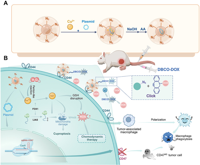
Targeted cancer therapy remains a significant challenge due to the complexity of tumor microenvironments and the need for precise drug delivery systems that can overcome these obstacles. To address this challenge, a nanoparticle platform based on prodrug activation and immune enhancement for "two-missile" targeting of TNBC has been developed: Azido-HA@Cu2O@DNA. This nanoparticle platform utilises azido hyaluronic acid (Azido-HA) as the primary carrier to enhance specificity to tumor tissues and targeting to tumor cells. The bioorthogonal reaction between the azido moiety and dibenzocyclooctyne-doxorubicin (DBCO-DOX) has been employed to improve the tumor targeting efficiency of chemotherapy is significantly enhanced, while the addition of copper ions promotes a Fenton-like reaction that induces cellular copper death. This mechanism not only spawns reactive oxygen species (ROS) for potent chemokinetic therapy but also facilitates the transition of macrophages from the M2 to the M1 phenotype, thereby bolstering anti-tumor immune responses. In addition, the platform's accompanying CRISPR/Cas9 system can reprogram tumor cells by down-regulating CD47 expression, enhancing macrophage recognition and phagocytosis activity, which in turn amplifies the efficacy of ensuing immunotherapy. The Azido-HA@Cu2O@DNA nanoplatform demonstrates excellent biocompatibility and safety, offering a promising strategy for enhancing tumor treatment. This methodology integrates the precise activation of prodrugs, immune system modulation, and macrophage-mediated tumor eradication, offering a holistic and potent paradigm for cancer therapy.

Keywords: Bioorthogonal reaction; CD47; CRISPR/Cas9; Cuproptosis; Macrophage M1 polarization; Macrophage phagocytosis.


Conflict of interest statement

The authors declare that they have no known competing financial interests or personal relationships that could have appeared to influence the work reported in this paper.

Figures

Image 1
Graphical abstract
Fig. 1
Fig. 1
This study presents a novel approach to the therapeutic treatment of triple-negative tumors by enhancing macrophage phagocytosis. The method employs gene-editable copper-based bioorthogonal nanoplatforms, which have been designed to facilitate the targeted delivery of therapeutic agents to tumor sites. (A) Schematic illustration of Azido-HA@Cu2O@DNA preparation. Azido-HA@Cu2O@DNA via a biomineralization method, was synthesized using a biomineralization method. This process involved the initial binding of copper ions to plasmid DNA, followed by the reduction of the copper ions using ascorbic acid and then the functionalization of the resulting complex with azido-HA. (B) The mechanism of Azido-HA@Cu2O@DNA -mediated anticancer therapy. Azido-HA surface modification endows Azido-HA@Cu2O@DNA with the capacity for selective tumor targeting and enhanced cellular uptake. Once Azido-HA@Cu2O@DNA has been targeted to tumor cells, DBCO-DOX is subsequently targeted to tumor cells. Following the entry of Azido-HA@Cu2O@DNA into tumor cells, it undergoes copper death and a Fenton-like reaction that generates ROS for CDT and macrophage M1 polarization. In addition, Azido-HA@Cu2O@DNA inhibits the expression of CD47 in tumor cells through CRISPR/Cas9 gene editing, which blocks the 'don't eat me' signal and enhances macrophage recognition and phagocytosis, thereby providing additional benefit to anti-tumor therapy.
Fig. 2
Fig. 2
Characterization of Azido-HA@Cu2O@DNA. (A) TEM image of Cu2O. (B) TEM image of HA@Cu2O@DNA. (C) TEM image of Azido-HA@Cu2O@DNA. (D) Enlarged section of (C). (E) Size distribution of Cu2O, HA@Cu2O@DNA, and Azido-HA@Cu2O@DNA measured by DLS. (F) Zeta potential of Cu2O, HA@Cu2O@DNA, and Azido-HA@Cu2O@DNA was measured by DLS. (G) XRD spectrum of Azido-HA@Cu2O@DNA. (H) FT-IR spectrum of Azido-HA@Cu2O@DNA, and Azido-HA. (I) TEM image of the variation in particle size of Azido-HA@Cu2O@DNA nanoparticles in the presence of 1 mM H2O2. The data were expressed as the mean ± standard deviation (n = 3).
Fig. 3
Fig. 3
Characterization of Azido-HA@Cu2O@DNA performance. (A) The ability of Azido-HA@Cu2O@DNA to bind DBCO-AF488 in vitro was investigated. (B) Azido-HA@Cu2O@DNA exhibits a concentration-dependent binding affinity for DBCO-AF488. (C) The following is a data analysis of Fig. 3B. (D) The Fenton-like catalytic activity of Azido-HA@Cu2O@DNA was evaluated at varying concentrations (0, 10, 20, 40, 60, and 80 μg/mL). (E) Fenton-like catalytic activities of Azido-HA@Cu2O@DNA under different pH conditions (pH = 5.5, 6.5, and 7.2). (F) Time-dependent Fenton-like catalytic activities of Azido-HA@Cu2O@DNA The data were expressed as the mean ± standard deviation (n = 3), ∗∗∗p < 0.001, two-tailed Student's t-test.
Fig. 4
Fig. 4
In vitro anticancer analysis of Azido-HA@Cu2O@DNA. (A) Cellular uptake profile of Azido-HA@Cu2O@DNA in 4T1 cells. 4T1 cells were incubated with DBCO-AF488-labeled Azido-HA@Cu2O@DNA, and then their uptake was monitored by laser confocal for 1 h and 4 h profiles. (B) Endosomal escape of Azido-HA@Cu2O@DNA in 4T1 cells. 1: 30 min. 2: 1.5 h. 3: 3 h. 4: 4 h. (C) Anticancer effects evaluated by the CCK-8 assay. The 4T1 cells were incubated with different concentrations of DOX, or DBCO-DOX, and Azido-HA@Cu2O@DNA alone, or in combination with DBCO-DOX (10 μM), followed by measuring 24 h cell viability with the CCK-8 assay. (D) Changes in mRNA expression levels of DLAT, FDX1, LTAS genes in 4T1 cells treated with different nanoparticles. (E) Intracellular uptake of Cu ion by 4T1 cells after incubation with Cu2O, Azido-HA@Cu2O, HA@Cu2O@DNA, or Azido-HA@Cu2O@DNA (40 μg/mL) for 12 h. (F) Fluorescence images of intracellular ROS in 4T1 cells. Cells were incubated with Cu2O, HA@Cu2O@DNA or Azido-HA@Cu2O@DNA (40 μg/mL). (G) Mitochondrial membrane potential measured by the JC-1 assay. Cells were incubated with Cu2O, Azido-HA@Cu2O, HA@Cu2O@DNA or Azido-HA@Cu2O@DNA (40 μg/mL). (H) Measurement of intracellular ATP levels. Cells were incubated with Cu2O, Azido-HA@Cu2O, HA@Cu2O@DNA, or Azido-HA@Cu2O@DNA (40 μg/mL) for 12 h, and then intracellular ATP was measured using a commercially available assay kit. The data are presented as the mean ± standard deviation (n = 3). One-way ANOVA with a Tukey post hoc test, ∗P < 0.05, ∗∗P < 0.01, ∗∗∗P < 0.001.
Fig. 5
Fig. 5
In vitro gene editing efficiency and TAM polarization and phagocytosis induced by Azido-HA@Cu2O@DNA. (A) Schematic representation of the polarization assay. (B–D) Levels of TNF-α, IL-6 and IL-10 in cell supernatants. 4T1 and RAW 264.7 cells were co-cultured and then treated with Azido-HA@Cu2O@DNA (40 μg/mL) for 12 h. The levels of cytokine markers were then measured. (E–G) Representative flow diagram of macrophage repolarization from the M2-type to the M1-type. (H) Schematic diagram of Cas9/sgCD47 gene editing. (I, J) Identification of CD47 protein expression in 4T1 cells by protein blotting. 4T1 cells were incubated with Azido-HA@Cu2O@DNA for 12 h and then analyzed by protein blotting. (K) Schematic representation of the phagocytosis assay. (L) Representative images of CMTPX-stained 4T1 cells phagocytosed by CMFDA-stained BMDM cells. (M) Representative images of RAW264.7 cells phagocytosing 4T1-Luc tumor cells following treatment with Azido-HA@Cu2O@DNA (40 μg/mL) for 24 h. (N) The percentage of tumor cells eliminated was calculated based on bioluminescence intensity. The data were expressed as the mean ± standard deviation (n = 3), ∗P < 0.05; two-tailed Student's t-test.
Fig. 6
Fig. 6
In vivo targeting effect of Azido-HA@Cu2O@DNA. (A) Schematic diagram of in vivo targeting experiment. (B) DBCO-Cy5 was injected 1h after the intravenous injection of Azido-HA@Cu2O@DNA nanoparticles, and 24 h later in vivo fluorescence images of 4T1 tumor-bearing mice. (C) Quantification of fluorescence intensity over time in the tumor region after 24 h of intravenous injection of DBCO-Cy5. (D) An intravenous injection of Azido-HA@Cu2O@DNA was administered, followed 1 h later by an injection of DBCO-Cy5. Ex vivo fluorescence images of major organs and tumors were obtained 24 h later. (E, F) Intravenous injection of Azido-HA@Cu2O@DNA nanocomplexes was followed 1h later by injection of DBCO-Cy5, and then fluorescence intensity in major organs and tumors was quantified 24 h later. The data were expressed as the mean ± standard deviation (n = 3), One-way ANOVA with a Tukey post hoc test, ∗∗∗P < 0.001.
Fig. 7
Fig. 7
In vivo anti-tumor effect of combination therapy. (A) The treatment regimen for 4T1 tumor-bearing BALB/c mice. (B) Tumor picture on the 21st day of mice throughout the course of therapy. (C) Tumor weight. (D) Tumor volume. (E) Histological analysis of tumor tissues after 21 days of treatment, including H&E, TUNEL, CD86, and CD206 staining. (F) survival curve. (G) Changes in body weight of mice. (H) Tumor necrosis area ratio. The data were expressed as the mean ± standard deviation (n = 5), One-way ANOVA with a Tukey post hoc test, ∗P < 0.05, ∗∗P < 0.01, ∗∗∗P < 0.001.
Fig. 8
Fig. 8
Immune response modulation induced by combination therapy. (A) Representative flow cytometric analysis images and (E) relative quantification of M1 macrophage cells (F4/80+CD86+) (n = 4). (B) Representative flow cytometric analysis images and (F) relative quantification of T cells (CD3+CD8+) (n = 4). (C, D) Representative flow cytometric analysis images and (G, H) relative quantification of CD8+IFN-γ+ and CD8+TNF-α+ cells in tumors (n = 4). (J) Relative levels of inflammatory cytokines in tumors after the various treatment regimens (n = 3). The data were expressed as the mean ± standard deviation, One-way ANOVA with a Tukey post hoc test, ∗P < 0.05, ∗∗P < 0.01, ∗∗∗P < 0.001.
Fig. 9
Fig. 9
In vivo anti-metastasis effect of combination therapy. (A) Photographs and (B) quantification of lung metastases (n = 3). (C) H&E staining and (D) quantification for metastasis area percentages of the lung slices (n = 3). (E) In vivo bioluminescence imaging for 4T1-luc tumor after various treatments on day 21 (n = 5). The data were expressed as the mean ± standard deviation, One-way ANOVA with a Tukey post hoc test, ∗P < 0.05, ∗∗P < 0.01, ∗∗∗P < 0.001.

Similar articles

References

    1. So J.Y., Ohm J., Lipkowitz S., Yang L. Triple negative breast cancer (TNBC): non-genetic tumor heterogeneity and immune microenvironment: emerging treatment options. Pharmacol. Therapeut. 2022;237 doi: 10.1016/j.pharmthera.2022.108253. - DOI - PMC - PubMed
    1. Sun X., Tang H., Chen Y., Chen Z., Hu Z., Cui Z., Tao Y., Yuan J., Fu Y., Zhuang Z., et al. Loss of the receptors ER, PR and HER2 promotes USP15-dependent stabilization of PARP1 in triple-negative breast cancer. Nat. Cancer. 2023;4(5):716–733. doi: 10.1038/s43018-023-00535-w. - DOI - PubMed
    1. Li Y., Zhang H., Merkher Y., Chen L., Liu N., Leonov S., Chen Y. Recent advances in therapeutic strategies for triple-negative breast cancer. J. Hematol. Oncol. 2022;15(1) doi: 10.1186/s13045-022-01341-0. - DOI - PMC - PubMed
    1. Gordon S., Martinez F.O. Alternative activation of macrophages: mechanism and functions. Immunity. 2010;32(5):593–604. doi: 10.1016/j.immuni.2010.05.007. - DOI - PubMed
    1. Subramanian S., Parthasarathy R., Sen S., Boder E.T., Discher D.E. Species- and cell type-specific interactions between CD47 and human SIRPα. Blood. 2006;107(6):2548–2556. doi: 10.1182/blood-2005-04-1463. - DOI - PMC - PubMed

LinkOut - more resources