Review
. 2025 May 15:32:101863.
doi: 10.1016/j.mtbio.2025.101863. eCollection 2025 Jun.

Regulating periodontal disease with smart stimuli-responsive systems: Antimicrobial activity, immunomodulation, periodontium regeneration

Affiliations
Review

Regulating periodontal disease with smart stimuli-responsive systems: Antimicrobial activity, immunomodulation, periodontium regeneration

Guang-Liang Su et al. Mater Today Bio. .

Abstract

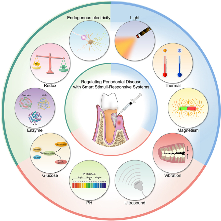
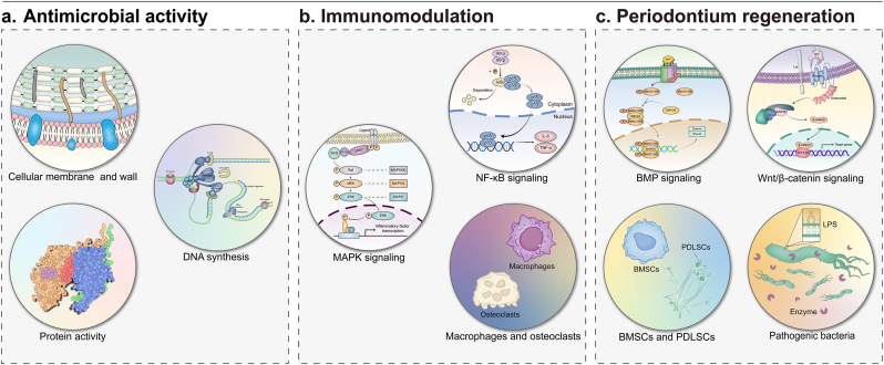
Periodontal disease is a worldwide inflammatory condition that seriously affects both oral and systemic health. The presence of microbial biofilms and the dysregulation of the host immune response are considered crucial factors in the initiation and progression of periodontal disease. Mechanical debridement combined with antibiotic therapy is the standard non-surgical treatment for periodontal disease; however, this approach faces limitations in deep bacterial clearance and resistance to antibiotics. Although some new drugs and accessible nanodelivery systems have been developed, their targeting accuracy and drug utilization still require improvement in the complex oral environment. In recent years, intelligent biomaterials with stimuli-responsive characteristics have garnered more attention due to their unique advantages. These materials can respond to specific physiological signals or external stimuli, enabling precise release of functional agents. However, existing studies focus on the optimization of the single material system, lacking the horizontal comparisons and clinical evaluations of different stimulus-responsive materials. This review aims to address this gap by systematically examining the roles of endogenous and exogenous stimuli in regulating the periodontal disease progression and activating responsive substances. While various stimulus-regulated systems have their respective advantages, the complex oral environment necessitates synergistic action among multiple signals. The review further explores the applications of smart responsive materials in eradicating periodontal pathogens, regulating the inflammatory microenvironment, and promoting periodontium regeneration. Coordinated integration of functional mechanisms is crucial to achieving periodontal disease recovery. Moreover, the challenges faced by intelligent responsive materials in periodontal disease treatment are examined, along with outlining potential directions for future research. It outlines potential research directions to prioritize personalized material design, safety evaluations, and production quality control to advance clinical application.

Keywords: Antimicrobial activity; Immunomodulation; Periodontal disease; Periodontium regeneration; Stimuli-responsive.


Conflict of interest statement

The authors declare that they have no competing interests.

Figures

Image 1
Graphical abstract
Fig. 1
Fig. 1
Regulating periodontal disease with smart stimuli-responsive systems. Smart responsive materials possess the capability to react precisely to specific endogenous or exogenous stimuli, enabling targeted delivery of active molecules. Endogenous stimuli include changes in pH, increased glucose concentration, upregulated enzyme expression, elevated redox potential, and bioelectric signals, whereas exogenous stimuli encompass light, thermal energy, magnetic fields, mechanical vibrations, and ultrasound.
Fig. 2
Fig. 2
PH responsive materials. (A) pH-responsive releasing mechanism of Au@MPN@BMP2. (B) BMP2 release profiles of Au@MPN-BMP2 under different pH environments. (A, B) Reproduced with permission. [20] Copyright 2024, American Chemical Society. (C) The Mg-GA releasing profile of CSBDX@10MOF in different condition. Reproduced with permission. [21] Copyright 2024, The Author(s). (D) Fourier-transform infrared test of GelBA. (E) Release behavior of H2O2 and (F)Mg2+ at different pH values. (D, E, F) Reproduced with permission. [22] Copyright 2024, American Chemical Society. (G)In vitro release studies of TCS and (H)DFO under PH5.5 and PH7.4. (G, H) Reproduced with permission. [23] Copyright 2025, The Author(s).
Fig. 3
Fig. 3
Glucose responsive materials. (A) Transmission electron microscope (TEM) images of CMGCZ under 0, 5, 15, and 35 mM glucose. (B) The pyroptosis of RAW264.7 cells under various conditions was detected via flow cytometric analysis. (C) Western Blotting assay revealed the expression levels of p16, p21 and β - actin in HGFs which had been cultured in the supernatant of RAW264.7's different medium for 24 h (A, B, C) Reproduced with permission. [29] Copyright 2023, The Author(s). (D) Diagrammatic illustration depicting the cascade reaction process of Au/Pt NCs@GOx. Reproduced with permission. [30] Copyright 2023, Elsevier. (E) Schematic representation of the cascade reaction mechanism for MSN-Au@CO. Reproduced with permission. [34] Copyright 2024, The Author(s). (F) The in - vitro cumulative insulin release from the GRI - MN patch at different glucose concentrations. (G) The in - vitro cumulative release of GCA from the GRG - MN patch at different glucose concentrations. (H) The release ratio of insulin to GCA from GRD-MN under different glucose concentrations. (F, G, H) Reproduced with permission. [32] Copyright 2022, The Author(s).
Fig. 4
Fig. 4
Enzyme responsive materials. (A) Schematic diagram of the enzyme-responsive mechanism of PEGPD@SDF-1. (B) The release profiles of SAMP from the hydrogels PEGP and PEGPD over a 10 - day period in various solutions. (A, B) Reproduced with permission. [38] Copyright 2021, American Chemical Society. (C) In vitro, the release rate patterns of CuTA NSs from the TM/BHT/CuTA hydrogel within PBS at 37 °C, with and without MMP - 9. Reproduced with permission. [39] Copyright 2023, American Chemical Society. (D) Descriptive schematic of activatable therapy. The Michaelis - Menten master curve used to describe the reaction rates of the enzyme - substrate systems of (E) SAP9 and (F) RgpB as the substrate concentration increased. (D, E, F) Reproduced with permission. [40] Copyright 2024, The Author(s). (G) Photoemission spectral data of (a) TA-CuO NP-AAP-ALP and (b) TA-CuO NP-AAP-ALP following the incorporation of L-phenylalanine (ALP inhibitor) into the CuO NP-AAP-ALP group. (H) The electron spin resonance (ESR) spectra of CuO NP-AAP-ALP in (a) and CuO NP-AAP in (b) were obtained in a phosphate buffer (20 mM, PH = 7.4). The signal representing DMPO-OH is not observed in the (b) group. (I) Images of colonies with different ALP expression properties under various treatment conditions. [41] Copyright 2024, The Author(s).
Fig. 5
Fig. 5
Redox responsive materials. (A) The remaining mass and drug release profile of MNs in artificial saliva with or without H2O2 under 37 °C. Reproduced with permission. [54] Copyright 2023, The Authors. (B) Gel permeation chromatography of PEG-ss-PCL treated with or without ROS. (C) Osteogenic differentiation of hPDLSCs treated with different concentrations of LPS (0, 5, 10 μg/mL) in the absence or presence of NAC and PssL-NAC. Schematic diagrams of the obtained ALP activity, BMP-2 mRNA expression, Runx2 mRNA expression and PKA mRNA expression. (B, C) Reproduced with permission. [55] Copyright 2021, Elsevier. (D) The release patterns of RB with a loading efficiency of 5.2 % in PBS solutions featuring various GSH concentrations (0, 1, and 10 mM). Reproduced with permission. [55] Copyright 2022, Elsevier. (E) PLGA/MSNs-PMS immobilized PLLA spongy nanofibrous micro - scaffold and the related dual - response mechanism. (F) Release kinetics of the micro-scaffold carrying rhodamine-labeled BMP-2 (using fluorescently labeled BSA as a model) and CEL (using coumarin-6 as a model) complex with a dual-dependence on matrix metallo proteinases (MMP) and GSH. (E, F) Reproduced with permission. [58] Copyright 2021, Elsevier.
Fig. 6
Fig. 6
Endogenous electricity responsive materials. (A) Conductance properties of scaffolds with diverse contents of PGO. (B)Immunofluorescence staining of osteocalcin (OCN) and Runx2 (green) in defect areas, and normalized fluorescence intensity. (A, B) Reproduced with permission. [66] Copyright 2022, The Author(s). (C) Electrical conductivities of hydrogels containing varying amounts of PEDOT – PSF. (D) Conductivity of the BNP-PEDOT-PSF-AG hydrogel following compression. (E) Osteogenesis-related gene expressions of OCN (a) and ALP (b), as well as the ALP activity, of PDLSCs on various hydrogels under different ES potentials. (C, D, E) Reproduced with permission. [67] Copyright 2024, The Author(s). (For interpretation of the references to colour in this figure legend, the reader is referred to the Web version of this article.)
Fig. 7
Fig. 7
Light responsive materials. (A) Thermal images of AuAg, AuAg@PC-FeI, AuAg@PC-FeII, and AuAg@PC-FeIII solution (100 μg/mL) under irradiation for 808 nm 10 min at 2.5 W/cm2. (B) temperature profile. (A, B) Reproduced with permission. [80] Copyright 2022, The Authors. (C) Release of gallium porphyrin from GLR in PBS or the P. gingivalis supernatant. (D) Flow analysis of intracellular ROS produced in P. gingivalis after treatment with PBS, LP (liposome), LR (liposome-containing red blood cell membranes), GLP (liposome loaded with gallium porphyrins), or GLR (liposome-containing red blood cell membranes loaded with gallium porphyrins) under blue light. (C, D) Reproduced with permission. [85] Copyright 2024, American Chemical Society. (E) Electron spin resonance spectra of ·O2− trapped by DMPO in the presence of MoO3 or I-MoO3−x under dark or irradiation of NIR laser (808 nm, 1 W/cm2). DMPO, 5,5-dimethyl-1-pyrroline-N-oxide; DPBF, 1,3-diphenylisobenzofuran. NIR, near-infrared. (F) Survival rate of S. aureus. (E, F) Reproduced with permission. [86] Copyright 2023, The Authors. (G) Schematic illustration of photoinduced redox imbalance in anaerobic bacteria through multi-step electron transfer of TBSMSPy+. (H) Quantitative analysis of NADH level in bacterial suspension treated with TBSMP and TBSMSPy+ at laser irradiation and without irradiation. (G, H) Reproduced with permission. [88] Copyright 2024, The Authors. (For interpretation of the references to colour in this figure legend, the reader is referred to the Web version of this article.)
Fig. 8
Fig. 8
Thermal responsive materials. (A) Synesis process and schematic illustration of CS hydrogel. (B) The release curves of DPSC-Exo from the CS hydrogel. (A, B) Reproduced with permission. [92] Copyright 2020, The Authors. (C) Contour plots for drug release at 1hr. (D) Contour plots for time required to release 90 % of drug. (C, D) Reproduced with permission. [94] Copyright 2019, Craniofacial Research Foundation. (E) Ultra violet (UV) absorbance of triggered release. (F) The control and CHX@CP3 groups with and without NIR irradiation of S. aureus. (G) The bacterial viability of the control and CHX@CP3 with and without NIR irradiation of S. aureus. (E, F, G) Reproduced with permission. [95] Copyright 2020, Elsevier. (For interpretation of the references to colour in this figure legend, the reader is referred to the Web version of this article.)
Fig. 9
Fig. 9
Magnetism responsive materials. (A, B) Penetrating effect of FPM on biofilms. Transverse cross-sectional CLSM images of periodontal biofilms treated with FPM before and after magnetic motivation. (C) Representative live/dead and 3D reconstruction images of P. gingivalis on HA disks (dead bacteria stained red; live bacteria stained green). (A, B, C) Reproduced with permission. [102] Copyright 2023, The Author(s). (D) Images of 3D reconstruction and sectional view of the effects of FMSs on bone defect healing at 3 months post-surgery. (E) The quantitative analyses of the regenerated bone height along the mesial surface of M1 and the bone healing area percentage. (D, E) Reproduced with permission. [104] Copyright 2023, The Authors. (F) Time-lapse microscopy images illustrating tracking lines of swimming of Fe3O4@PEI/BiVO4 magnetic microrobots under a transversal rotating magnetic field using the predefined clockwise circular propulsion mode. (G) Antibiofilm activity of Fe3O4@PEI/BiVO4 photoactive magnetic microrobots under different experimental conditions. (F, G) Reproduced with permission. [105] Copyright 2022, American Chemical Society. (For interpretation of the references to colour in this figure legend, the reader is referred to the Web version of this article.)
Fig. 10
Fig. 10
Vibration responsive materials. (A, B, C) Osteogenic differentiation evaluation of GelMA and PiezoGEL hydrogels in vitro, evaluated by the factors including (A) RUNX2, (B) COL1A1, (C) ALP. (D) Normalized absorbance values of Alizarin Red S staining of the formed ECM minerals formed after 14 days of cell culture. (A, B, C, D) Reproduced with permission. [113] Copyright 2023, American Chemical Society. (E) The expression of Arg-1 after treatments. (F) Salivary inflammatory factors (IL-1β, IL-6, and TNF-α) after 12 weeks of post-surgery. (E, F) Reproduced with permission. [114] Copyright 2024, The Authors. (G) The scheme of ZnO@Bdello to eradicate plaque biofilm in periodontitis. Reproduced with permission. [117] Copyright 2022, Elsevier. (H) Electrochemical impedance spectroscopy (EIS) analysis for different prepared samples. (I) Decomposition ratio for the different piezocatalyst. (H, I) Reproduced with permission. [116] Copyright 2022, The Author(s). (For interpretation of the references to colour in this figure legend, the reader is referred to the Web version of this article.)
Fig. 11
Fig. 11
Ultrasound responsive materials. (A, B, C) The fluorescence intensities of DCF in the solutions of DT-Ag-CS upon US irradiation with different power intensities. (D) The bacterial viability rate of P. gingivalis after incubation with different concentrations of DT-Ag-CS with or without US irradiation. (A, B, C, D) Reproduced with permission. [128] Copyright 2022, Elsevier. (E) The cyclic voltammogram of TPP-TeV at different scan rates in DMF solution with tetrabutylammonium hexafluorophosphate. (F) EPR spectra of the radical species of TPP-TeV and TPP-V after addition of ultrasonic. (G) H&E, Masson, and the immunohistochemical analysis of MMP-9 and TNF-α staining images of the periodontium after the treatment in decalcified maxilla sections. (E, F, G) Reproduced with permission. [130] Copyright 2023, Elsevier. (H) Magnified views of periodontal tissue in the high-intensity ultrasound-microbubble (HUM-Sc) group. Scale bar = 10 μm. Reproduced with permission. [134] Copyright 2017, The Author(s).
Fig. 12
Fig. 12
Multi-stimuli-responsive materials. (A) The stimulus-responsive mechanism of pGM/cPL@NI. (B) Cumulative release of MCC950 from pGM/cPL@NI under different pH and H2O2 environments. (C) IF staining of CD206 (red), Runx2 (green), IL-1β (red), and DAPI (blue) in periodontal tissue. (A, B, C) Reproduced with permission. [138] Copyright 2024, The Author(s). (D) MZ@PNM release curves of MZ@PNM@GCP under different pH conditions with time. (E) Representative fluorescence and TEM images of the destruction of P.g. by MZ@PNM. (F) Serum levels of IL1-β, IL-6 and TNF-α. (G) H&E staining of major organs in rats after MZ@PNM@GCP treatment. (D, E, F, G) Reproduced with permission. [140] Copyright 2022, American Chemical Society. (For interpretation of the references to colour in this figure legend, the reader is referred to the Web version of this article.)
Fig. 13
Fig. 13
The active substances delivered by smart responsive materials modulate periodontal disease through three primary mechanisms: 1) Eradication of pathogenic microorganisms by disrupting bacterial cell membrane or wall structures, interfering DNA synthesis, and inhibiting protein function; 2) Immune modulation by regulating inflammatory signaling pathways (such as NF-κB and MAPK pathways) and controlling the activity of inflammatory cells (such as macrophages and osteoclasts); 3) Promoting regeneration of periodontal tissues by eliminating pathogenic bacteria and activating specific growth signaling pathways (such as BMP and Wnt/β-catenin pathways) and enhancing the proliferation and differentiation of BMSCs and PDLSCs. Abbreviation: NF-κB, Nuclear Factor kappa-light-chain-enhancer of activated B cells; MAPK, Mitogen-Activated Protein Kinase; BMP, bone morphogenetic protein; Wnt/β-catenin, Wingless-Int/β-catenin; BMSCs, bone marrow stem cells; PDLSCs, periodontal ligament stem cells.

Similar articles

Cited by

References

    1. WHO Oral health. 2024. https://www.who.int/zh/news-room/fact-sheets/detail/oral-health
    1. Reynolds I., Duane B. Periodontal disease has an impact on patients' quality of life. Evid Based Dent. 2018;19:14–15. doi: 10.1038/sj.ebd.6401287. - DOI - PubMed
    1. Isola G., Polizzi A., Serra S., Boato M., Sculean A. Relationship between periodontitis and systemic diseases: a bibliometric and visual study. Periodontol. 2025;2000 doi: 10.1111/prd.12621. - DOI - PubMed
    1. Kinane D.F., Hajishengallis G. Polymicrobial infections, biofilms, and beyond. J. Clin. Periodontol. 2009;36:404–405. doi: 10.1111/j.1600-051X.2009.01396.x. - DOI - PubMed
    1. Isola G., Polizzi A., Santagati M., Alibrandi A., Iorio-Siciliano V., Ramaglia L. Effect of nonsurgical mechanical debridement with or without chlorhexidine formulations in the treatment of peri-implant mucositis. A randomized placebo-controlled clinical trial. Clin. Oral Implants Res. 2025 doi: 10.1111/clr.14405. - DOI - PMC - PubMed

LinkOut - more resources