. 2025 May 15:32:101875.
doi: 10.1016/j.mtbio.2025.101875. eCollection 2025 Jun.

A triple-targeting "nano-brake" remodeling the impaired immune microenvironment in skin lesions for psoriasis treatment

Affiliations

A triple-targeting "nano-brake" remodeling the impaired immune microenvironment in skin lesions for psoriasis treatment

Ruijie Chen et al. Mater Today Bio. .

Abstract

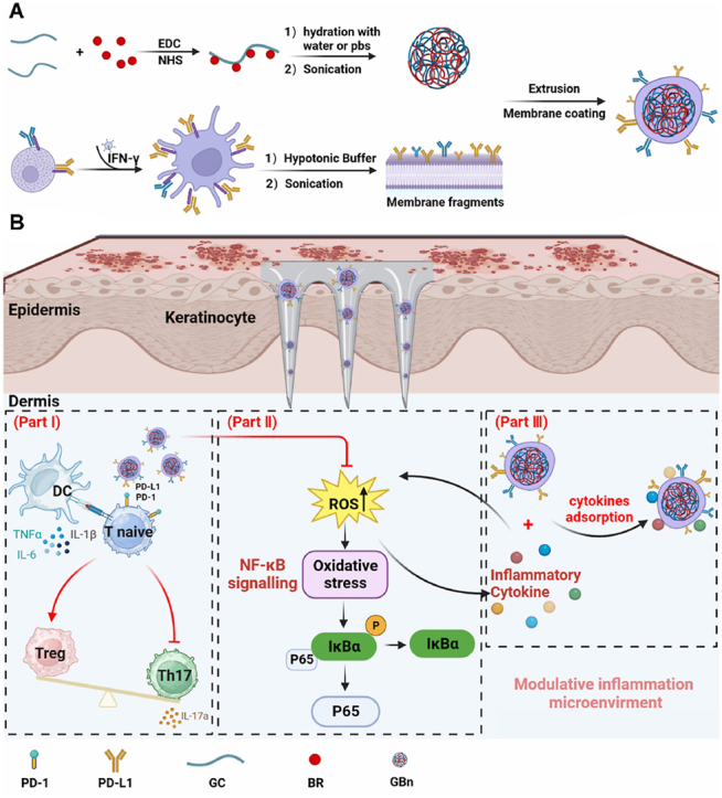
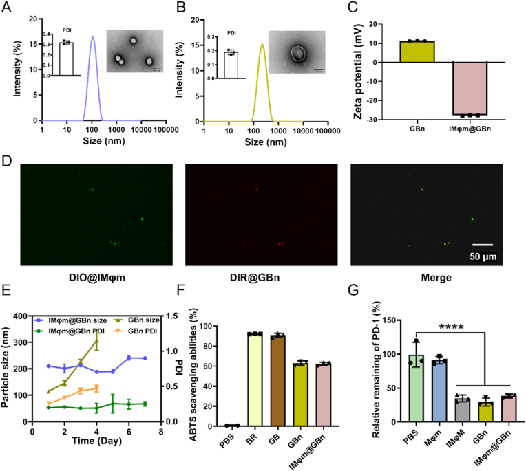
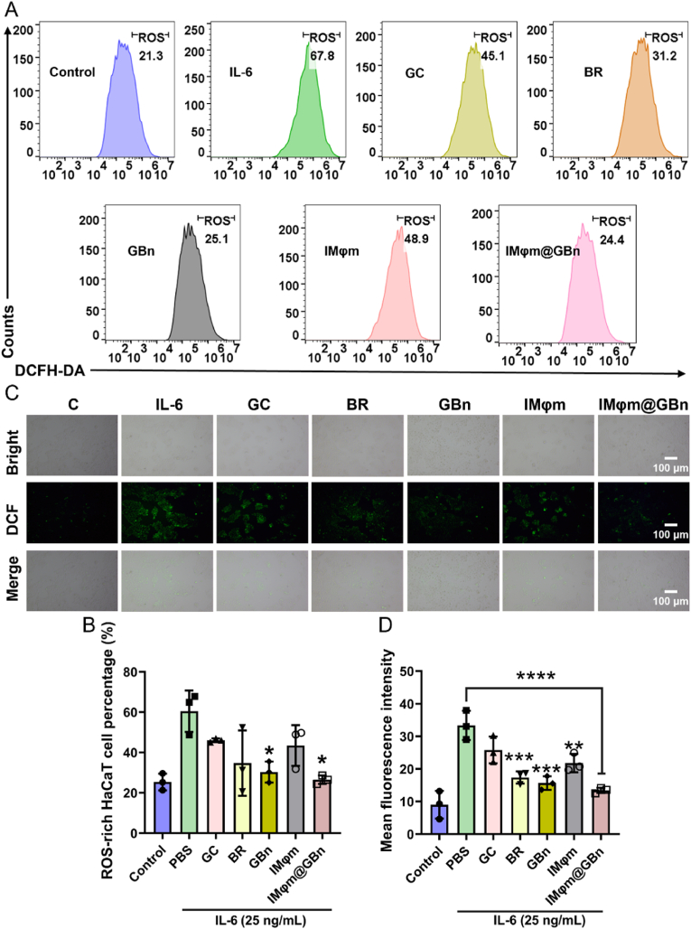
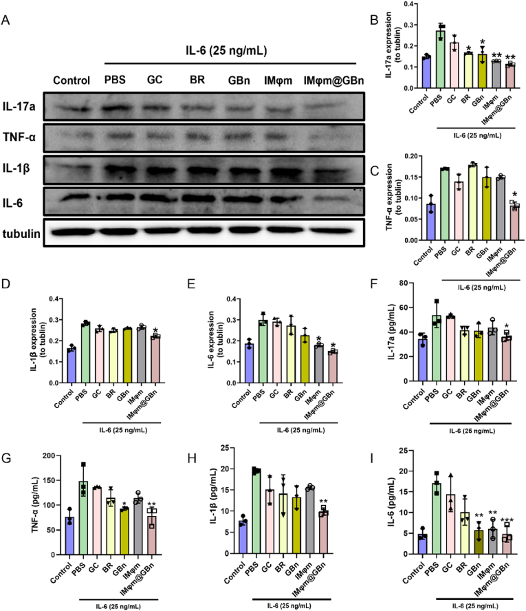
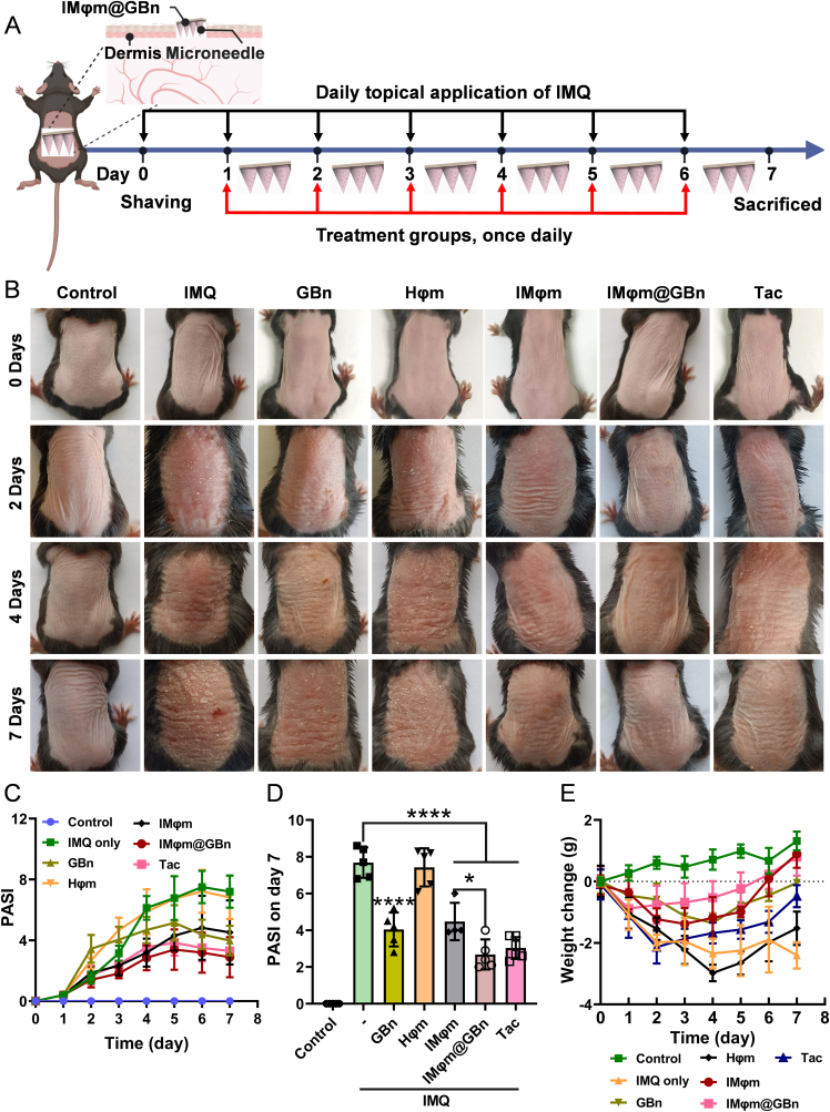
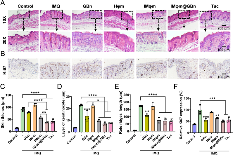
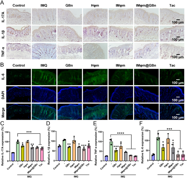
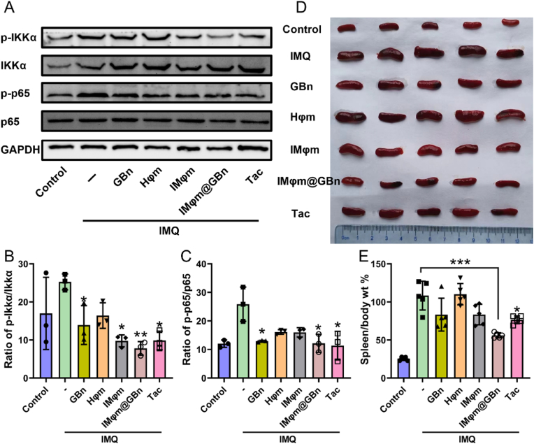
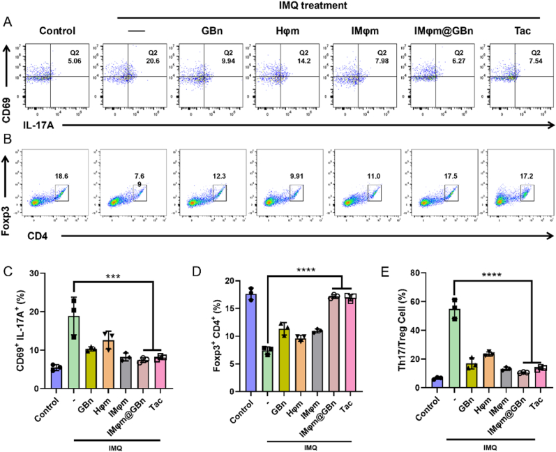
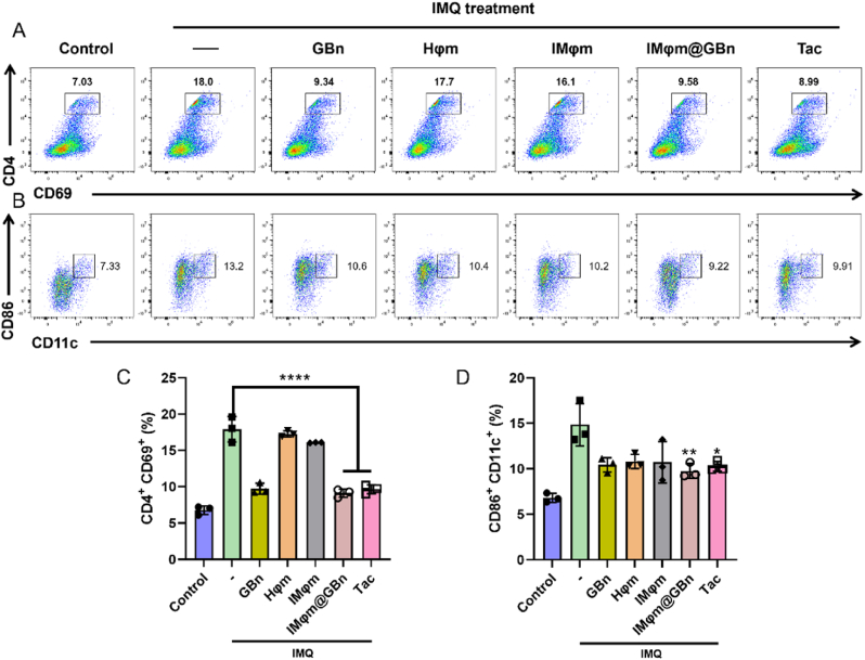
Psoriasis, a prevalent immune-mediated chronic inflammatory skin ailment, has been linked to heightened oxidative stress and compromised immune tolerance. Immune checkpoint pathways, particularly the programmed cell death-1 (PD-1)/programmed cell death-ligand 1 (PD-L1) signaling axis, are instrumental in establishing and sustaining self-tolerance and regulating immune responses. Augmenting PD-1/PD-L1 interaction holds promise for curtailing the proliferation and activation of infiltrating T cells and curbing the release of inflammatory cytokines, thereby mitigating psoriasis induced by impaired immune tolerance. Consequently, neutralizing the surplus reactive oxygen species (ROS) in the affected skin and revitalizing local immune tolerance could represent a beneficial approach to psoriasis treatment. In light of these insights, this study introduced a bilirubin-based nanoparticle cloaked in IFN-γ-stimulated macrophage membrane (designated as IMφm@GBn or "nano-brake"). Treatment with IFN-γ conferred the macrophage membrane with heightened expression of pro-inflammatory cytokine receptor and PD-L1. As a result, the engineered IMφm@GBn not only scavenged excessive ROS in psoriatic lesions but crucially also absorbed a wide spectrum of pro-inflammatory cytokines. Furthermore, it inhibited the proliferation and activation of infiltrating T cells through augmented PD-1/PD-L1 interactions, thereby rebalancing the Th17/Treg ratio. In an in vivo psoriasis mouse model, the "nano-brake" was locally and accurately delivered to the dermis via microneedle, orchestrating the immune microenvironment of psoriasis and mitigating autoimmune damage linked to impaired immune tolerance. This approach significantly enhanced the therapeutic efficacy in ameliorating psoriasis-associated symptoms. This study sets an illuminating precedent for cell membrane-based biomimetic nano-formulations, holding broad implications for psoriasis treatment through multi-pronged immunomodulation.

Keywords: Bilirubin; PD-1/PDL-1; Psoriasis; ROS; Th17/Treg.


Conflict of interest statement

The authors declare that they have no known competing financial interests or personal relationships that could have appeared to influence the work reported in this paper.

Figures

Image 1
Graphical abstract
Scheme 1
Scheme 1
The schematic diagram illustrates the preparation of IFN-γ treated RAW 264.7 cell membrane-coated nano-brake (IMφm@GBn) and elucidates their immunomodulatory actions for treating psoriasis. (A) The preparation of IMφm@GBn. (B) The mode of action by which IMφm@GBn addressed psoriasis. Part I: In the context of immune dysregulation characteristic of psoriasis, T cells experience abnormal activation and differentiation towards Th17 cells, resulting in an imbalanced Th17/Treg cell ratio. The dysregulated immune response would be regulated by the administered IMφm@GBn through the PD-1/PD-L1 signaling pathway. Part II: IMφm@GBn could scavenge the overloaded ROS in the inflammatory milieu and suppress the activation of the NF-κB pathway, thereby mitigating the local inflammation. Part III: IMφm@GBn could adsorb and neutralize proinflammatory cytokines, alleviating the continued inflammatory stimulus. Herein, IMφm@GBn acted as a nano-brake by presenting triple actions to shut down the excessively aberrant immune response and remodel the impaired immune microenvironment in skin lesions for psoriasis treatment.
Fig. 1
Fig. 1
Characterization of IMφm@GBn. The particle size, polydispersity index (PDI) and TEM image (scale bar = 100 nm) of (A) GBn, (B) IMφm@GBn. (C) Zeta potential of GBn and IMφm@GBn. (D) Fluorescence microscope images of IMφm@GBn (DiR, Red, loaded in GBn; DiO, green, loaded in IMφm). (E) Changes in the particle size and PDI of GBn and IMφm@GBn in pH 7.4 PBS at 4 °C. (F) ABTS radical scavenging ratio of the BR, GB, GBn and IMφm@GBn. (G) Remaining PD-1 after incubation with PBS, Mφm, IMφm, GBn or IMφm@GBn (n = 3). Mφm: Macrophage membrane without IFN-γ stimulation. IMφm: Macrophage membrane with IFN-γ stimulation. Data presented as mean ± SD. ∗∗∗∗P < 0.0001 indicate significant difference compared to the PBS group. (For interpretation of the references to colour in this figure legend, the reader is referred to the Web version of this article.)
Fig. 2
Fig. 2
Uptake properties of IMφm@GBn in HaCaT cells. Fluorescence microscopy was used to visualize cellular uptake following incubation with free Coumarin-6 (fC6) or C6-loaded IMφm@GBn (C6@ IMφm@GBn) for 2 h in (A) normal HaCaT cells and (B) IL-6 stimulated inflammatory HaCaT cells (Scale bar = 100 μm). (C) Quantification of fluorescence in (A) and (B); n = 3. Data presented as mean ± SD. ∗∗∗P < 0.001, ∗∗∗∗P < 0.0001 indicating significant difference between groups.
Fig. 3
Fig. 3
IMφm@GBn inhibiting inflammatory keratinocyte hyperproliferation. (A) The viability of normal HaCaT cells following treatment with different concentrations of IMφm@GBn (2–80 μM) for 24 h (n = 3). (B) Viability of IL-6-induced HaCaT cells following treatment with different concentrations (1–80 μM) of IMφm@GBn for 24 h (n = 3). (C) The colony formation assay and (D) the quantification (n = 3). (E) IL-6-induced hyperplasia morphology and alterations following treatment with GC, BR, GBn, IMφm, and IMφm@GBn for 24 h under the microscope (Scale bar = 500 μm). Data presented as mean ± SD. ∗∗∗∗P < 0.0001 indicate significant difference between groups.
Fig. 4
Fig. 4
IMφm@GBn protecting cells from oxidative stress by downregulating intracellular ROS levels. (A) Flow cytometry and (C) Fluorescence microscopy analysis of ROS levels in HaCaT cells after various treatments (Scale bar = 100 μm). (B) and (D) are quantitative analyses of the fluorescence signals in (A) and (C) (n = 3). Data presented as mean ± SD. ∗P < 0.05, ∗∗P < 0.01, ∗∗∗P < 0.001, ∗∗∗∗P < 0.0001 indicate significant differences compared to the PBS group.
Fig. 5
Fig. 5
IMφm@GBn inhibiting the expression of various pro-inflammatory cytokine to exhibit anti-inflammatory effects. (A) Expression pattern of IL-17a, TNF-α, IL-1β, and IL-6 under various treatment conditions. Quantification of the protein of (B) relative IL-17a/Tubulin, (C) relative TNF-α/Tubulin, (D) relative IL-1β/Tubulin, and (E) relative IL-6/Tubulin. (F)–(I) The levels of IL-17a (F), TNF-α (G), IL-1β (H) and IL-6 (I) in the cell supernatant were determined by ELISA kits. Data presented as ± SD (n = 3). ∗P < 0.05; ∗∗P < 0.01; ∗∗∗P < 0.001 indicating statistical difference compared to the PBS group.
Fig. 6
Fig. 6
Psoriatic symptoms of the skin lesion after topical application of imiquimod (IMQ) and intradermal injection of different preparations. (A) The experimental scheme. (B) Representative gross images of mouse backs on days 0, 2, 4, and 7 after various treatments. (C) The Psoriatic Area and Severity Index (PASI) score of skin lesions, were recorded for 7 days (n = 5). (D) the PASI score on day 7. (E) Body weight changes of the mice in the experimental groups (n = 5). Data presented as mean ± SD. ∗P < 0.05 and ∗∗∗∗P < 0.0001, indicating statistical difference between indicated groups.
Fig. 7
Fig. 7
IMφm@GBn inhibiting epidermal proliferation in psoriasis-like skin lesion in mouse model. (A) Representative H&E staining of skin sections on day 7 (Scale bar = 100 μm). (B) Skin sections with immunohistochemistry staining of Ki67 on day 7. Quantitative analysis of (C) skin thickness, (D) keratinocyte layer, (E) rete ridges length and (F) relative Ki67 expression (n = 3). Data presented as mean ± SD. ∗P < 0.05, ∗∗P < 0.01, ∗∗∗P < 0.001, and ∗∗∗∗P < 0.0001 indicating significant differences compared to the IMQ group or between groups.
Fig. 8
Fig. 8
IMφm@GBn decreased the secretion of inflammatory factors in the psoriatic skin. (A) Immunohistochemistry staining of IL-17A, IL-1β, and TNF-α and (B) immunofluorescence of IL-6 in skin sections on day 7. (Scale bar = 100 μm). (C ∼ F) Quantitative analysis of relative expression of (C) IL-17A, (D) IL-1β, (E) TNF-α, and (F) IL-6 (n = 3). Data presented as mean ± SD. ∗P < 0.05, ∗∗P < 0.01, ∗∗∗P < 0.001, and ∗∗∗∗P < 0.0001 indicating significant differences compared to the IMQ group or between groups.
Fig. 9
Fig. 9
IMφm@GBn effectively inhibit NF-κB signaling pathways to relieve psoriasis symptoms as well as the immune reaction in spleen. (A) A representative Western blot image of p-IKKα, IKKα, p-p65 and p65 expressions on day 7 after various treatments in the skin of IMQ-induced psoriasis-like mice. Quantification of the protein relative expression of (B) p-IKKα/IKKα, (C) p-p65/p65. GAPDH was used as an internal control (n = 3). (D) Images of spleens in different groups. (E) The relative spleen/body (%, w/w) was recorded and calculated on Day 7 (n = 5). Data presented as mean ± SD. ∗P < 0.05, ∗∗P < 0.01, and ∗∗∗P < 0.001 indicating significant differences compared to the IMQ group.
Fig. 10
Fig. 10
IMφm@GBn rebalanced Th17/Treg ratio in the psoriatic mice. Following various treatments, flow cytometry was used to evaluate the (A) Th17 (IL-17+CD69+) and the (B) Treg (Foxp3+CD4+) population in the CD4+ splenocyte fraction. Histogram presented the percentage of (C) Th17 (IL-17+CD69+) and (D) Treg (Foxp3+CD4+) in various groups. (E) The ratio of Th17/Treg in various groups. Data presented as mean ± SD. ∗∗∗P < 0.001 and ∗∗∗∗P < 0.0001, indicating significant differences compared to the IMQ group.
Fig. 11
Fig. 11
IMφm@GBn modulating the immune microenvironment by inhibiting T-cell activation and DC maturation. (A) CD69 was used to measure the activation of CD4+ naive T cells and (C) the quantitative presentation. (B) The flow cytometric analysis of mature DCs (CD86+/CD11c+, gated on CD11c+ cells) and (D) the quantitative presentation. Data presented as mean ± SD (n = 3). ∗P < 0.05, ∗∗P < 0.01 and ∗∗∗∗P < 0.0001, indicating differences compared to the IMQ group.

Similar articles

Cited by

References

    1. Kurpet K., Chwatko G. S100 proteins as novel therapeutic targets in psoriasis and other autoimmune diseases. Molecules. 2022;27 - PMC - PubMed
    1. Langley R.G., Krueger G.G., Griffiths C.E. Psoriasis: epidemiology, clinical features, and quality of life. Ann. Rheum. Dis. 2005;64 Suppl 2, ii18-23;discussion ii24-15. - PMC - PubMed
    1. Nellikode D.A.S. Effiffifficacy of shodhana and sarvang takradhara therapy in the management of psoriasis A case study. International Journal of Preclinical & Clinical Research. 2023;4(1):15–20.
    1. Xu Q., Du J., Cao W., Sun S. Demographic and epidemiological drivers of global burden of psoriasis. Australas. J. Dermatol. 2021;62:e554–e558. - PubMed
    1. Le A.M., Torres T. New topical therapies for psoriasis. Am. J. Clin. Dermatol. 2022;23:13–24. - PubMed

LinkOut - more resources