. 2025 May 14:32:101871.
doi: 10.1016/j.mtbio.2025.101871. eCollection 2025 Jun.

Recent advances in NIR-II photothermal and photodynamic therapies for drug-resistant wound infections

Affiliations

Recent advances in NIR-II photothermal and photodynamic therapies for drug-resistant wound infections

Xiang Chen et al. Mater Today Bio. .

Abstract

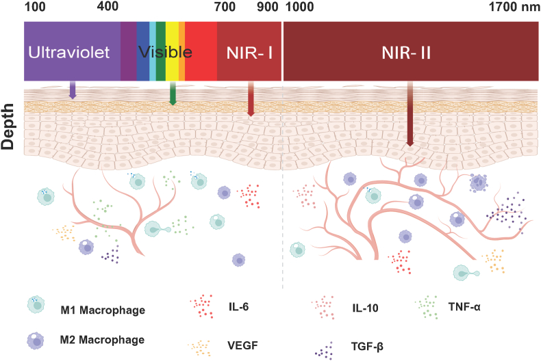
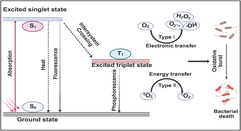
Bacterial infection can delay wound healing, while drug resistance further complicates the treatment of wound infection. Phototherapy, including photothermal therapy (PTT) and photodynamic therapy (PDT), is a non/mini-invasive and efficient antibacterial strategy that rarely induces bacterial resistance. This treatment relies on specific wavelengths of light to activate photothermal agents (PTAs) or photosensitizers, killing bacteria by generating local heats or reactive oxygen species (ROS), respectively. However, the light for traditional PTT/PDT mainly falls in the visible and near-infrared I light (Vis/NIR-I light, 400-900 nm) regions, which significantly limits further clinical translations due to its low tissue permeability. The near-infrared II (NIR-II,1000-1700 nm) light is increasingly utilized in antibacterial PTT/PDT to improve tissue penetration and ameliorate the immune microenvironment of deeper wounds. Meanwhile, NIR-II light offers a higher maximum permissible exposure (MPE) for PTT/PDT in treating wound infections, thereby facilitating the security, in comparison to Vis/NIR-I light. This review highlights recent advancements in NIR-II PTT/PDT for drug-resistant wound infections, focusing on mechanisms, therapeutic outcomes, challenges, and prospects.

Keywords: Near-infrared II; Photodynamic therapy; Photothermal therapy; Wound healing; Wound infections.


Conflict of interest statement

The authors declare that they have no known competing financial interests or personal relationships that could have appeared to influence the work reported in this paper.

Figures

Image 1
Schematic representation of NIR-II photothermal therapy (PTT) and photodynamic therapy (PDT) for treating drug-resistant wound infections.
Fig. 1
Fig. 1
Schematic illustration of the process of infected wound healing. (A) Inflammation. The wound becomes infected with bacteria, and proinflammatory macrophages (M1) produce proinflammatory factors such as interleukin-1 (IL-1), interleukin-6 (IL-6), and tumor necrosis factor-α (TNF-α). Neutrophils are recruited to remove bacteria and produce pus. (B) Proliferation. Macrophages transition to M2 type and release healing cytokines such as transforming growth factor β (TGF-β), epidermal growth factor (EGF), vascular endothelial growth factor (VEGF), accompanying granulation tissue formation, angiogenesis, and epithelial regeneration. (C) Remodeling. Cells rearrange, and disorganized collagen is degraded and absorbed. Created with BioRender.com.
Fig. 2
Fig. 2
Wavelength ranges and tissue penetration depth of Ultraviolet, Visible light, and near-infrared I (NIR-I) and near-infrared II (NIR-II).
Fig. 3
Fig. 3
NIR-II photothermal therapy and photodynamic therapy in wound infections by bacteria.
Fig. 4
Fig. 4
Schematic illustration of the mechanism of antibacterial photothermal therapy (PTT).
Fig. 5
Fig. 5
NIR-II photothermal therapeutic effects of Au/Ag core/shell nanorods (NRs) for treating MRSA infections and promoting wound healing. (A) Activatable NIR-II photothermal and photoacoustic properties of Au/Ag core/shell NRs. (B) Uv–Vis–NIR absorption spectra of Au/Ag NRs, Au/Ag NRs (Intermediate), Au/Ag NRs (Activated). (Insert: photograph of the corresponding samples). (C) TEM images of Au/Ag core/shell NRs, scale bar: 20 nm. The insert shows the clear core/shell structure of the corresponding NRs, scale bar: 5 nm. (D) Scanning electron microscopy (SEM) images of MRSA treated with PBS, Au/Ag, and Au/Ag + NIR-II laser, scale bar: 500 nm. White arrows indicate the wrinkled surface of MRSA after synergistic therapy. (E) Representative photos of MRSA colonies on agar plates after different treatments. (F) Schematic illustration of Au/Ag NRs for treating mice with infected wounds. (G) The counting Forming Unit of MRSA was harvested from the cured tissue and infected tissue. (H) Quantitative curves of wound area over time for each treatment group. Reproduced with permission [64].Copyright 2020, Elsevier.
Fig. 6
Fig. 6
NIR-II photothermal therapy with infection microenvironment-activated Cu2O nanoparticles for the treatment of wound infections and the promotion of wound healing. (A) Scheme for the infection microenvironment-activated Cu2O nanoparticles for NIR-II photoacoustic imaging-guided photothermal/chemodynamic synergistic anti-infective therapy. (B) UV–Vis–NIR absorption spectra of Cu2O NPs and Cu9S8 NPs. (C–D) SEM images and photos of LB agar plates of MRSA after the following treatments: Control, Cu2O NPs + H2O2, Cu2O NPs + NaHS + NIR, and Cu2O NPs + NaHS + NIR + H2O2. (E) Representative skin images after different treatments. (F) Quantitative analysis of bacterial colonies of day 7 in (E). ∗Indicates significant difference (∗∗p < 0.01). Reproduced with permission [61]. Copyright 2021, Elsevier.
Fig. 7
Fig. 7
NIR-II photothermal properties of N-GQDs for treating wound infections and promoting healing. (A) Schematic illustration of the synthesis procedures of N-GQDs. (B) NIR absorption of the N-GQDs. (C) Temperature variation of the N-GQDs over ten cycles of laser irradiation. (D) Temperature elevation of the N-GQDs (0, 50, 100, 150, and 200 μg/ml) under 1064 nm (1 W/cm2) laser irradiation. (E) Infrared thermographic images of the N-GQDs under continuous 1064 nm laser irradiation. (F) Live/dead SEM images of MRSA in each group. (G) Representative culture images of colonies of MRSA after receiving treatments with various concentrations of N-GQD aqueous solution without or with laser irradiation. (H) Photographs of MRSA-infected skin on days 0, 2, 4, 6, 9, and 12 after different treatments. (I) Photographs of bacterial colonies from the tissues in (H) Reproduced with permission [44]. Copyright 2022, Royal Society of Chemistry.
Fig. 8
Fig. 8
An acidity-responsive polyoxometalate with inflammatory retention for NIR-II photothermal-enhanced chemodynamic antibacterial therapy. (A) Preparation of acidity-aggregated POM clusters for photothermal-enhanced CDT in the NIR-II window. (B) Absorbance spectra of POM solutions at various concentrations or pH values. (C) Photothermal performance of POM with different pH and concentrations under 1060 nm laser irradiation (1 W/cm2). (D) Antibacterial effect of POM under different conditions. (I) PBS, (II) H2O2, (III) POM, (IV) POM + H2O2, (V) POM + NIR, (VI) POM + H2O2 + NIR. Scar bar: 4.5 cm. (E) SEM images of drug-resistant S. aureus after receiving different treatments. (I) PBS, (II) H2O2, (III) POM, (IV) POM + H2O2, (V) POM + NIR, (VI) POM + H2O2 + NIR. (F) Live/dead cell assay using AM and PI double staining after receiving different treatments of drug-resistant S. aureus. (G) Relative cell viability of HUVEC incubated with different concentrations of POM. (H) In vivo abscess treatment images after being treated with (I) PBS, (II) H2O2, (III) POM, (IV) POM + H2O2, (V) POM + NIR, (VI) POM + H2O2 + NIR, respectively [71]. Copyright 2021, American Chemical Society.
Fig. 9
Fig. 9
NIR-II photothermal properties of CNs nanoparticles for treating wound infections and promoting wound healing. (A) Chemical structure of CNs. (B) Absorption of CNs in CHCl3. (C) Photos of bacterial colonies and bacterial viability of (D) S. aureus, (E) E. coli treated with CN3 NPs (5∼20 μg/mL) in the dark and under 1064 nm laser irradiation (1.0 W/cm2, 10 min). (F) Bacterial viability of MRSA treated with CN3 NPs (5–20 μg/mL) in the dark and under 1064 nm laser irradiation (1.0 W/cm2, 10 min). (G) Photos of the infected wound within 5 days. Reproduced with permission [74]. Copyright 2023, Elsevier.
Fig. 10
Fig. 10
Schematic illustration of the mechanism of antibacterial photodynamic therapy (PDT). S0, ground state; S1, excited singlet state; T1, excited triplet state.
Fig. 11
Fig. 11
NIR-II photodynamic therapy using Ag/BMO nanozymes to treat wound infections and promote wound healing. (A) Preparation of Ag/BMO nanozyme and NIR-enhanced catalytic activity mechanisms for synergistic bacterial therapy. (B) Plate photographs demonstrating the antibacterial activity of Ag/BMO NPs. (C) The singlet oxygen (1O2) detection using the SOSG probe. (D) Confocal Laser Scanning Microscopy (CLSM) images of MRSA stained by Live/dead dye following incubation with Ag/BMO NPs with or without NIR laser irradiation. (PI emits red fluorescence and SYTO9 emits green fluorescence). (E) Corresponding quantitative survival ratio of MRSA in (B). (F) Photographs of MRSA-infected wounds in various groups after treatments. (G) The change of wound areas for 7 days. ∗∗p < 0.01, ∗∗∗p < 0.001. (H) H&E and Masson-stained tissue slices of infected wounds. Reproduced with permission [129]. Copyright 2022, The Author(s).
Fig. 12
Fig. 12
Drug-dye antimicrobial nanomotor for precise treatment of multidrug-resistant bacterial infections. (A)NIR-II light-triggered synchronous autonomous movement and synergistic photothermal/photocatalytic antibacterial of the nanomotors for enhancing transdermal penetration and effectively treating MRSA infections. (B) UV–Vis–NIR absorption spectra of different samples. (C) Thermal images and (D) corresponding temperature changes at the skin abscess site in mice undergoing different treatments (1064 nm,0.75W/cm2): (i) PBS, (ii)AuNR-SiO2, (iii)AuNR-SiO2-Cu2O, and (iv) AuNR-SiO2-Cu7S4 groups. (E) ROS generation as detected by methylene blue (MB) upon exposure to 1064 nm light irradiation (0.75W/cm2). (F) Live/dead stained images of MRSA and bacterial morphology observed by SEM after different treatments. (G) Antibacterial efficiency of MRSA. (H) Photographs of MRSA-infected skin abscess model mice undergoing different treatments under NIR-II light irradiation. The dashed circle indicates the MRSA-infected skin abscess site. Reproduced with permission [145].Copyright 2023, American Chemical Society.

Similar articles

References

    1. Pormohammad A., Monych N.K., Ghosh S., Turner D.L., Turner R.J. Nanomaterials in wound healing and infection control. Antibiotics (Basel) 2021;10:473. doi: 10.3390/antibiotics10050473. - DOI - PMC - PubMed
    1. Canchy L., Kerob D., Demessant A., Amici J.-M. Wound healing and microbiome, an unexpected relationship. J. Eur. Acad. Dermatol. Venereol. 2023;37:7–15. doi: 10.1111/jdv.18854. - DOI - PubMed
    1. Huang F., Lu X., Yang Y., Li Y., Kuai L., Li B., Dong H., Shi J. Microenvironment-based diabetic toot ulcer nanomedicine. Adv. Sci. (Weinh.) 2023;10 doi: 10.1002/advs.202203308. - DOI - PMC - PubMed
    1. Meng S., Wu H., Xiao D., Lan S., Dong A. Recent advances in bacterial cellulose-based antibacterial composites for infected wound therapy. Carbohydr. Polym. 2023;316 doi: 10.1016/j.carbpol.2023.121082. - DOI - PubMed
    1. Li L., Ma Y., He G., Ma S., Wang Y., Sun Y. Pilose antler extract restores type I and III collagen to accelerate wound healing. Biomed. Pharmacother. 2023;161 doi: 10.1016/j.biopha.2023.114510. - DOI - PubMed

LinkOut - more resources