. 2025 May 27;30(1):146.
doi: 10.3892/etm.2025.12896. eCollection 2025 Jul.

Plasmon-activated water enhances gut-barrier function and alleviates inflammation in a mouse model of ulcerative colitis

Affiliations

Plasmon-activated water enhances gut-barrier function and alleviates inflammation in a mouse model of ulcerative colitis

Chun-Chao Chang et al. Exp Ther Med. .

Abstract

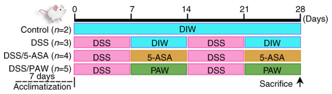
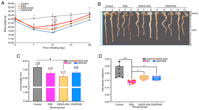
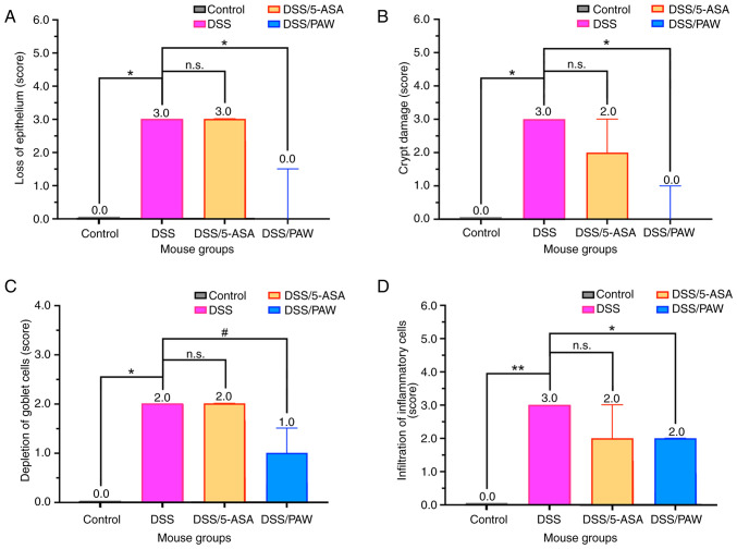
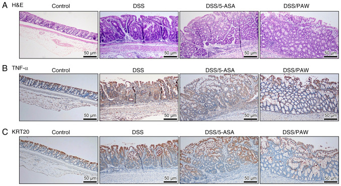
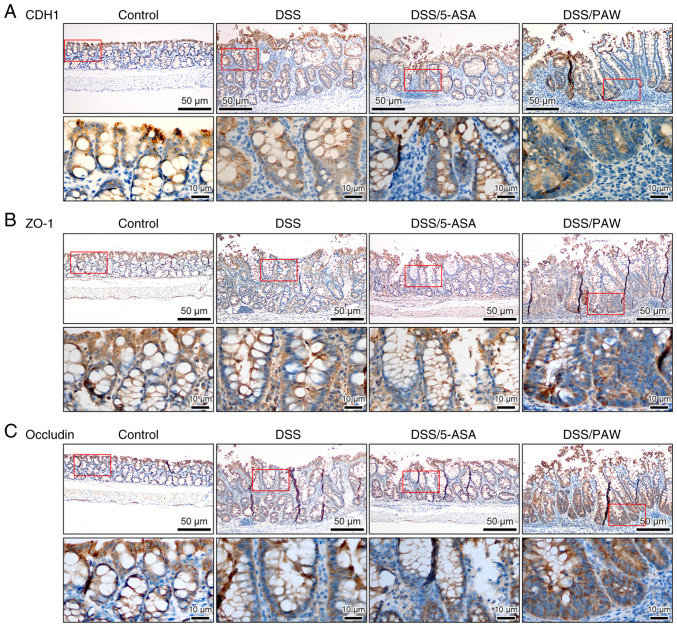
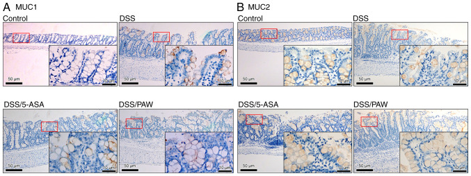
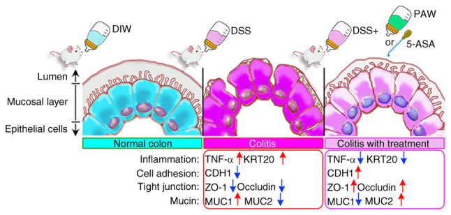
Ulcerative colitis (UC) is a chronic, relapsing inflammatory bowel disease characterized by disruption of the intestinal epithelial barrier and alterations in mucosal gene expression associated with intestinal integrity. Given the risks associated with UC, novel therapies capable of restoring intestinal barrier function and inhibiting inflammation are needed. Plasmon-activated water (PAW) is a nontoxic form of water with potential in the treatment of inflammatory diseases. The aim of the present study was to evaluate the therapeutic effects of PAW in a murine model of UC. Histological and immunohistochemical analyses were performed on colon tissues from mice with dextran sodium sulfate (DSS)-induced UC treated with either 5-aminosalicylic acid (5-ASA) or PAW. Epithelial cell density was decreased in the DSS model mice compared with that in the normal control mice, whereas treatment with 5-ASA or PAW attenuated this DSS-induced reduction. Microscopy revealed that the DSS/PAW group exhibited significantly reduced epithelial loss, crypt damage and inflammatory cell infiltration compared with that in the DSS group. In addition, immunohistochemical analysis demonstrated that PAW downregulated the DSS-induced expression of tumor necrosis factor-α and keratin 20 in epithelial cells and the lamina propria. Furthermore, PAW also attenuated the DSS-induced loss of expression of three proteins essential for cell adhesion and tight junctions, namely E-cadherin (CDH1), tight junction protein 1 (ZO-1) and occludin, in the colonic epithelium, particularly in intestinal crypts. In addition, mucin 1 (MUC1) expression was decreased and MUC2 expression increased in the mucosal layer of the colons of the DSS/PAW group compared with those in the DSS group. In conclusion, the colonic mucosa is a reliable site for evaluating epithelial damage and inflammatory infiltration. PAW ameliorated DSS-induced UC in mice by modulating the expression of key barrier-associated proteins, including CDH1, occludin, ZO-1, MUC1 and MUC2. These findings highlight the therapeutic potential of PAW in the treatment of colitis.

Keywords: 5-ASA; PAW; UC; gut barrier; inflammation.


Conflict of interest statement

The authors declare that they have no competing interests.

Figures

Figure 1
Figure 1
Experimental timeline for DSS induction, 5-ASA administration and PAW consumption. Mice were acclimatized to the environment for 7 days before random assignment into four groups based on body weight: Control, DSS, DSS/5-ASA and DSS/PAW. Each treatment was administered for two one-week periods as shown. The control group drank deionized water; the DSS group was treated with 3% DSS; the DSS/5-ASA group was treated with 3% DSS and 200 mg/kg/day 5-ASA; and the DSS/PAW group was treated with 3% DSS and received PAW to drink. DSS, dextran sodium sulfate; 5-ASA, 5-aminosalicylic acid; PAW, plasmon-activated water; DIW, deionized water.
Figure 2
Figure 2
Analysis of body weight and epithelial cell density in the inflamed intestines of mice with DSS-induced ulcerative colitis. (A) Body weights, (B) photographs of the colons, (C) colon lengths and (D) average epithelial cell density in four groups of mice: Control group, deionized water; DSS group, 3% DSS; DSS/5-ASA group, 3% DSS + 200 mg/kg 5-ASA group; DSS/PAW 3% DSS + PAW. Mean values and the positive SD are shown. #0.05<P<0.1, *P<0.05, **P<0.01 and n.s. as indicated. DSS, dextran sodium sulfate; 5-ASA, 5-aminosalicylic acid; PAW, plasmon-activated water; SD, standard deviation; n.s., not significant.
Figure 3
Figure 3
Histological scores for indicators of colonic inflammation in mice with DSS-induced ulcerative colitis. (A) Loss of epithelium, (B) crypt damage, (C) depletion of goblet cells and (D) infiltration of inflammatory cells in the four groups of mice: Control group, deionized water; DSS group, 3% DSS; DSS/5-ASA group, 3% DSS + 200 mg/kg 5-ASA; DSS/PAW group, 3% DSS + PAW. Median values with interquartile range are shown for each group. #0.05<P<0.1, *P<0.05, **P<0.01 and n.s. as indicated. DSS, dextran sodium sulfate; 5-ASA, 5-aminosalicylic acid; PAW, plasmon-activated water; n.s., not significant.
Figure 4
Figure 4
Histological and immunohistochemical analysis of the colonic tissues of mice with DSS-induced ulcerative colitis. (A) Representative H&E staining images and immunostaining of (B) TNF-α and (C) KRT20 in the four groups: Control group, deionized water; DSS group, 3% DSS; DSS/5-ASA group, 3% DSS + 200 mg/kg 5-ASA; and DSS/PAW group, 3% DSS + PAW. Scale bar, 50 µm. H&E, hematoxylin and eosin; DSS, dextran sodium sulfate; TNF-α, tumor necrosis factor-α; KRT20, keratin 20; 5-ASA, 5-aminosalicylic acid; PAW, plasmon-activated water.
Figure 5
Figure 5
Immunohistochemical analysis of cell adhesion and tight junction molecules in the colonic tissues of mice with DSS-induced ulcerative colitis. (A) CDH1, (B) ZO-1 and (C) occludin expression in the four groups: Control group, deionized water; DSS group, 3% DSS; DSS/5-ASA group, 3% DSS + 200 mg/kg 5-ASA; DSS/PAW group, 3% DSS + PAW. Scale bar, 50 µm. The lower image (scale bar, 10 µm) in each panel represents a closer view of the area in the red box of the upper image. DSS, dextran sodium sulfate; CDH1, E-cadherin; ZO-1, tight junction protein 1; 5-ASA, 5-aminosalicylic acid; PAW, plasmon-activated water.
Figure 6
Figure 6
Immunohistochemical analysis of mucins in the colonic tissues of mice with DSS-induced ulcerative colitis. (A) MUC1 and (B) MUC2 expression in the four groups: Control group, deionized water; DSS group, 3% DSS; DSS/5-ASA group, 3% DSS + 200 mg/kg 5-ASA; DSS/PAW group, 3% DSS + PAW. DSS, dextran sodium sulfate; Scale bar, 50 µm. The inset (scale bar, 10 µm) in each panel represents a closer view of the area in the red box. MUC1, mucin 1; MUC2, mucin 2; 5-ASA, 5-aminosalicylic acid; PAW, plasmon-activated water.
Figure 7
Figure 7
Molecular mechanisms underlying the PAW-mediated enhancement of gut barrier integrity in mice with DSS-induced ulcerative colitis. DSS, dextran sodium sulfate; plasmon-activated water; DIW, deionized water; 5-ASA, 5-aminosalicylic acid; TNF-a, tumor necrosis factor-a; KRT20, keratin 20; CDH1, E-cadherin; ZO-1, tight junction protein 1; MUC1, mucin 1; MUC2, mucin 2.

Similar articles

References

    1. Le Berre C, Ananthakrishnan AN, Danese S, Singh S, Peyrin-Biroulet L. Ulcerative colitis and Crohn's disease have similar burden and goals for treatment. Clin Gastroenterol Hepatol. 2020;18:14–23. doi: 10.1016/j.cgh.2019.07.005. - DOI - PubMed
    1. Li XX, Liu Y, Luo J, Huang ZD, Zhang C, Fu Y. Vitamin D deficiency associated with Crohn's disease and ulcerative colitis: A meta-analysis of 55 observational studies. J Transl Med. 2019;17(323) doi: 10.1186/s12967-019-2070-5. - DOI - PMC - PubMed
    1. Alarfaj SJ, Mostafa SA, Negm WA, El-Masry TA, Kamal M, Elsaeed M, El Nakib AM. Mucosal genes expression in inflammatory bowel disease patients: New insights. Pharmaceuticals (Basel) 2023;16(324) doi: 10.3390/ph16020324. - DOI - PMC - PubMed
    1. Neurath MF, Vieth M. Different levels of healing in inflammatory bowel diseases: Mucosal, histological, transmural, barrier and complete healing. Gut. 2023;72:2164–2183. doi: 10.1136/gutjnl-2023-329964. - DOI - PubMed
    1. Sanchez de Medina F, Romero-Calvo I, Mascaraque C, Martinez-Augustin O. Intestinal inflammation and mucosal barrier function. Inflamm Bowel Dis. 2014;20:2394–2404. doi: 10.1097/MIB.0000000000000204. - DOI - PubMed

LinkOut - more resources