Review
. 2025 Jun 10:8:0728.
doi: 10.34133/research.0728. eCollection 2025.

Aberrant Tryptophan Metabolism Manipulates Osteochondral Homeostasis

Affiliations
Review

Aberrant Tryptophan Metabolism Manipulates Osteochondral Homeostasis

Tingwen Xiang et al. Research (Wash D C). .

Abstract

Tryptophan (Trp), an essential amino acid, performs as a precursor for synthesizing various bioactive molecules primarily metabolized through the kynurenine (Kyn), serotonin, and indole pathways. The diverse metabolites were deeply implicated in multiple physiological processes. Emerging research has revealed the multifaceted contribution of Trp in skeletal health and pathophysiology of bone-related disease with the involvement of specific receptors including aryl hydrocarbon receptor (AhR), which modulated the downstream signaling pathways to manage the expression of pivotal genes and thereby altered cellular biological processes, such as proliferation and differentiation. Accompanied by distinct alterations in immune function, inflammatory responses, endocrine balance, and other physiological aspects, their impact and efficacy in osteochondrogenic disorders have also been well documented. Nevertheless, a thorough understanding of Trp metabolism within bone biology is currently lacking. In this review, we elucidate the complexities of Trp metabolic pathway and several metabolites, delineating their versatile modulatory roles in the physiology and pathology of osteoblasts (OBs), osteoclasts (OCs), chondrocytes, and intercellular coupling effects, as well as in the progression of osteochondral disorder. Moreover, we comprehensively delineate the regulatory mechanisms by which gut microbiota-generated indole derivatives mediate bidirectional crosstalk along the gut-bone axis. The establishment of an elaborate governing network about bone homeostasis provides a novel insight on therapeutic interventions.


Conflict of interest statement

Competing interests: The authors declare that they have no competing interests.

Figures

Fig. 1.
Fig. 1.
Three pathways of Trp metabolism in the human body. (A) Kyn pathway: Trp undergoes conversion to NFK via 3 rate-limiting enzymes—IDO1, IDO2, and TDO—and then is deacylated to Kyn by arylformamidase. Kyn can be converted to KYNA by KAT, AA by KYNU, or 3-HK by KMO. KAT catalyzes the conversion of 3-HK to XANA, whereas KYNU catalyzes it to 3-HAA and alanine, and then the former becomes QA, and the latter is converted to pyruvate through transamination. (B) 5-HT pathway: TpH catalyzes Trp into 5-HTP, which is decarboxylated to form 5-HT under AADC. 5-HT produces NAS by AANAT, then to melatonin by ASMT. Alternatively, 5-HT can also be catalyzed into 5-HIAA by MAO. (C) Indole pathway: TrpD catalyzes Trp to tryptamine, further processed into IE, indole-3-acetate, and IAA. Skatole and IAld are identified as the metabolites of IAA. Tryptophanase converts Trp into IAld, IAA, IPA, and indole. Indole can be further metabolized into IS and indole-3-carboxylic acid. Trp converts into IPγA mediated by ArAT, which performs as a precursor of ILA, IAcr, and IAA. [All original elements used in the schematic figures are acquired from Servier Medical Art (http://smart.servier.com/).]
Fig. 2.
Fig. 2.
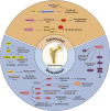
The bioactive effect of Trp metabolites in bone-related cells. Osteoblastic cell: Trp improved mRNA levels of Nanog and Oct-4 to maintain stemness, up-regulated osteogenic markers OPN and OCN, and decreased PPAR-γ and LPL to promote osteogenesis. Kyn up-regulated CYP1A1 and CYP1B1 expression to generate ROS. Kyn inhibited Runx2 expression and generated calcified matrix by damaging ATP production. Kyn could up-regulate miR-29b-1-5p to decrease CXCL12, CXCR4, and ACKR3 partially through the AhR pathway to inhibit osteogenic differentiation. PICA promoted the expression of osteogenic genes Runx2 and OCN to protect bone formation. 5-HT activated HTR1B to diminish phosphorylation of CREB and suppress CyclinD1. 5-HT also targets HTR2a and HTR2b to activate the PLC-IP3/DAG-PKC signaling pathway to enhance OB proliferation. Melatonin conversed the inhibitory effects of TNF-α on osteogenic differentiation, curbed the MT2-mediated NF-κB signaling pathway, and activated AMPK signaling, up-regulating Foxo3a and Runx2. Melatonin facilitated Osterix expression through PKA and PKC signaling pathways and up-regulated ZIP-1 to promote bone mineralization. Melatonin could suppress the expression of circ_0003865 to regulate GAS1 translationally by sponging miR-3653-3p to enhance osteogenic differentiation. Melatonin also promoted NSD2 expression by targeting MT1/2 to manage H3K36me2 and H3K27me3 modification, increasing Runx2 and BGLAP expression. IS inhibited ERK and p38 MAPK pathway through the AhR signaling. Osteoclastic cell: Kyn activated AhR to up-regulate c-Fos and NFATc1, as well as Blimp1, Cyp1b1, and Cyp1a2 expression, and enhanced NF-κB pathways to promote OC differentiation. Melatonin inhibited osteoclastogenesis through the miR-882/Rev-erbα axis and induced ROS. IPA enhanced PXR/P65 complex synthesis. IS affected the NFATc1 expression mediated by AhR signaling pathways in a time- and dose-dependent manner. Chondrocyte: 5-HT facilitated CCN2 production engaged by 5-HTR2A in the growth plates and reduced CCN2 generation through 5-HTR2B in articular cartilage. 5-HT can stimulate phospholipase A2 to increase collagenase type II activity, causing cartilage damage. Melatonin up-regulated Aanat, Mt1, Mt2, and Pthrp expression, subsequently followed by increased levels of Sox9 and Ihh. Besides, Bmal1 expression was enhanced, whereas Per1 expression was reduced. Melatonin facilitated the cartilage matrix synthesis via the TGF-β signaling pathway. Melatonin down-regulated the expression of MMP-13 and ADAMTS4 and promoted Col2A1 expression. Melatonin also inhibited the MAPK signaling pathway to repress IL-1β and TNF-α and was involved in activating the Wnt/β-catenin signaling pathway and inactivating the NF-κB signaling pathway. Melatonin maintained mitochondrial redox balance and repressed oxidative stress by modulating the AMPK/Foxo3 pathways. Melatonin enhanced the expression of Sirt1, inhibiting IRE1α-XBP1S-CHOP to alleviate ERS and curtailing EPC calcification. High melatonin concentrations impeded the chondrocyte proliferation and differentiation by down-regulating the Col2 and aggrecan expression and reduced the protein expression levels of PCNA, Sox9, and Smad4. Melatonin also up-regulated the expression of miR-526b-3p and miR-590-5p, which in turn boosted the phosphorylation of SMAD1 by targeting SMAD7, ultimately promoting chondrogenic differentiation. [All original elements used in the schematic figures are acquired from Servier Medical Art (http://smart.servier.com/).]
Fig. 3.
Fig. 3.
Interaction between Trp metabolites and OA. IPA targeted AhR to inhibit the expression of inflammatory factors (NO, PGE2, TNF-α, IL-6, iNOS, and COX-2) and matrix-degrading enzymes (MMP-3, MMP-13, and ADAMTS-5) by inactivating the NF-κB pathway. 5-HT activated Wnt/β-catenin signaling to inhibit gene expression of Sox9 and promote Mmp13. Melatonin inhibited the phosphorylation of PI3K/Akt, p38, ERK, JNK, and MAPK. Melatonin maintained mitochondrial functions, decreased mitochondrial oxidative stress, and inhibited NOX4 expression on mitochondria to mitigate ferroptosis. Melatonin significantly up-regulated HO-1 by enhancing the protein levels of NRF2 by suppressing miR-146a. Melatonin mitigated matrix degradation with enhanced SIRT1 expression by blocking the NF-κB signaling pathway and preserved chondrocytes through activation of the TGF-β1/Smad2 pathway, which inhibited the expression levels of MMP-3, MMP-13, ADAMTS-4, iNOS, NO, PGE2, and COX-2 and accelerated Col2 expression. [All original elements used in the schematic figures are acquired from Servier Medical Art (http://smart.servier.com/).]
Fig. 4.
Fig. 4.
Interaction between Trp metabolites and OP. Osteoblastogenesis: Kyn inhibited osteogenic marker expression including OCN and Runx2. The kyn-AhR axis decreased LC3B-II and autophagolysosomal production, with increased p62 levels. Kyn also induced a senescent phenotype with up-regulated p21, as well as enhanced aggregation of nuclear H3K9me3. KYNA could reduce NF-κB p65 phosphorylation by activating the Gpr35 receptor to up-regulate Runx2 expression. Melatonin suppressed OB differentiation through activation of the PI3K/AKT and BMP/Smad signaling and restrained the autophagy-targeted miR-224-5p/SIRT3/AMPK/mTOR axis. Melatonin also enhanced osteogenic differentiation and delayed bone loss via the HGF/PTEN/Wnt/β-catenin axis and inhibited the activation of the NLRP3 inflammasome mediated by the Wnt/β-catenin signaling pathway. Melatonin decreased the level of mitochondrial superoxide by activating SIRT1 and its downstream SOD2. SIRT1 could also enhance SIRT3 and suppress p66Shc expression. Melatonin promoted NSD2 expression, leading to a rebalancing of H3K36me2 and H3K27me3 modifications to enhance the expressions of Runx2 and BGLAP. Melatonin reversed oxidation level and led to elevated ALP activity, and up-regulated expression of BMP2, Runx2, and OPN. Melatonin down-regulated SMURF1 expression, consequently reducing ubiquitination and degradation of SMAD1 protein. Melatonin significantly relieved ERS by inhibiting the cascade of the PERK–eIF2α–ATF4–CHOP signaling axis. Osteoclastogenesis: KYNA could reduce NF-κB p65 phosphorylation by activating the Gpr35 receptor to inhibit NFATc1 expression. Melatonin increased BMAL1 expression to inhibit the activation of ROS and phosphorylation of MAPK-p38. It also obstructed osteoclastogenesis by inhibiting PRMT1 and ADMA expression, as well as TRAF6 and the phosphorylation of JNK. Melatonin also restrained the transcriptional activity of NF-κB by disturbing the binding of PRMT1 and NF-κB subunit p65. [All original elements used in the schematic figures are acquired from Servier Medical Art (http://smart.servier.com/).]
Fig. 5.
Fig. 5.
Interaction between Trp metabolites and RA. In the Kyn pathway, QUDA can restrain both the proliferation and motility of synoviocytes. QA can promote the proliferation of human fibroblast-like synoviocytes and stimulate mitochondrial respiration and glycolysis. In the 5-HT pathway, melatonin inhibited Cry1 expression, leading to enhanced cAMP levels and activation of PKA and NF-κB. Melatonin mitigated inflammation with the G protein–adenyl cyclase–cAMP pathway and Met-Enk inhibition. Melatonin significantly reduced the production of TNF-α and IL-1β by inhibiting PI3K/AKT, ERK, and NF-κB pathways. Besides, melatonin elevated IL-1β and IL-6 levels and reduced oxidative stress. [All original elements used in the schematic figures are acquired from Servier Medical Art (http://smart.servier.com/).]
Fig. 6.
Fig. 6.
Interaction between Trp metabolites and AS. Kyn significantly boosted OPG and OCN expressions by enhancing bone mineralization and suppressing RANKL-mediated OC differentiation. There was an inverse relationship between 5-HT levels and pCREB activation. Melatonin showed a positive correlation with OCN and IL-1β expression, as well as BASDAI, BASFI, the duration of morning stiffness, and CRP levels. IAA derived from gut microbiota down-regulated cytokine levels including TNF-α, IL-6, IL-17A, and IL-23, as well as improved the generation of anti-inflammatory cytokine IL-10. IAA could also enhance the intestinal barrier, relieve pathological changes, and reshape the gut microbiota. IAA stimulated the AhR pathway, up-regulating FoxP3 and increased Treg cells, down-regulating RORγt and STAT3, and decreasing Th17 cells. [All original elements used in the schematic figures are acquired from Servier Medical Art (http://smart.servier.com/).]

Similar articles

References

    1. Wu G. Amino acids: Metabolism, functions, and nutrition. Amino Acids. 2009;37(1):1–17. - PubMed
    1. Yusufu I, Ding K, Smith K, Wankhade UD, Sahay B, Patterson GT, Pacholczyk R, Adusumilli S, Hamrick MW, Hill WD, et al. A tryptophan-deficient diet induces gut microbiota dysbiosis and increases systemic inflammation in aged mice. Int J Mol Sci. 2021;22(9):5005. - PMC - PubMed
    1. Vécsei L, Szalárdy L, Fülöp F, Toldi J. Kynurenines in the CNS: Recent advances and new questions. Nat Rev Drug Discov. 2013;12(1):64–82. - PubMed
    1. Yano JM, Yu K, Donaldson GP, Shastri GG, Ann P, Ma L, Nagler CR, Ismagilov RF, Mazmanian SK, Hsiao EY. Indigenous bacteria from the gut microbiota regulate host serotonin biosynthesis. Cell. 2015;161(2):264–276. - PMC - PubMed
    1. Agus A, Planchais J, Sokol H. Gut microbiota regulation of tryptophan metabolism in health and disease. Cell Host Microbe. 2018;23(6):716–724. - PubMed

LinkOut - more resources