Multicenter Study
. 2025 May 27:16:1540341.
doi: 10.3389/fimmu.2025.1540341. eCollection 2025.

Long COVID-19 autoantibodies and their potential effect on fertility

Affiliations
Multicenter Study

Long COVID-19 autoantibodies and their potential effect on fertility

Laura Talamini et al. Front Immunol. .

Abstract

Impaired spermatogenesis has been reported in coronavirus disease 2019 (COVID-19) patients. However, the impact of severe acute respiratory syndrome coronavirus 2 (SARS-CoV-2) on male fertility remains unclear. The purpose of this multicenter study was to investigate the possible impact of SARS-CoV-2 infection on male fertility and determine the potential reasons leading to impaired male reproductive functions. In silico approach identified ~60 amino acid sequences containing at least five continuous residues shared by SARS-CoV-2 Spike glycoprotein and spermatogenesis-linked proteins. Four synthetic peptides were tested with sera from independent cohorts of patients with acute and long COVID-19 syndrome (LCS), and naïve vaccinated subjects. Immunogenicity and pathogenicity studies were performed by immunizing mice with two selected peptides and testing the antigenicity of induced antibodies. While none of four peptides were recognized by antibodies from vaccinated people, infected patients exhibited high reactivity to peptide 4, and LCS patients, especially women, showed elevated antibody levels against peptide 2. Women with LCS and chronic fatigue syndrome had higher levels of peptide 2-reacting antibodies than those with idiopathic chronic fatigue syndrome. Noteworthy, peptide 2 antibodies showed, in in vitro experiment, a specific interaction with mouse testicular tissue antigens. These findings raise the possibility that cross-reactive epitopes between SARS-CoV-2 Spike protein and spermatogenesis-related antigens may affect infected patients' fertility, suggesting a potential for autoimmune responses with human consequences.

Keywords: autoantibodies; coronavirus infection; male reproductive system; peptide sequence identity; post-COVID-19 condition.


Conflict of interest statement

The authors declare that the research was conducted in the absence of any commercial or financial relationships that could be construed as a potential conflict of interest. The author(s) declared that they were an editorial board member of Frontiers, at the time of submission. This had no impact on the peer review process and the final decision.

Figures
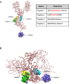
Figure 1
Figure 1
Molecular models of SARS-CoV-2 Spike protein and relative location of peptide sequences targets. (A) Side view of the SARS-CoV-2 Spike protein chain A backbone shown in pink pale ribbons from the Protein Data Bank file 6vxx code. Peptides 1 to 4 (the sequence of each is shown) are highlighted in the Spike protein 3D structure. Peptides 2, 3, and 4 (green, cyan, and yellow in solid surface volume representations). A molecular predicted model for peptide 1 from PepFold is shown in a blue solid surface volume and superimposed to the chain A 3D structure for its relative localization. (B) A Spike peptide view from the bottom highlighting peptides 1 to 4.
Figure 2
Figure 2
Levels of serum reactivity with peptides 1 to 4 of the SARS-CoV-2 Spike protein categorized by country and disease groups. (A) Schematic representation of the study cohort showing the number of individuals by disease group for each country. (B) Dot plot showing the levels of serum reactivity to peptides 1 to 4 of all infected individuals. (C) Heatmaps showing the levels of serum reactivity to peptides 1 to 4 per country of each patient. The intensity of the reaction is indicated by colors as indicated. (D) Bar graphics showing the levels (ELISA absorbance values) of peptide-reacting antibodies by country. (E) Dot plot showing the levels of serum reactivity to peptides 1 to 4 distinguishing patients with acute versus long COVID-19. NHS (normal human sera from healthy donors) were used as controls. The dashed line in each bar graphic denotes the cutoff value for each peptide. Data are reported as mean ± SD. Statistical analysis is determined by non-parametric Kruskal-Wallis following Dunn’s test. Differences were considered significant when P < 0.05.
Figure 3
Figure 3
Levels of serum reactivity with peptides 2 and 4 of the SARS-CoV-2 Spike protein according to groups of age, gender, and disease status (acute or long COVID-19 indication). Donut graphs showing the distribution of serum reactivity levels of (A) peptide 2 and (B) peptide 4 between acute and long COVID-19 patients classified by age (left panels). Scatter plot showing the age distribution of serum levels. Blue lines represent acute COVID-19, and red lines show long COVID-19 group (right panels). The R-squared and adjusted P-values from the Kendall correlation are displayed at the top of the graphs. (C) Dot plot reporting serum level of groups described above according to gender. Results are reported as mean ± SD. A dashed line denotes the cutoff value determined for each peptide. Statistical analysis is determined by the Mann–Whitney non-parametric test. Differences were considered significant when P < 0.05. M, men; W, women; yo, year-old.
Figure 4
Figure 4
Levels of serum reactivity with peptides 2 and 4 of the SARS-CoV-2 Spike protein according to clinical features of infected patients. (A) Dot plot showing serum levels in long COVID-19 patients affected by chronic fatigue syndrome (with CFS) or unaffected (w/o CFS). (B) Data in (A) according to gender. (C) Dot plot showing serum levels in uninfected individuals presenting idiopathic CFS (non–COVID-19 subjects) and in long COVID-19 patients who developed CFS after virus infection. (D) Results in (C) according to gender. Results are reported as mean ± SD. The cutoff value of each peptide is denoted as a dashed line. Statistical analysis is determined by Mann–Whitney non-parametric test. Differences were considered significant when P < 0.05. M, men; W, women; w/o, without.
Figure 5
Figure 5
Characterization of IgG antibodies generated in normal mice against peptides 2 and 4. (A) Coomassie blue staining gel showing IgG antibodies to peptides 2 and 4 before and after purification on protein G magnetic beads. (B) Reactivity in ELISA of IgG antibodies to peptides 2 and 4 towards their homologous (immunogen) peptide. Plates were coated with 2µM of Cys-peptide 2 or Cys-peptide 4 in bicarbonate/carbonate buffer (pH 9.6), and the reactivity of purified IgG antibodies was tested with increasing concentrations (0 to 1.2 µg/mL) of each of them. Normal mouse IgG was used as a control. Data are reported as mean ± SD of absorbance measured at 450 nm. (C) Western blots showing the reactivity of purified IgG to peptides 2 and 4 with the His tag-SARS-CoV-2 Spike protein. A control protein (CTR), lacking the His tag and produced in another species, has been added to the gel as a negative control. Membranes were blotted for IgG antibodies at 1 µg/mL, or for anti-6x His tag at 0.7 µg/mL. IgG anti-mouse and anti-rabbit HRP at 0.05 µg/mL were used as secondary antibodies. (D) Representative histological images of mouse testicular tissue stained with mouse anti-peptide 2 IgG or normal mouse IgG used as control, and revealed with AF488-labeled goat anti-mouse Ig (magenta). Nuclei were stained with Hoechst (grey). Scale bar = 50 µm or 15 µm (higher magnification).

Similar articles

References

    1. Wildner NH, Ahmadi P, Schulte S, Brauneck F, Kohsar M, Lütgehetmann M, et al. B cell analysis in SARS-CoV-2 versus malaria: Increased frequencies of plasmablasts and atypical memory B cells in COVID-19. J Leukoc Biol. (2021) 109:77–90. doi: 10.1002/JLB.5COVA0620-370RR - DOI - PMC - PubMed
    1. Chang R, Yen-Ting Chen T, Wang S-I, Hung Y-M, Chen H-Y, Wei C-CJ. Risk of autoimmune diseases in patients with COVID-19: a retrospective cohort study. eClinicalMedicine. (2023) 56:101783. doi: 10.1016/j.eclinm.2022.101783 - DOI - PMC - PubMed
    1. Dotan A, Muller S, Kanduc D, David P, Halpert G, Shoenfeld Y. The SARS-CoV-2 as an instrumental trigger of autoimmunity. Autoimmun Rev. (2021) 20:102792. doi: 10.1016/j.autrev.2021.102792 - DOI - PMC - PubMed
    1. Knight JS, Caricchio R, Casanova J-L, Combes AJ, Diamond B, Fox SE, et al. The intersection of COVID-19 and autoimmunity. J Clin Invest. (2021) 131:e154886. doi: 10.1172/JCI154886 - DOI - PMC - PubMed
    1. Wang EY, Mao T, Klein J, Dai Y, Huck JD, Jaycox JR, et al. Diverse functional autoantibodies in patients with COVID-19. Nature. (2021) 595:283–8. doi: 10.1038/s41586-021-03631-y - DOI - PubMed

Publication types

LinkOut - more resources