. 2025 Dec;17(1):2517380.
doi: 10.1080/19490976.2025.2517380. Epub 2025 Jun 11.

Human gut commensal Alistipes timonensis modulates the host lipidome and delivers anti-inflammatory outer membrane vesicles to suppress colitis in an Il10-deficient mouse model

Affiliations

Human gut commensal Alistipes timonensis modulates the host lipidome and delivers anti-inflammatory outer membrane vesicles to suppress colitis in an Il10-deficient mouse model

Ethan A Older et al. Gut Microbes. 2025 Dec.

Abstract

Correlative studies have linked human gut microbes to specific health conditions. Alistipes is one such microbial genus negatively linked to inflammatory bowel disease (IBD). However, the protective role of Alistipes in IBD is understudied, and the underlying molecular mechanisms remain unknown. In this study, colonization of Il10-deficient mice with Alistipes timonensis DSM 27924 delays colitis development. Colonization does not significantly alter the gut microbiome composition, but instead shifts the host plasma lipidome, increasing phosphatidic acids while decreasing triglycerides. Outer membrane vesicles (OMVs) derived from Alistipes are detected in the plasma of colonized mice, carrying potentially immunomodulatory metabolites into the host circulatory system. Fractions of A. timonensis OMVs suppress LPS-induced Il6, Il1b, and Tnfa expression in vitro in murine macrophages. We detect putative bioactive lipids in the OMVs, including immunomodulatory sulfonolipids (SoLs) in the active fraction, which are also increased in the blood of colonized mice. Treating Il10-deficient mice with purified SoL B, a representative SoL, suppresses colitis development, suggesting its contribution to the anti-inflammatory phenotype observed with A. timonensis colonization. Thus, A. timonensis OMVs represent a potential mechanism for Alistipes-mediated delay of colitis in Il10-deficient mice via delivery of immunomodulatory lipids and modulation of the host plasma lipidome.

Keywords: Alistipes; IBD; Il10-deficient mice; OMVs; colitis; gut microbes; inflammation; sulfonolipids.


Conflict of interest statement

No potential conflict of interest was reported by the author(s).

Figures

Figure 1.
Figure 1.
A. timonensis colonization delays piroxicam-accelerated colitis development in IL10 KO mice. (a) Representative H&E images of uncolonized (uncol.) and A. timonensis-colonized (A. tim.) mouse colons showing less inflammation in colonized mice. One-sided student’s t-test confirmed significantly decreased gross pathology and histology scores in uncol. (n = 9 mice) compared to A. tim. (n = 9 mice) mice. (b-e) The expression of pro-inflammatory factors Il1b, Tnfa, Il6, and Nos2 were significantly decreased in the distal colons of A. timonensis-colonized (A. tim., n = 9 mice) mice compared to uncolonized (Uncol., n = 9 mice) mice, supporting that Alistipes colonization mediated a suppressive effect against colitis progression. Significance was determined using two-sided student’s t-test. For all p-values, * 0.05 > p > 0.0 and **** 0.0001 > p.
Figure 2.
Figure 2.
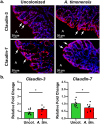
A. timonensis colonization modulates tight junction protein morphology and gene expression in IL10 KO mouse colons. (a) Immunofluorescent staining of tight junction proteins claudin-3 and claudin-7 (red) illustrating restoration of tight junction morphology and reduction of tight junction repair with A. timonensis colonization, respectively. Nuclei were stained with DAPI (blue). A: apical side of colon tissue; arrow: tight junction morphologies. (b) Gene expression of claudin-3 and claudin-7 in uncolonized (uncol., n = 9 mice) and A. timonensis-colonized (A. tim., n = 9 mice) mice, which is consistent with immunofluorescence staining. Significance was determined using two-sided student’s t-test. For all p-values, * 0.05 > p > 0.01.
Figure 3.
Figure 3.
Microbiome composition analysis indicates A. timonensis colonization does not reverse dysbiosis. (a) Beta diversity measured by principal coordinate analysis (PCoA) using Jaccard distances shows clear separation between both independent cohorts (red and green triangles and circles) and the control group (blue squares) but no separation between A. timonensis-colonized (red) versus uncolonized (green) microbiome profiles. (b) Alpha diversity measured by Shannon diversity index indicates a significant loss of microbial diversity from control (n = 17 mice) to uncolonized (n = 11 mice) and A. timonensis-colonized (n = 10 mice) mice that was not restored by A. timonensis colonization. Significance was determined using two-sided student’s t-test. For all p-values, * 0.05 > p > 0.01, ** 0.01 > p > 0.001, and ns: not significant. (c) Heatmap of microbiome composition at the family level showing a clear difference in control samples compared to uncolonized and A. timonensis-colonized mice. (d) Genera composition of the phylum Bacteroidetes highlights the major expansion of A. timonensis in colonized compared to uncolonized samples.
Figure 4.
Figure 4.
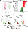
Plasma lipidomics reveals colonization-driven change in circulating lipid profile. (a) Orthogonal partial least squares-discriminant analysis (OPLS-DA) shows clear difference in plasma lipid profiles between uncolonized (n = 9 mice) and A. timonensis-colonized (n = 9 mice) mice. (b) The top 20 significantly differentially abundant lipids reveal a marked depletion of triglycerides (TGs) and accumulation of phosphatidic acids (PAs) in A. timonensis-colonized mice compared to uncolonized mice. (c,d) A. timonensis colonization affected the balance of two pools of arachidonic acid-containing lipids: triglycerides (c), which were less abundant in A. timonensis-colonized mice, and phospholipids (d), which were more abundant in A. timonensis-colonized mice. Significance was determined using two-sided student’s t-test. For all p-values, * 0.05 > p > 0.01 and ** 0.01 > p > 0.001.
Figure 5.
Figure 5.
A. timonensis-derived OMVs are abundant in the plasma of A. timonensis-colonized mice. (a) TEM was used to visually confirm the presence of OMVs after negative staining. x30.0k magnification was used to confirm the diameter of OMVs in the expected range. (b) DLS was used to measure the size distribution of plasma OMVs from uncolonized (green) and A. timonensis-colonized (red) mice alongside OMVs purified from A. timonensis culture (black). DLS analysis showed two clear vesicle populations with diameter ranges of 10–55 nm and 55–320 nm. The rightmost larger diameter population corresponds with A. timonensis culture OMVs and reflects the increased abundance of OMVs derived from A. timonensis colonization. (c) Lipids were extracted from purified blood OMVs, measured using targeted metabolomics, and SoL B was found to be significantly increased in plasma OMVs from A. timonensis-colonized (n = 4 mice) compared to uncolonized (n = 4 mice) mice. Significance was determined using two-sided student’s t-test. For all p-values, ** 0.01 > p > 0.001.
Figure 6.
Figure 6.
Sulfonolipid abundance significantly increased in the plasma of A. timonensis-colonized mice. (a-c) abundance of SoL B (a), SoL a (b), and SoL F (c) measured in uncolonized (n = 10 mice) compared to A. timonensis-colonized (n = 10 mice) mouse plasma by targeted metabolomics. (d) Corresponding chemical structures of SoL B, A, and F. (e) Retention time comparison of in-house SoL B standard and observed SoL B peak in A. tim. samples supporting identification as SoL B. (f) MS/MS fragmentation used to positively identify SoL B based on characteristic fragmentation including the ~81 m/z fragment corresponding to the loss of the HSO3 headgroup. (g) The plasma-to-feces ratio of SoL B abundance was significantly greater in A. timonensis-colonized (n = 7) compared to uncolonized (n = 7) samples, suggesting that the increased production of SoL B by A. timonensis in the gut led to increased SoL B abundance in the plasma via active translocation of SoL B from the gut to the plasma by OMVs. Significance was determined using two-sided student’s t-test. For all p-values, * 0.05 > p > 0.01, ** 0.01 > p > 0.001, *** 0.001 > p > 0.0001, and **** 0.0001 > p.
Figure 7.
Figure 7.
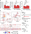
Bioactive molecular networking of A. timonensis OMV fractions suggests multiple compound classes contributing to an overall anti-inflammatory effect against LPS-induced inflammation. (a) Relative Fold change expression pro-inflammatory cytokines Il1b, Il6, and tnfa after treating mouse peritoneal macrophages 1 μg/mL LPS, or 1 μg/mL LPS with fractions of A. timonensis OMVs at 20 μg/mL (n = 3 wells per treatment). Fraction 2 (F2) showed the strongest suppression among other fractions and was further investigated for its bioactive components. Significance was determined using two-sided student’s t-test. For all p-values, * 0.05 > p > 0.01, *** 0.001 > p > 0.0001, and ns: not significant. (b) Untargeted metabolomics and subsequent bioactive molecular networking analysis of fraction 2 of A. timonensis OMVs revealed multiple clusters of molecules including SoLs which were abundant in this active fraction. (c) Predicted chemical structures of the major clusters, suggesting other potential contributors to the biological activity of fraction 2.
Figure 8.
Figure 8.
IL10 KO mice treated with purified SoL B by I.P. injection exhibit delayed colitis progression. (a) Representative histology images illustrate the difference in colon pathology associated with purified SoL B treatment (SoL B) compared to vehicle control (vehicle). Gross pathological analysis revealed significantly lower levels of inflammation in SoL B-treated mice. Histological analysis also supports lower inflammation in the SoL B-treated mice. (b-e) RT-qPCR showed expression of pro-inflammatory markers were decreased in SoL B-treated mice (SoL B) compared to vehicle-treated mice (veh.), supporting that SoL B treatment suppresses colitis development.

Update of

References

    1. FitzGerald R, Smith SM.. 2021. An overview of helicobacter pylori infection. In: Smith SM, editor. Helicobacter pylori. Vol. 2283. New York (NY): Springer; p. 1–22. - PubMed
    1. Cani PD, de Vos WM. Next-generation beneficial microbes: the case of akkermansia muciniphila. Front Microbiol. 2017;8. doi: 10.3389/fmicb.2017.01765. - DOI - PMC - PubMed
    1. Lau K, Srivatsav V, Rizwan A, Nashed A, Liu R, Shen R, Akhtar M. Bridging the gap between gut microbial dysbiosis and cardiovascular diseases. Nutrients. 2017;9(8):859. doi: 10.3390/nu9080859. - DOI - PMC - PubMed
    1. Sheflin AM, Whitney AK, Weir TL. Cancer-promoting effects of microbial dysbiosis. Curr Oncol Rep. 2014;16(10):406. doi: 10.1007/s11912-014-0406-0. - DOI - PMC - PubMed
    1. Sultan S, El-Mowafy M, Elgaml A, Ahmed TAE, Hassan H, Mottawea W. Metabolic influences of gut microbiota dysbiosis on inflammatory bowel disease. Front Physiol. 2021;12. doi: 10.3389/fphys.2021.715506. - DOI - PMC - PubMed

LinkOut - more resources