Review
. 2025 Oct 1;105(4):2305-2376.
doi: 10.1152/physrev.00039.2024. Epub 2025 Jun 11.

The human placenta and its role in reproductive outcomes revisited

Affiliations
Review

The human placenta and its role in reproductive outcomes revisited

Irving L M H Aye et al. Physiol Rev. .

Abstract

The placenta performs many key tasks that are essential for the healthy growth and development of the human fetus. Placental dysfunction has multiple manifestations, but they share the common property of lacking a mechanistic understanding of etiology. The clinical consequences of placental dysfunction are a major determinant of the global burden of disease. Currently, the primary clinical method for assessing placental function is ultrasonic Doppler flow velocimetry of the umbilical and uterine arteries. More recently, some biomarkers have emerged that can predict or diagnose placentally related complications of pregnancy. However, methods for identifying and characterizing placental dysfunction have developed relatively little over the last 20 years and perform poorly, and there remains an absence of disease-modifying therapies targeted at the placenta. Understanding disease mechanisms is made more difficult due to the profound differences in pregnancy and placentation comparing humans and the most commonly used laboratory animals, limiting the utility of animal models. The use of omics methods in human samples may yield progress: omics analyses of maternal blood show promise in identifying better predictors of disease, and single-cell analyses, including spatial omics of healthy and abnormal placentas, could identify therapeutic targets. Limitations in cellular models of the placenta have been significantly overcome in the last 5 to 10 years by the development of human cell models, including human trophoblast stem cells and organoids, and the use of these model systems may allow hypothesis testing experiments in a more clinically relevant context than animal models or immortalized cell lines.

Keywords: development; fetal growth restriction; preeclampsia; pregnancy; trophoblast.


Conflict of interest statement

Disclosures

The authors declare the following competing interests: GS and DC-J have received research support from Roche Diagnostics Ltd for studies of diagnostics and screening for fetal growth restriction and preeclampsia. GS’s department has received payment from Roche for a talk given by GS (fetal growth restriction). GS has been a paid consultant to GSK (preterm birth) and has been a member of a Data Monitoring Committee for GSK trials of RSV vaccination in pregnancy. GS is currently a member of a Data Monitoring Committee for RSV vaccination in pregnancy (Moderna) and chairs a Data Monitoring Committee for a hyperemesis gravidarum therapeutic trial (NG Biopharmaceuticals). GS and DC-J have received nonfinancial support from Illumina. GS and DC-J have received support from Pfizer and Sera Prognostics. Cambridge Enterprise (UK) have filed a patent relating to the prediction of pregnancy outcome with GS, DC-J and IA as named inventors. ST is a paid consultant to Diamedica Therapeutics and Avilar Therapeutics. He is on the scientific advisory board of Diamedica Therapeutics. ST is named on patents related to SPINT1 as a blood test for placental insufficiency and fetal growth restriction.

Figures

Figure 1
Figure 1. Cross-section of a post-implantation embryo.
Histological section of a post-implantation embryo (Carnegie stage 5c, 11-12 dpf) following Hematoxylin and Eosin staining. The cytotrophoblasts have begun to differentiate into the primitive syncytium which invades into the maternal decidua forming lacunae. Images reprinted with permission from the Virtual Human Embryo Project at Louisiana State University (http://virtualhumanembryo.lsuhsc.edu).
Figure 2
Figure 2. Cross-section of the maternal-fetal interface in early pregnancy.
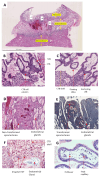
Hematoxylin and eosinstained sections from the Boyd collection of [A-D, F-G] a 6-week gestational age specimen (H710) and [E] a 10-week gestational age specimen (H630) obtained from pregnant women where hysterectomies were performed. A Overview of the uterus with attached conceptus. B High magnification image showing cytotrophoblast cell columns migrating towards the decidua. C High magnification image showing floating villi and anchoring villi with cytotrophoblasts spreading to form the cytotrophoblast shell. D Low magnification image showing non-transformed spiral arteries with narrow bores and surrounding endometrial glands. E Low magnification image of transformed spiral arteries showing dilatated vessels with large bores. F High magnification image of invasive EVTs migrating from CTB shell. G High magnification image showing a villus with a Hofbauer cell in a stromal channel formed by fibroblasts and a fetal capillary with nucleated red blood cells. CTB, cytotrophoblast; EVT, extravillous trophoblast; STB, syncytiotrophoblast. Placenta-in-situ slides from the Loke Centre for Trophoblast Research Boyd Collection, funded by Loke Centre for Trophoblast Research and the Wellcome Trust (215361/Z/19/Z). Licensed under CC BY-NC-SA 4.0. Retrieved 23-Apr-2025 from https://www.trophoblast.cam.ac.uk/Resources/boyd-collection.
Figure 3
Figure 3
Overview of molecular pathways regulating trophoblast self-renewal or differentiation. The illustration provides an overview of the molecular pathways discussed in the review and is not an exhaustive list. CTB, cytotrophoblast; EVT, extravillous trophoblast; STB, syncytiotrophoblast. Created using images from a licensed Biorender.com.
Figure 4
Figure 4. Human placental terminal villi at term.
A) Scanning electron micrograph of a freeze-cracked human placental villus. The villus has been cracked open to reveal the capillaries inside. The syncytiotrophoblast surface in contact with maternal blood is covered in microvilli. The thin barrier at the vasculosyncytial membrane is arrowed. B) Transmission electron micrograph of a terminal villus. The microvilli on the surface of the syncytiotrophoblast are visible. C) Confocal images of perfused human villi; syncytiotrophoblast is stained red, endothelial cells green and nuclei blue. MBS maternal blood space; MV micro villi; FC fetal capillary; fRBC fetal red blood cell; E endothelial cell nucleus and P pericyte. Image A is from Charnock-Jones and Burton 2000 (16) and B was provided by GJ Burton, samples in C are those described in Mayo et al 2016 (125).
Figure 5
Figure 5
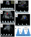
A. Low resistance flow in the umbilical artery, as evidenced by a high rate of forward flow at the end of diastole B. High resistance flow in the umbilical artery as evidenced by the absence of forward flow at the end of diastole, C. Very high resistance flow in the umbilical artery, as evidenced by the reversal of the direction of flow at end of diastole, D. Low resistance flow in the maternal uterine artery, as evidenced by a high rate of flow at the end of diastole and the absence of a notch after the initial systolic peak, E. High resistance flow in the maternal uterine artery, as evidenced by a low rate of flow at the end of diastole and the presence of a notch after the initial systolic peak, F. Calculation of the pulsatility index, a measure of downstream vascular resistance, from the Doppler flow velocity waveform.
Figure 6
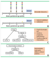
Figure 6. Schematic illustration of clinical study design for the implementation of a biomarker following omic discovery.
A. In the original discovery study a candidate test is identified in a study which has a large number of hypothesis tests. B. Having identified the candidate, its predictive utility needs to be externally validated. This rules out the possibility that the initial findings were a false discovery due to a large number of hypothesis tests. It also assesses the test in separate and, ideally, diverse populations (confirming external validity) and the absolute risk of the outcome in relation to the level of the test (calibration). C. A proof of principle study randomizes high risk women to intervention or routine care, and this allows both validation of the screening test and assessment of the effect of the intervention. This study design also requires a much smaller sample size than the design below (253). D. Randomization to screen or no screen gives the best information about the likely effect of implementation of screening into a healthcare system. Implementation of the screening program into a group of hospitals can be both performed and assessed by randomization at the level of the hospital, using a stepped wedged cluster randomized controlled trial (254).
Figure 7
Figure 7. Overview of placental lesions associated with fetal growth restriction.
PAPPA, pregnancy associated plasma protein-A; hCG, human chorionic gonadotropin; PlGF, placental growth factor. Created using images from a licensed Biorender.com.
Figure 8
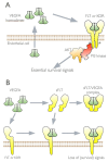
Figure 8. Schematic illustration of VEGFA signaling and the dominant negative action of sFLT1.
VEGFA is secreted from endothelial and binds with high affinity to the VEGF receptors (FLT1 or KDR). This leads to dimerization and autophosphorylation of the receptor (-P). A signaling cascade is triggered which activates PI3 kinase and AKT, promoting endothelial cell survival. B) In the presence of sFLT, VEGFA (and PlGF) are bound in a complex which prevents their binding to cell surface receptors. Thus, sFLT can act as a competitive inhibitor. However, sFLT can also bind to the cell surface receptors and form a heterodimer. As sFLT lacks the intracellular kinase domain, autophosphorylation cannot occur and activation of the full-length receptor is blocked and the survival signals are not generated. Figure modified from Charnock-Jones 2016 (406).
Figure 9
Figure 9. Overview of the pathogenesis of preeclampsia.
sFLT1, soluble fms-like tyrosine kinase 1; VCAM1, vascular cell adhesion molecule 1; PlGF, Placental growth factor. Created using images from a licensed Biorender.com.
Figure 10
Figure 10. Schematic representation of the metabolic effects of maternal gestational diabetes on placental function and fetal growth.
Maternal insulin resistance diverts circulating nutrients (amino acids, fatty acids and glucose) across the placenta. Maternal hyperinsulinemia promotes placental endothelial vascularization, trophoblast amino acid transport and fatty acid esterification. The resulting increase in nutrient accumulation in the fetus promotes fetal insulin secretion leading to excess fetal growth. PGH, placental growth hormone; PL, placental lactogen; Lep, leptin; E2, estrogen; P4, progesterone.
Figure 11
Figure 11. The dashed lines represent the 3rd, 50th and 97th percentile of observed birth weight across the range of gestational age.
The solid lines represent the 3rd, 50th and 97th percentile of ultrasonically estimated fetal weight. Whereas the distributions are very similar at term, the distribution of observed birth weights is lower than the ultrasonic estimated fetal weights of on-going pregnancies at the same gestational age in the preterm period. Figure modified from Smith et al. Best Pract Res Clin Obstet Gynaecol 2018; 49: 478-486 (476).
Figure 12
Figure 12
Schematic representation of integrated pathways of steroid biosynthesis in the fetus and placenta. DHEA denotes dehydroepiandrosterone and DHEA denotes dehydroepiandrosterone sulfate.
Figure 13
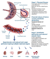
Figure 13. Schematic overview indicating the link between placental dysfunction, fetal stress and the initiation of labor and delivery.
Maternal factors associated with an increased risk of stillbirth are highlighted (blue box) as potentially inhibiting the effect of activation of the fetal hypothalamo-pituitary adrenal axis to promote labor (broken line as mechanisms less well understood in humans than other model species, e.g. sheep). Hence, the above model could explain the relationship between these factors and the risk of stillbirth.

References

    1. Roth GA, Abate D, Abate KH, Abay SM, Abbafati C, Abbasi N, Abbastabar H, Abd-Allah F, Abdela J, Abdelalim A, Abdollahpour I, et al. Global, regional, and national age-sex-specific mortality for 282 causes of death in 195 countries and territories, 1980–2017: a systematic analysis for the Global Burden of Disease Study 2017. The Lancet. 2018;392:1736–1788. doi: 10.1016/S0140-6736(18)32203-7. - DOI - PMC - PubMed
    1. Brosens I, Pijnenborg R, Vercruysse L, Romero R. The “great Obstetrical Syndromes” are associated with disorders of deep placentation. Am J Obstet Gynecol. 2011;204:193–201. doi: 10.1016/j.ajog.2010.08.009. - DOI - PMC - PubMed
    1. Smith GCS, Pell JP, Walsh D. Pregnancy complications and maternal risk of ischaemic heart disease: a retrospective cohort study of 129 290 births. The Lancet. 2001;357:2002–2006. - PubMed
    1. Pell JP, Smith GCS, Walsh D. Pregnancy Complications and Subsequent Maternal Cerebrovascular Events: A Retrospective Cohort Study of 119,668 Births. Am J Epidemiol. 2004;159:336–342. - PubMed
    1. Poorthuis MHF, Algra AM, Algra A, Kappelle LJ, Klijn CJM. Female- and Male-Specific Risk Factors for Stroke: A Systematic Review and Meta-analysis. JAMA Neurol. 2017;74:75–81. - PubMed

LinkOut - more resources