. 2025 Dec;11(1):2476922.
doi: 10.1080/20565623.2025.2476922. Epub 2025 Jun 11.

Amniotic fluid MSCs for scaffold-free cartilage repair: spheroid fusion and chondrogenic microtissue development

Affiliations

Amniotic fluid MSCs for scaffold-free cartilage repair: spheroid fusion and chondrogenic microtissue development

Carolina Coli Zuliani et al. Future Sci OA. 2025 Dec.

Abstract

Background: Articular cartilage injuries are challenging due to limited regenerative capacity, causing chronic pain and impaired mobility. Current treatments are often inadequate, necessitating novel cartilage repair approaches. This study investigates amniotic fluid-derived mesenchymal stromal cells (AF-MSC) as a promising cell source for tissue engineering.

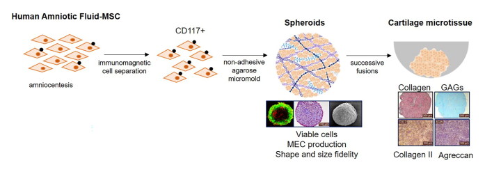
Research design and method: Cartilage-like microtissues were produced by differentiating AF-MSC into chondrocytes within a 3D culture system. Using a 3D-printed non-adhesive micromold, AF-MSC spheroids were formed and fused into larger microtissues. Spheroids were characterized for morphology, viability, and extracellular matrix (ECM) production. The mechanical properties of resulting microtissues were compared to native cartilage and agarose hydrogel.

Results: AF-MSC proved a viable, scalable cell source for cartilage microtissues. Spheroid fusion created structures with mechanical properties and ECM components resembling native cartilage.

Conclusions: AF-MSCs differentiated into chondrocytes when stimulated with TGF-β3 in a 3D micromolded culture, forming uniform, viable spheroids with robust ECM production and mechanical properties. These spheroids fused into neocartilage microtissue, showing potential for regenerative medicine, especially osteoarthritis treatment and drug testing. Further research should optimize conditions and evaluate long-term biomechanical performance.

Keywords: Mesenchymal stem cells; amniotic fluid; chondrogenesis; tissue engineering and cartilage regeneration.

Plain language summary

Cartilage injuries are hard to heal because cartilage does not repair itself easily. This can cause long-term pain and make movement difficult. Current treatments do not always work well, so scientists are looking for new ways to help cartilage heal. In this study, researchers used special cells from amniotic fluid, called mesenchymal stromal cells (AF-MSCs). These cells were grown in a 3D system, where they formed small, round clusters called spheroids. Over time, the spheroids merged to create tiny tissue structures similar to cartilage. The results showed that the cells stayed healthy, produced important cartilage materials, and had properties similar to natural cartilage. This method could help treat cartilage damage, including conditions like osteoarthritis. It could also be useful for testing new medicines. However, more research is needed to improve the process and make sure these engineered tissues remain stable over time.


Conflict of interest statement

No potential conflict of interest was reported by the authors.

Figures

None
Graphical abstract
Figure 1.
Figure 1.
Morphological characterization, differentiation test, and immunophenotyping of AF-MSC. Representative image of human amniotic fluid cells in cellular expansion exhibiting fibroblast-like colony-forming morphology. Phase contrast microscope (400X) (A); Cells differentiated into adipocytes with fat vesicles stained orange by Oil Red (B); osteocytes with calcium crystals stained red by Alizarin Red (C); chondrocytes with glycosaminoglycan production stained blue by Alcian Blue (D); Immunophenotypic analysis by flow cytometry of mesenchymal markers (E), pluripotency markers (F), and indicators of higher chondrogenic potential markers (G). Histograms represent the fluorescence intensity of each marker by the number of counted events.
Figure 2.
Figure 2.
Characterization of produced spheroids regarding morphology, viability, and spontaneous fusion capacity. SEM images of a uniform group of spheroids (A); morphology of a single spheroid (B); internal porous structure of a spheroid in cross-section (C); detailed surface view showing a compact extracellular matrix (ECM) with a high number of cells (arrows) (D). Viability over the cultivation period. Viable cells were stained green, and dead cells were stained red using the LIVE/DEAD technique (E, F, G, H). Images were obtained using a confocal microscope. Fusion dynamics. Spheroids in the micromold (I); spheroids grouped in a single non-adhesive well after 24 hours (J); completely fused spheroids after 72 hours. Images were obtained using an inverted optical microscope (40X magnification).
Figure 3.
Figure 3.
Microtissue production through sequential spheroid fusion: macroscopic and histological analysis. The images depict the structural evolution during the culture process, with histological staining at different stages. Hematoxylin and eosin staining highlights cell nuclei (purple) and extracellular matrix (pink) (A–C). Alcian Blue staining indicates glycosaminoglycans (light blue) (D–F), while Masson’s trichrome reveals collagen fibers (blue) (G–I). Picrosirius Red staining further highlights collagen fibers (red) throughout the tissue (J–O). Black arrows mark spheroid adhesion regions (M), with cellular migration contributing to fusion (N) and extensive extracellular matrix deposition (O).
Figure 4.
Figure 4.
Quantification of matrix components using immunostaining. Semi-quantitative analysis of immunohistochemical data for collagen type II and aggrecan in each group, represented as a percentage of the labeled area using ImageJ software, is presented in the graph. (A) In the immunohistochemical staining, positive labeling for type II collagen antibodies (B, C, D) and aggrecan (E, F, G) is shown in brown, while nuclei were counterstained in purple with hematoxylin. A negative control was included to assess immunoreactivity (H, I, J). The antibodies used were: Rabbit Polyclonal Anti-Collagen II Antibody, BIOSS, and Rabbit Polyclonal Anti-Aggrecan Antibody, BIOSS.
Figure 5.
Figure 5.
Mechanical Properties of Cartilaginous Microtissue. Comparative analysis between the microtissue and human knee cartilage samples (A). The shape and dimensions of each sample are described (B), and the mechanical properties are shown (C). The compressive behavior of each material is represented in the graph during testing. Samples were subjected to compressive loading until 100% deformation, at which point the maximum compressive stress (MCS) was recorded (D).

Similar articles

References

References: Papers of special note have been highlighted as either of interest (*) or of considerable interest (**) to readers.: * The article provides a good background on regenerative medicine and cartilage injuries.

    1. Hunter DJ, Bierma-Zeinstra S.. Osteoarthritis. Lancet. 2019;393(10182):1745–1759. - PubMed
    1. Chiang H, Jiang CC.. Repair of articular cartilage defects: Review and perspectives. J Formos Med Assoc. 2009;108(2):87–101. doi: 10.1016/S0929-6646(09)60039-5 - DOI - PubMed
    1. Loo SJQ, Wong NK.. Advantages and challenges of stem cell therapy for osteoarthritis (review). Biomed Rep. 2021;15(2):67. doi: 10.3892/br.2021.1443 - DOI - PMC - PubMed
    1. Davies RL, Kuiper NJ.. Regenerative medicine: a review of the evolution of autologous chondrocyte implantation (aci) therapy. Bioengineering (Basel). 2019;6(1):22. doi: 10.3390/bioengineering6010022 - DOI - PMC - PubMed
    1. Martínez-Moreno D, Jiménez G, Gálvez-Martín P, et al. Cartilage biomechanics: a key factor for osteoarthritis regenerative medicine. Biochim Biophys Acta Mol Basis Dis. 2019;1865(6):1067–1075. doi: 10.1016/j.bbadis.2019.03.011 - DOI - PubMed

*The article introduces the potential use of amniotic fluid mesenchymal cells in regenerative therapies.

    1. Prusa AR, Marton E, Rosner M, et al. Oct-4-expressing cells in human amniotic fluid: a new source for stem cell research? Hum Reprod. 2003;18(7):1489–1493. - PubMed
    1. De Coppi P, Bartsch G, Siddiqui MM, et al. Isolation of amniotic stem cell lines with potential for therapy. Nat Biotechnol. 2007;25(1):100–106. doi: 10.1038/nbt1274 - DOI - PubMed
    1. Stultz B, Mcginnis K, Thompson E, et al. Chromosomal stability of mesenchymal stromal cells during in vitro culture. Cytotherapy. 2016;18(3):336–343. doi: 10.1016/j.jcyt.2015.11.017 - DOI - PMC - PubMed
    1. Loukogeorgakis SP, De Coppi P.. Stem cells from amniotic fluid–potential for regenerative medicine. Best Pract Res Clin Obstet Gynaecol. 2016;31:45–57. doi: 10.1016/j.bpobgyn.2015.08.009 - DOI - PubMed
    1. Ramasamy TS, Velaithan V, Yeow Y, et al. Stem cells derived from amniotic fluid: a potential pluripotent-like cell source for cellular therapy? Curr Stem Cell Res Ther. 2018;13(4):252–264. doi: 10.2174/1574888X13666180115093800 - DOI - PubMed

**The article provides a basic understanding of 3D cell culture for spheroid formation.

    1. Lin R-Z, Chou L-F, Chien C-CM, et al. Dynamic analysis of hepatoma spheroid formation: roles of e-cadherin and β1-integrin. Cell Tissue Res. 2006;324(3):411–422. doi: 10.1007/s00441-005-0148-2 - DOI - PubMed
    1. Côrtes I, Matsui RAM, Azevedo MS, et al. A scaffold- and serum-free method to mimic human stable cartilage validated by secretome. Tissue Eng Part A. 2021;27(5–6):311–327. doi: 10.1089/ten.TEA.2018.0311 - DOI - PubMed
    1. Souza G, Molina J, Raphael R, et al. Three-dimensional tissue culture based on magnetic cell levitation. Nat Nanotechnol. 2010;5(4):291–296. doi: 10.1038/nnano.2010.23 - DOI - PMC - PubMed
    1. Brittberg M, Lindahl A, Nilsson A, et al. Treatment of deep cartilage defects in the knee with autologous chondrocyte transplantation. N Engl J Med. 1994;331(14):889–895. doi: 10.1056/NEJM199410063311401 - DOI - PubMed
    1. Dominici M, Le Blanc K, Mueller I, et al. Minimal criteria for defining multipotent mesenchymal stromal cells. The international society for cellular therapy position statement. Cytotherapy. 2006;8(4):315–317. doi: 10.1080/14653240600855905 - DOI - PubMed

**These articles discuss the potential formation of viable microtissues resembling cartilage using mesenchymal cells.

    1. Saris TFF, de Windt TS, Kester EC, et al. Five-year outcome of 1-stage cell-based cartilage repair using recycled autologous chondrons and allogenic mesenchymal stromal cells: a first-in-human clinical trial. Am J Sports Med. 2021;49(4):941–947. Saris doi: 10.1177/0363546520988069 - DOI - PubMed
    1. Shamsnajafabadi H, Soheili ZS.. Amniotic fluid characteristics and its application in stem cell therapy: a review. Int J Reprod Biomed. 2022;20(8):627–643. doi: 10.18502/ijrm.v20i8.11752 - DOI - PMC - PubMed
    1. Vennila R, Sundaram RSM, Selvaraj S, et al. Effect of human platelet lysate in differentiation of wharton’s jelly derived mesenchymal stem cells, Endocr Metab Immune Disord Drug Targets. 2019;19(8):1177–1191. doi: 10.2174/1871530319666190226165910 - DOI - PubMed
    1. Ylostalo JH, Bartosh TJ, Tiblow A, et al. Unique characteristics of human mesenchymal stromal/progenitor cells pre-activated in 3-dimensional cultures under different conditions. Cytotherapy. 2014;16(11):1486–1500. doi: 10.1016/j.jcyt.2014.07.010 - DOI - PMC - PubMed
    1. Arnhold S, Glüer S, Hartmann K, et al. Amniotic-fluid stem cells: growth dynamics and differentiation potential after a cd-117-based selection procedure. Stem Cells Int. 2011;2011:715341. doi: 10.4061/2011/715341 - DOI - PMC - PubMed

LinkOut - more resources