. 2025 Jul 9;16(7):e0123725.
doi: 10.1128/mbio.01237-25. Epub 2025 Jun 11.

CCR2 recruits monocytes to the lung, while CX3CR1 modulates positioning of CD11cpos cells in the lymph node during pulmonary tuberculosis

Affiliations

CCR2 recruits monocytes to the lung, while CX3CR1 modulates positioning of CD11cpos cells in the lymph node during pulmonary tuberculosis

Alexander Mohapatra et al. mBio. .

Abstract

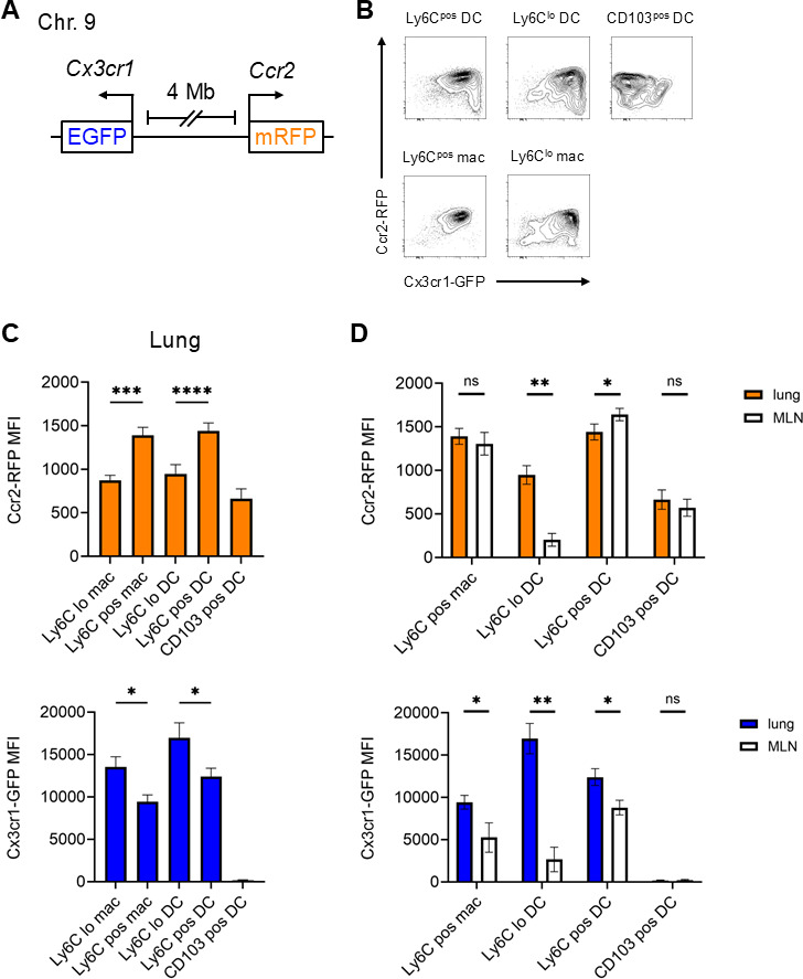
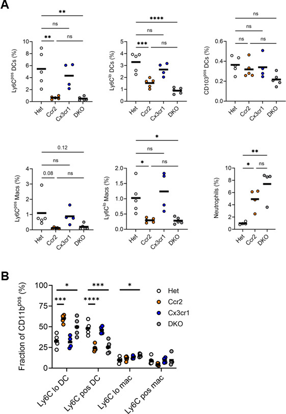
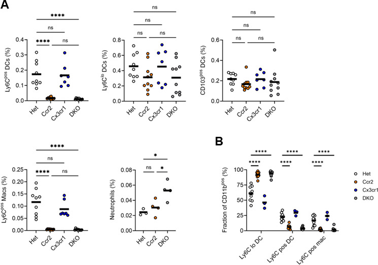
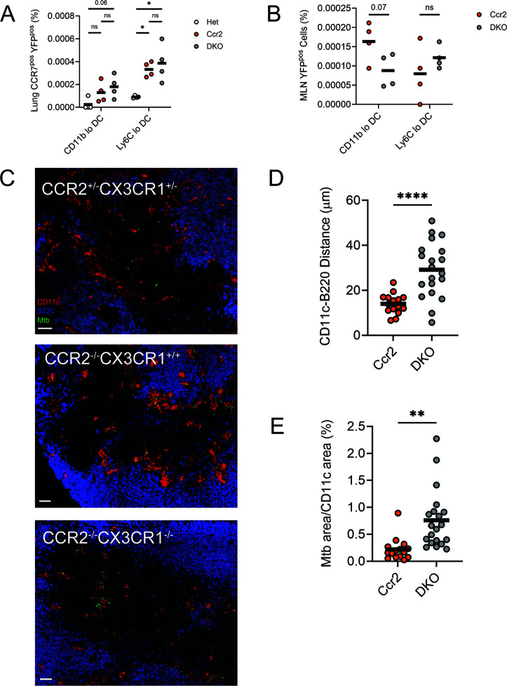
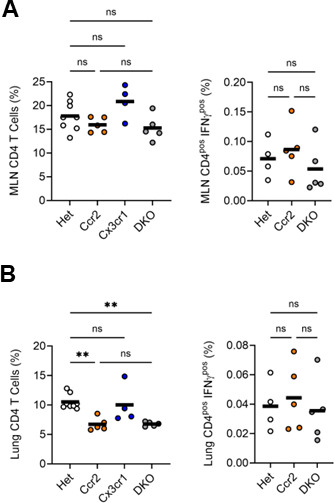
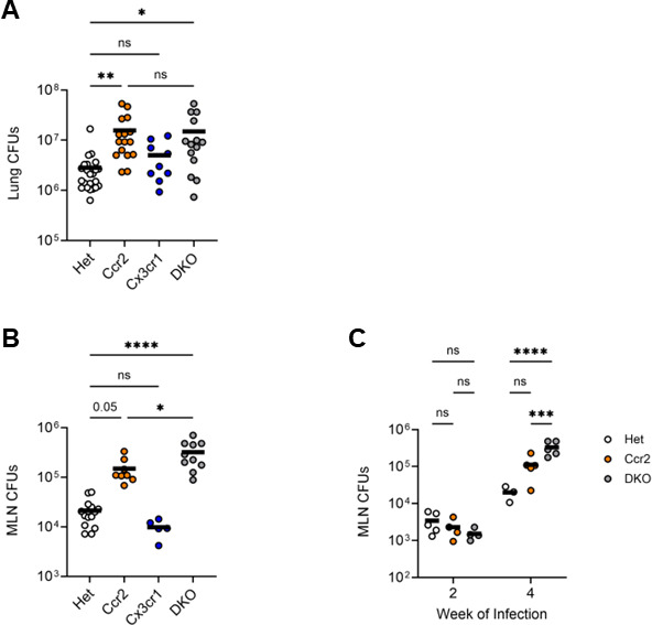
Infection by Mycobacterium tuberculosis (Mtb) continues to cause more than 1 million deaths annually, due to pathogen persistence in lung macrophages and dendritic cells derived from blood monocytes. While the accumulation of monocyte-derived cells in the Mtb-infected lung partially depends on the chemokine receptor CCR2, the other chemoattractant receptors regulating trafficking remain undefined. We used mice expressing knock-in/knockout reporter alleles of Ccr2 and Cx3cr1 to interrogate their expression and function in monocyte-derived populations of the lungs and draining mediastinal lymph nodes during Mtb infection. CCR2 and CX3CR1 expression varied across monocyte-derived subsets stratified by cell surface Ly6C expression in both organs. We found that the expression of CCR2 predicted the dependence of monocyte-derived cells on the receptor for lung and lymph node accumulation. CCR2-deficient mice were also observed to have worsened lung and lymph node Mtb burden. While CX3CR1 deficiency, alone or in combination with CCR2 deficiency, did not affect cell frequencies or lung Mtb control, its absence was associated with altered positioning of monocyte-derived dendritic cells in mediastinal lymph nodes. We found that the combined loss of Ccr2 and Cx3cr1 also worsened Mtb control in the mediastinal lymph node, suggesting a rationale for the persistent expression of CX3CR1 among monocyte-derived cells in pulmonary tuberculosis.IMPORTANCEMycobacterium tuberculosis (Mtb) is the respiratory pathogen responsible for the deadliest infectious disease worldwide. Susceptible humans exhibit ineffective immune responses, in which infected phagocytes are not able to eliminate the pathogen. Since recruited monocyte-derived cells serve as reservoirs for persistent infection, understanding how these phagocytes accumulate in the lung and why they are unable to eliminate Mtb can inform the development of therapies that can synergize with antimicrobials to achieve faster and more durable Mtb elimination. Monocyte-derived cells express the chemokine receptors CCR2 and CX3CR1, but the role of the latter in Mtb infection remains poorly defined. The significance of our study is in elucidating the roles of these receptors in the trafficking of monocyte-derived cells in the infected lung and mediastinal lymph node. These data shed light on the host response in tuberculosis and other pulmonary infections.

Keywords: Mycobacterium tuberculosis; cell trafficking; immunity; monocytes.


Conflict of interest statement

The authors declare no conflict of interest.

Figures

Fig 1
Fig 1
A dual Ccr2-Cx3cr1 reporter mouse demonstrates varied receptor expression by cell type and location. (A) Schematic of the dual reporter genes on chromosome 9. (B through D) Ccr2RFP/+; Cx3cr1GFP/+ mice were infected with a target dose of 100 CFUs of Mtb, then euthanized at 4 weeks post-infection for analysis of lung and MLN cells by flow cytometry (gating in Fig. S1). (B) Representative contour plots of the indicated populations from the lungs of infected mice (“mac”: monocyte-derived macrophage). (C) MFIs of RFP (top) and GFP (bottom) in lung cells. (D) Comparison of RFP (top) and GFP (bottom) MFIs among equivalent lung and MLN populations (Ly6Clo macrophages were not observed in significant numbers in the MLN). Data in panels B through D are from 1 of the 2 experiments with similar results, each conducted with 4 mice per genotype. All results are presented as arithmetic mean + SD. *, P < 0.05; **, P < 0.005; ***, P < 0.0005; ****, P < 0.00005; ns, not significant; by one-way analysis of variance (ANOVA) with multiple comparisons (C) or t test (D).
Fig 2
Fig 2
Phagocyte frequencies in the lungs of Mtb-infected CCR2-deficient, CX3CR1-deficient, and DKO mice. Ccr2RFP/+; Cx3cr1GFP/+ (Het), Ccr2RFP/RFP (Ccr2), Cx3cr1GFP/GFP (Cx3cr1), and DKO mice were infected with ∼100 CFUs Mtb for 4 weeks, and lungs were harvested for analysis. (A) Frequencies of the indicated lung populations among live cells by flow cytometry (live cell gate defined in Fig. S1). (B) Proportions of the indicated populations among CD11bpos cells (parent gate defined in Fig. S1). Data are from 1 of the 2 experiments with similar results, each conducted with four mice per genotype. The horizontal bar represents the arithmetic mean in all graphs. Significance assessed by one-way ANOVA with multiple comparisons.
Fig 3
Fig 3
Phagocyte frequencies in the MLNs of Mtb-infected CCR2-deficient, CX3CR1-deficient, and DKO mice. Mice were infected, as in Fig. 2, and MLNs were harvested for analysis. (A) Frequencies of the indicated MLN populations among live cells by flow cytometry (live cell gate defined in Fig. S1). (B) Proportions of the indicated populations among CD11bpos cells (parent gate defined in Fig. S1). Data are from 1 of the 2 experiments with similar results, each conducted with four mice per genotype. The horizontal bar represents the arithmetic mean in all graphs. Significance assessed by one-way ANOVA with multiple comparisons.
Fig 4
Fig 4
Mtb-infected cells exhibit altered positioning in the MLNs of CCR2–CX3CR1 DKO mice. Lungs and MLNs were harvested from mice infected with ∼100 CFUs of YFP-expressing Mtb for 4 weeks. (A) Frequency of CCR7pos YFPpos cells of the indicated subsets among total lung cells. (B) Frequency of YFPpos cells of the indicated subsets among total MLN cells. Data in panels A and B are from 1 of the 2 experiments with similar results, each conducted with four mice per genotype. (C) Representative immunofluorescence staining of non-follicular zones in MLN sections from Het, Ccr2, or DKO mice. Scale bar is 70 µm. (D) For each CD11cpos cell in non-follicular zones, the distance to the closest B220pos cell was measured. (E) Mtb-positive staining area was measured as a percentage of the total CD11c-positive staining area in non-follicular zones. Graphs in panels D and E show the average distance by non-follicular zone from sections of the indicated genotype and are compiled from three experiments. Horizontal bar represents the arithmetic mean in all graphs. Significance assessed by one-way ANOVA with multiple comparisons (A) or t test (B, D, and E).
Fig 5
Fig 5
Accumulation and polarization of CD4 T cells in MLNs and lungs of CCR2-deficient, CX3CR1-deficient, and DKO mice. Frequencies of total and IFNγpos CD4 T cells in the MLN (A) and lung (B) 4 weeks after infection with ∼100 CFUs of Mtb. Data are from 1 of the 2 experiments with similar results, each conducted with four mice per genotype. The horizontal bar represents the arithmetic mean in all graphs. Significance assessed by one-way ANOVA with multiple comparisons.
Fig 6
Fig 6
DKO mice have a greater mycobacterial burden in MLNs than CCR2-deficient mice. Mice were infected for 2 or 4 weeks with ∼100 CFUs of Mtb, then lungs and MLNs were harvested. (A) Quantitation of Mtb bacilli in the lungs of infected mice by colony formation (CFUs) on agar. Data are compiled from four experiments. (B) Quantitation of Mtb bacilli in the MLNs of mice infected for 4 weeks. Data are compiled from three experiments. (C) MLN CFUs after 2 and 4 weeks of infection. Data are from 1 of the 2 experiments with similar results, each conducted with three mice per genotype. The horizontal bar represents the arithmetic mean in all graphs. Significance assessed by one-way (A and B) or two-way (C) ANOVA with multiple comparisons.

Update of

Similar articles

References

    1. World Health Organization . 2024. Global tuberculosis report. World Health Organization, Geneva
    1. Scriba TJ, Maseeme M, Young C, Taylor L, Leslie AJ. 2024. Immunopathology in human tuberculosis. Sci Immunol 9:eado5951. doi: 10.1126/sciimmunol.ado5951 - DOI - PubMed
    1. Lee J, Boyce S, Powers J, Baer C, Sassetti CM, Behar SM. 2020. CD11cHi monocyte-derived macrophages are a major cellular compartment infected by Mycobacterium tuberculosis. PLoS Pathog 16:e1008621. doi: 10.1371/journal.ppat.1008621 - DOI - PMC - PubMed
    1. Zheng W, Chang IC, Limberis J, Budzik JM, Zha BS, Howard Z, Chen L, Ernst JD. 2024. Mycobacterium tuberculosis resides in lysosome-poor monocyte-derived lung cells during chronic infection. PLoS Pathog 20:e1012205. doi: 10.1371/journal.ppat.1012205 - DOI - PMC - PubMed
    1. Lai R, Williams T, Rakib T, Lee J, Behar SM. 2024. Heterogeneity in lung macrophage control of Mycobacterium tuberculosis is modulated by T cells. Nat Commun 15:5710. doi: 10.1038/s41467-024-48515-7 - DOI - PMC - PubMed

LinkOut - more resources