Review
. 2025 May 23;14(11):767.
doi: 10.3390/cells14110767.

Vasohibins in Health and Disease: From Angiogenesis to Tumorigenesis, Multiorgan Dysfunction, and Brain-Heart Remodeling

Affiliations
Review

Vasohibins in Health and Disease: From Angiogenesis to Tumorigenesis, Multiorgan Dysfunction, and Brain-Heart Remodeling

Ghulam Abbas et al. Cells. .

Abstract

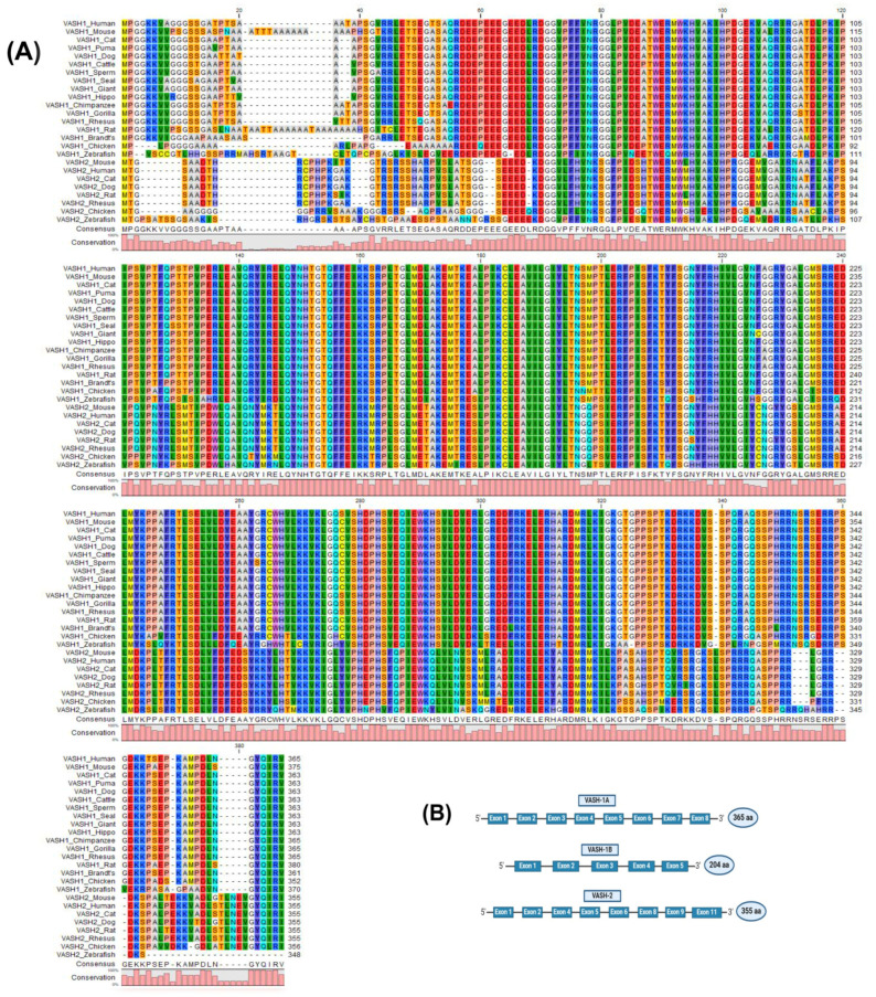
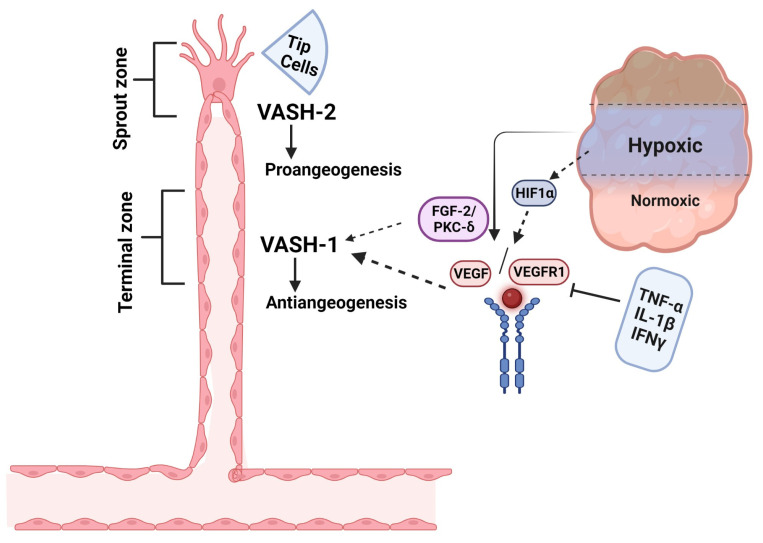
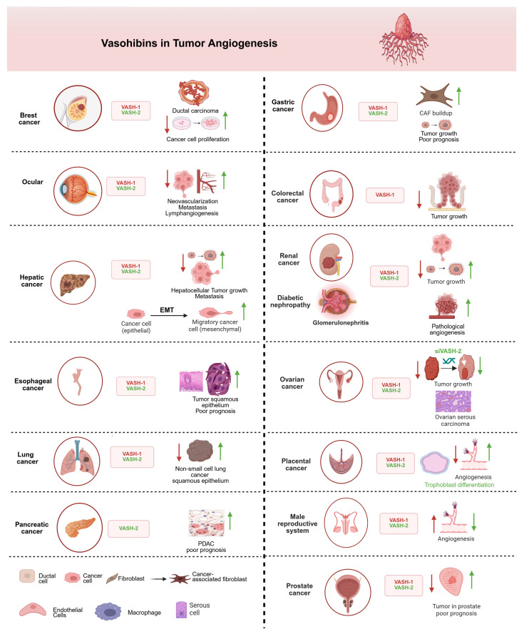
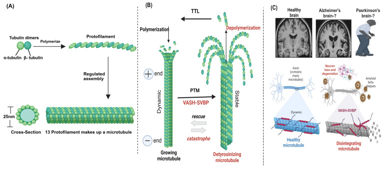
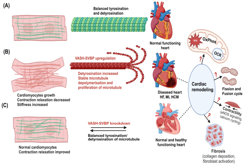
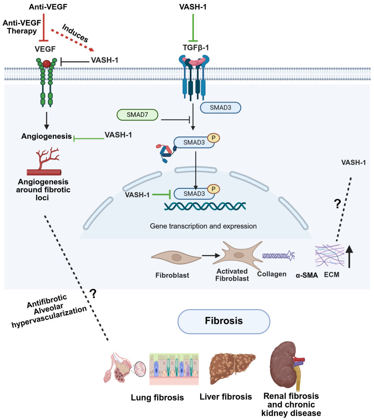
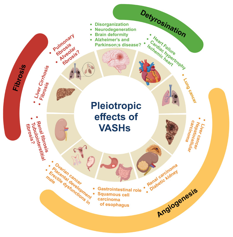
Vasohibins (VASHs), comprising VASH-1 and VASH-2, were initially identified as regulators of angiogenesis. Recent studies, however, have unveiled their novel role in fibrosis and microtubule detyrosination. The dysregulated expression of VASHs is associated with several pathological processes, such as angiogenesis dysfunction, microtubule detyrosination, and fibrosis, contributing to various diseases. These findings suggest the pleiotropic effects of VASHs in multiple organs and systems beyond angiogenesis. This review explores the molecular properties of VASHs and their emerging functions in tubulin carboxyl activity and microtubule detyrosination-key to brain and cardiac remodeling. We also discuss the potential therapeutic applications of their interference in diseases such as tumorigenesis, as well as renal-, reproductive-, and liver-related diseases.

Keywords: angiogenesis regulator; cardiac/brain remodeling; fibrosis TGF-β/SMAD pathway; microtubule detyrosination; vasohibins.


Conflict of interest statement

The authors declare no conflicts of interest.

Figures

Figure 1
Figure 1
Multiple sequence alignment structure of the vasohibin (VASH) proteins. (A) The protein sequences obtained from NCBI (accession numbers provided in Supplementary Materials) were aligned using CLC viewer 8.0. The bar graphs represent the degree of conservation among species, and the colors are in correspondence with the amino acids’ identity. (B) VASH-1A contains eight exons and 365 amino acids (aa), whereas VASH-1B contains five exons and 204 aa. VASH-2 has eight exons and consists of 355 aa.
Figure 2
Figure 2
Schematic representation of basic induction of VASH isoforms in angiogenesis regulation. VASH-1 is induced in the vascular termination zone, where it functions to stop angiogenesis, whereas VASH-2 localizes to the sprout zone to promote vessel growth. Under hypoxic conditions, HIF-1α-mediated upregulation of VEGF drives angiogenic activation, which is counterbalanced by VASH-1 through two key mechanisms: (1) FGF2-induced PKC-δ activation and (2) synergistic action with anti-angiogenic cytokines (TNF-α, IL-1β, IFN-γ) to suppress VEGF signaling. This spatial and functional segregation of VASH isoforms creates a dynamic regulatory system for controlled vascular patterning. This figure is adopted from Du et al. [21], licensed under CC BY-NC 3.0. Created in biorender.com.
Figure 3
Figure 3
The involvement of VASHs in different types of tumors and organ systems. VASH-1 acts as an anti-angiogenetic, inhibiting/downregulating factor (red arrow), and VASH-2 acts as pro-angiogenic, activating/upregulating factor (green arrow). VASH-1 consistently suppresses tumor progression through angiogenesis inhibition in most cancers, while VASH-2 promotes vascular growth and tumor development, except for the male reproductive system, where VASH-1 paradoxically enhances angiogenic processes to rescue erectile dysfunction. In renal cancer, VASH-2 promotes glomerular damage via aberrant angiogenesis and primary tumor growth, whereas VASH-1 downregulation enables renal cancer metastasis—a duality suggesting isoform-specific therapeutic targeting. The epithelial–mesenchymal transition (EMT) panel highlights VASH-2’s role in promoting cancer cell plasticity. All depicted interactions are supported by experimental evidence discussed in the main text. Created in biorender.com.
Figure 4
Figure 4
Microtubule structure, assembly, and post-translational modifications (PTMs) in the brain. (A) Microtubules are hollow cylindrical structures composed of α- and β-tubulin heterodimers [4]. (B) VASHs detyrosinate the microtubule through PTMs. (C) VASHs detyrosinate and disintegrate microtubules in the brain, leading to disease pathogenesis. VASH-mediated detyrosinated microtubules are involved in brain deformity [98], which may play a role in Alzheimer’s and Parkinson’s diseases. Created in biorender.com.
Figure 5
Figure 5
Potential mechanisms of VASHs in the cardiac system. (A) Normal cardiomyocytes with dynamic microtubules (green in color) and balanced tyrosination/detyrosination cycle in which the heart performs normal functions. (B) Cardiomyocyte growth occurs particularly in the diseased hypertrophic heart. VASH-SVBP may highly detyrosinate, stabilize, and proliferate microtubules. Denser microtubules (red) are seen in cardiomyocytes. Microtubules are in proximity to mitochondria in cardiomyocytes; therefore, upregulation of VASHs could lead to mitochondrial dysfunctions in response to cardiac remodeling. (C) VASH-SVBP suppression may improve contraction and relaxation, as well as decrease cardiac microtubule dysfunction, hence, balance between tyrosinated/detyrosinated states, representing the normal/healthy cardiomyocytes and heart. This remodeling could also alter both mitochondrial function and dysfunction. Thus, it is critical to study these alterations and mechanisms, including bioenergetics, fission and fusion, and fibrosis in the future. Created in biorender.com.
Figure 6
Figure 6
Potential mechanistic interplay between VASH-1, VEGF, and TGF-β signaling in fibrosis. Anti-VEGF therapy upregulates VASH-1, which subsequently inhibits both VEGF signaling (negative feedback) and TGF-β1/SMAD3 activation. (Right) This dual inhibition by VASH-1 reduces angiogenesis around fibrotic loci and could block fibroblast activation, collagen deposition, and ECM remodeling. While VASH-1 inhibits TGF-β signaling, the potential regulation of VASH-1 by TGF-β remains undefined. Created in biorender.com.
Figure 7
Figure 7
Pleotropic role of vasohibins (VASHs) in health and disease. VASHs play key roles in neurodegeneration and disorganization in neurons of the brain by inducing microtubule detyrosination. VASHs detyrosinate cardiac microtubules and impede contraction and relaxation. Therefore, the suppression of VASHs aids in the kinetics of contraction, which ultimately improves cardiac function and could have therapeutic use in heart failure, ischemic heart disease, and cardiac hypertrophy. In angiogenesis, VASH-1 plays an anti-angiogenic role, whereas VASH-2 is pro-angiogenic. The regulation of VASHs during angiogenesis plays a critical role in tumor growth and maintaining the functions of the gastrointestinal tract, as well as male and female reproductive organs. Moreover, the regulatory role of VASHs has been documented in liver, kidney, and pulmonary fibrosis. Created in biorender.com.

Similar articles

References

    1. Watanabe K., Hasegawa Y., Yamashita H., Shimizu K., Ding Y., Abe M., Ohta H., Imagawa K., Hojo K., Maki H. Vasohibin as an endothelium-derived negative feedback regulator of angiogenesis. J. Clin. Investig. 2004;114:898–907. doi: 10.1172/JCI200421152. - DOI - PMC - PubMed
    1. Nieuwenhuis J., Adamopoulos A., Bleijerveld O.B., Mazouzi A., Stickel E., Celie P., Altelaar M., Knipscheer P., Perrakis A., Blomen V.A. Vasohibins encode tubulin detyrosinating activity. Science. 2017;358:1453–1456. doi: 10.1126/science.aao5676. - DOI - PubMed
    1. Aillaud C., Bosc C., Peris L., Bosson A., Heemeryck P., Van Dijk J., Le Friec J., Boulan B., Vossier F., Sanman L.E. Vasohibins/SVBP are tubulin carboxypeptidases (TCPs) that regulate neuron differentiation. Science. 2017;358:1448–1453. doi: 10.1126/science.aao4165. - DOI - PubMed
    1. Dogterom M., Koenderink G.H. Actin-microtubule crosstalk in cell biology. Nat. Rev. Mol. Cell Biol. 2019;20:38–54. doi: 10.1038/s41580-018-0067-1. - DOI - PubMed
    1. Pimm M.L., Henty-Ridilla J.L. New twists in actin-microtubule interactions. Mol. Biol. Cell. 2021;32:211–310. doi: 10.1091/mbc.E19-09-0491. - DOI - PMC - PubMed

Publication types

LinkOut - more resources