. 2025 May 25;14(11):775.
doi: 10.3390/cells14110775.

RNF213 Acts as a Molecular Switch for Cav-1 Ubiquitination and Phosphorylation in Human Cells

Affiliations

RNF213 Acts as a Molecular Switch for Cav-1 Ubiquitination and Phosphorylation in Human Cells

Jungmi Choi et al. Cells. .

Abstract

RNF213 encodes a unique protein with AAA+ ATPase and E3 ubiquitin ligase activities that are critical for its diverse roles, which range from involvement in human vasculopathies, such as Moyamoya disease, to ubiquitination of viral and bacterial pathogens. Nevertheless, its primary functions in human signaling remain unclear due to the limited identification of direct substrates. Here, we investigated the interaction between RNF213 and caveolin-1 (Cav-1), a small scaffolding protein vital for caveolae formation and the regulation of a plethora of cellular processes. Cav-1 specifically binds within the two functional AAA+ domains of RNF213 in an ATP-dependent manner, highlighting the influence of cellular energy status on this interaction. Consequently, RNF213 ubiquitinates Cav-1 at several N-terminal lysine residues through K48 and K63 linkages, although several Moyamoya disease-associated RNF213 mutations greatly reduce this polyubiquitination. Moreover, RNF213 activity inhibits phosphorylation of a key regulatory residue of Cav-1, as RNF213 knockdown under oxidative stress markedly enhances Cav-1 Tyr14 phosphorylation and modifies nitric oxide bioavailability in endothelial cells. Collectively, our results indicate that RNF213 functions as a molecular switch modulating Cav-1 signaling based on RNF213 functionality and cellular conditions. These findings offer new insights into vascular pathogenesis and the vast signal pathways along the RNF213-Cav-1 axis.

Keywords: Cav-1; MMD; RNF213; caveolin-1; moyamoya disease.


Conflict of interest statement

The authors declare no conflicts of interest.

Figures
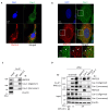
Figure 1
Figure 1
RNF213 co-localizes and interacts with Cav-1 in endothelial cells. (A) Confocal immunofluorescence microscopy using HUVECs, and (B) super-resolution confocal microscopy with HPAECs (white boxes represent magnified regions shown below). In both cases, nuclei were stained with DAPI (blue), Cav-1 was identified with Alexa-488 (green), and RNF213 was tagged with Alexa-555 (red). In the merged figures and inset, orange and yellow locations indicate co-localization of RNF213 and Cav-1 (white arrowheads). (C) Co-immunoprecipitation (co-IP) assay with an IgG negative control or RNF213 antibody from HUVEC lysates, followed by immunoblot (IB) analysis with the indicated antibodies. (D) Co-IP with an IgG control or RNF213 from HPAEC lysates transfected with scramble siRNA or siRNAs targeting RNF213, Cav-1, or both RNF213 and Cav-1, and treated with 100 ng mL−1 INFγ for 0 or 24 h to enhance RNF213 protein levels, followed by IB analysis with the indicated antibodies. The detected protein sizes are RNF213 (591 kDa), Cav-1 oligomer (>600 kDa) and Cav-1 monomer (~24 kDa). All analyses involving HUVECs and HPAECs were performed using a single donor-derived lot for each cell type, with a minimum of three independent experimental replicates to ensure consistency and reproducibility.
Figure 2
Figure 2
Identification of the RNF213 and Cav-1 domains necessary for their interaction. (A) HEK293T cells were co-transfected with combinations of FLAG-tagged full-length (WT) RNF213 and HA-tagged Cav-1 as indicated. Interactions between RNF213 and monomeric Cav-1 were detected by co-immunoprecipitation (co-IP) with anti-HA antibody, followed by immunoblot (IB) analysis with the indicated antibodies. (B) Schematic diagram of FLAG-tagged WT RNF213 and four different truncated RNF213 fragments (N1, N2, C3, and C5), and their use in co-IP experiments in HEK293T cells also overexpressing HA-tagged Cav-1. Co-IP with anti-HA antibodies followed by IB with the indicated antibodies were performed to identify the RNF213 regions interacting with monomeric Cav-1. (Right panel) The ratios of bound RNF213 to total input RNF213 were calculated (mean ± SEM, n = 3), with the mean WT ratio being arbitrarily defined as 1. Asterisks denote significant statistical difference from the WT, determined by one-sample t-tests with Holm–Šídák correction for multiple comparisons (* p < 0.05, ** p < 0.01, and *** p < 0.001). (C) Schematic diagram of WT RNF213, the N2 fragment, and the various N2 fragments with point mutations in the Walker A (WA) and/or Walker B (WB) of the A3 and/or A4 domains, or a complete deletion of the WA and WB region (ΔA) of the A3 domain of RNF213. “ΔA” refers to an internal deletion mutant of RNF213, in which amino acids 2365–2613 encompassing the A3 AAA+ domain [16] were deleted to assess the functional contributions of this region. Co-IP experiments were performed using FLAG-tagged RNF213 fragments and HA-tagged Cav-1 overexpressed in HEK293T cells, following the same co-IP procedure as in (B), to identify the RNF213 domains essential for binding Cav-1. (Right panel) The ratios of bound RNF213 to total input RNF213 were calculated (mean ± SEM, n = 4), with the mean WT ratio being arbitrarily defined as 1. Asterisks denote significant statistical difference from the N2 WT by one-way ANOVA with * p < 0.05, ** p < 0.01, *** p < 0.001 and **** p < 0.0001. (D) Schematic diagram of full-length Cav-1 and six truncated forms with serial deletions of domains D1–D6 (see Section 3 for full description). HEK293T cells were co-transfected with FLAG-tagged WT RNF213 and HA-tagged full-length or deleted Cav-1 fragments as above, followed by co-IP as in (B). (Right panel) The ratios of bound RNF213 to total input RNF213 were calculated (mean ± SEM, n = 3), with the mean WT Cav-1 ratio being arbitrarily defined as 1. For all immunoblots, β-actin was used as the protein loading control. All experiments were performed with a minimum of three independent experimental replicates to ensure consistency and reproducibility.
Figure 3
Figure 3
RNF213 binding to Cav-1 is ATP-dependent. Co-immunoprecipitation (co-IP) experiments using HEK293T cells overexpressing full-length FLAG-tagged WT RNF213 and HA-tagged Cav-1 under varying intracellular ATP concentrations and their effects on RNF213 and monomeric Cav-1 interactions. Cells were treated with 10 mM 2DG and 10 mM NaN3 for 30 min to modulate intracellular ATP levels, and the samples were then used for co-IP with anti-HA antibody, followed by immunoblotting (IB) with the indicated antibodies (upper panel). β-actin was used as the protein loading control. (Right panel) The ratios of bound RNF213 to total input RNF213 were calculated (mean ± SEM, n = 3), with the mean non-treatment control ratio being arbitrarily defined as 1. Asterisks denote significant statistical difference from the non-treatment control by one-sample t-tests with ** p < 0.01. Intracellular ATP levels were assessed using a luminescence-based ATP assay. The intracellular ATP levels were finely modulated by altering the levels of the inhibitors used, and the relationship between cellular ATP levels and the interactions between RNF213 and Cav-1 is shown by the regression line with an R2 value of 0.9475 (lower panel). The reference value of 1 on the Y-axis corresponds to 150.74 µM. Experiments were performed with three independent experimental replicates to ensure consistency and reproducibility.
Figure 4
Figure 4
Cav-1 suppresses enhanced NF-κB activation and apoptosis induced by RNF213 RING mutations in HEK293T cells. (A) HEK293T cells were co-transfected with firefly and Renilla luciferase constructs as well as with FLAG-tagged WT or mutant RNF213, including C3997A (CA), C3997Y (CY), D4013N (DN), R4019C (RC), P4033L (PL), R4810K (RK), and the third AAA+ deletion (ΔA), or with the pcDNA3.1 empty vector as control (EV), together with and without HA-tagged Cav-1. Lysed cells were then used for dual-luciferase reporter assays. Data are normalized to Renilla luciferase (mean ± SEM, n = 3). Asterisks (and red columns) denote significant statistical differences in a one-way ANOVA test with * p < 0.05. (Lower panel) Immunoblot analysis showing expression levels of FLAG-tagged WT and mutant RNF213 with and without HA-tagged Cav-1 co-transfection used in the luciferase assays. The CY samples shown on both the left and right panels are the same and were included as internal controls to allow comparison across membranes. (B) HEK293T cells were co-transfected with FLAG-tagged WT or mutant RNF213, together with and without HA-tagged WT Cav-1. The levels of cleaved-Caspase-3 induced by the mutants and suppressed by Cav-1 were then determined by immunoblot analysis. The arrow shows the specific cleaved-Caspase-3 bands of interest. All experiments were performed with a minimum of three independent experimental replicates to ensure consistency and reproducibility. For all immunoblots, β-actin was used as the protein loading control.
Figure 5
Figure 5
Ubiquitination of Cav-1 at multiple lysine residues by RNF213 and the effects of RNF213 MMD mutations. (A) Ubiquitination of Cav-1 in HEK293T cells overexpressing different combinations of FLAG-tagged RNF213 WT or mutant CA, HA-tagged Cav-1, and Myc-tagged ubiquitin (Ub). The extent of RNF213-mediated ubiquitination of Cav-1 was determined by immunoprecipitation (IP) with an anti-HA antibody and immunoblotting (IB) with the indicated antibodies. Arrows indicate ubiquitin-conjugated Cav-1. FLAG-tagged ZNRF1 was used as a control E3 ligase for Cav-1 ubiquitination. (B) Identification of lysine residues K26, K47, K57, and K65 in the N-terminal cytoplasmic region of Cav-1 as targets of RNF213-mediated ubiquitination (in Cav-1 sequence and image), as determined by LC-MS/MS comparisons of the effects of WT and CA mutant RNF213 (see Supplementary Tables S2 and S3). (C) Ubiquitination of Cav-1 was examined under the same conditions as in (A) but using WT Cav-1 or two independent Cav-1 KR mutants in which all four lysine (K) residues, identified by LC-MS/MS and shown in (B), were mutated to arginine (R) residues. Arrows indicate increased ubiquitin-conjugated Cav-1. (D) Tandem Ubiquitin-Binding Entity (TUBE) assays were performed using K63 and K48 TUBEs to determine the RNF213-mediated formation of K48- and/or K63-linked polyubiquitin chains on Cav-1. HEK293T cells overexpressing FLAG-tagged RNF213 WT or the CA mutant and HA-tagged Cav-1 were subjected to affinity pull-down with either K63 or K48 TUBEs, followed by IB with anti-HA antibody. Arrows indicate the increased K63- and K48-linked polyubiquitin chains formed on Cav-1 by WT RNF213. (E) (Upper panel) Schematic diagram of RNF213 and MMD mutations, with positions shown in red. (Lower panel) Co-immunoprecipitation (co-IP) experiments, to show the effects of MMD-associated RNF213 mutations on Cav-1 binding, were performed by overexpressing different combinations of FLAG-tagged WT or mutant RNF213 and HA-tagged Cav-1 in HEK293T cells, followed by co-IP with anti-HA antibody and IB with the indicated antibodies. β-actin was used as the protein loading control. (Right panel) The ratios of bound RNF213 to total input RNF213 were calculated (mean ± SED, n = 4), with the mean WT RNF213 ratio being arbitrarily defined as 1. An asterisk denotes a significant statistical difference from WT RNF213, determined by one-sample t-tests with Holm–Šídák correction for multiple comparisons (**** p < 0.0001). (F) Ubiquitination of Cav-1 was examined essentially as in (A) but using RNF213 WT and its various MMD-related mutations. Arrows indicate ubiquitin-conjugated Cav-1. All experiments were performed with a minimum of three independent experimental replicates to ensure consistency and reproducibility.
Figure 6
Figure 6
RNF213 knockdown enhances Cav-1 Y14 phosphorylation under H2O2 and affects nitric oxide stimulation in HPAECs. (A) Immunoblot analysis illustrating the effects of hydrogen peroxide (H2O2) and RNF213 knockdown on endogenous monomeric phosphorylated Y14 Cav-1 (pCav-1) levels in HPAECs. Cells were non-transfected (NT) or transfected with scramble or four different RNF213-targeted siRNAs, and then untreated (control) or treated with 1 mM H2O2 for 30 min to induce oxidative stress and lead to observable levels of monomeric pCav-1. Vinculin was used as a protein loading control. (B) Immunoblot analysis, used to investigate monomeric and oligomeric pCav-1 levels in HPAECs in response to H2O2, was essentially performed as in (A), but with siRNAs targeting RNF213 (siRNA 2), Cav-1, or both RNF213 and Cav-1, together with the indicated antibodies. (C) Fluorescence microscopy images of HPAECs incubated with DAF-FM DA dye for determining relative nitric oxide (NO) bioavailability. Cells were transfected with siRNAs targeting RNF213 (siRNA 2), Cav-1, or both RNF213 and Cav-1 and treated with H2O2 for 30 min as in (A). Photographs of sample cells were taken at 1, 15, and 30 min after treatment. (D) Quantitative fluorescence intensity of the NO production over time, determined in (C). Raw fluorescence intensity values for DAF-FM were measured every 5 min and normalized to the value at 1 min for each condition. Data are shown as means ± SEM (n = 7). Statistical comparisons between groups at each time point were performed using two-way ANOVA (* p < 0.05). All analyses involving HUVECs and HPAECs were performed using a single donor-derived lot for each cell type, with at least three independent experimental replicates for (A,C,D), and with two independent experimental replicates and three technical replicates for (B).

References

    1. Kamada F., Aoki Y., Narisawa A., Abe Y., Komatsuzaki S., Kikuchi A., Kanno J., Niihori T., Ono M., Ishii N., et al. A genome-wide association study identifies RNF213 as the first Moyamoya disease gene. J. Hum. Genet. 2011;56:34–40. doi: 10.1038/jhg.2010.132. - DOI - PubMed
    1. Liu W., Morito D., Takashima S., Mineharu Y., Kobayashi H., Hitomi T., Hashikata H., Matsuura N., Yamazaki S., Toyoda A., et al. Identification of RNF213 as a susceptibility gene for moyamoya disease and its possible role in vascular development. PLoS ONE. 2011;6:e22542. doi: 10.1371/journal.pone.0022542. - DOI - PMC - PubMed
    1. Koizumi A., Kobayashi H., Hitomi T., Harada K.H., Habu T., Youssefian S. A new horizon of moyamoya disease and associated health risks explored through RNF213. Environ. Health Prev. Med. 2016;21:55–70. doi: 10.1007/s12199-015-0498-7. - DOI - PMC - PubMed
    1. Ihara M., Yamamoto Y., Hattori Y., Liu W., Kobayashi H., Ishiyama H., Yoshimoto T., Miyawaki S., Clausen T., Bang O.Y., et al. Moyamoya disease: Diagnosis and interventions. Lancet Neurol. 2022;21:747–758. doi: 10.1016/S1474-4422(22)00165-X. - DOI - PubMed
    1. Fukushima H., Takenouchi T., Kosaki K. Homozygosity for moyamoya disease risk allele leads to moyamoya disease with extracranial systemic and pulmonary vasculopathy. Am. J. Med. Genet. A. 2016;170:2453–2456. doi: 10.1002/ajmg.a.37829. - DOI - PubMed

Publication types

LinkOut - more resources