. 2025 May 28;14(11):790.
doi: 10.3390/cells14110790.

SLFN11 Restricts LINE-1 Mobility

Affiliations

SLFN11 Restricts LINE-1 Mobility

Zhongjie Ye et al. Cells. .

Abstract

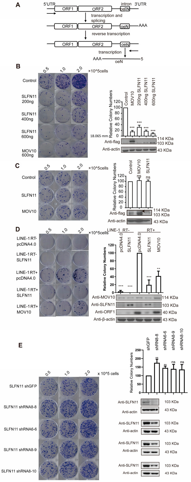
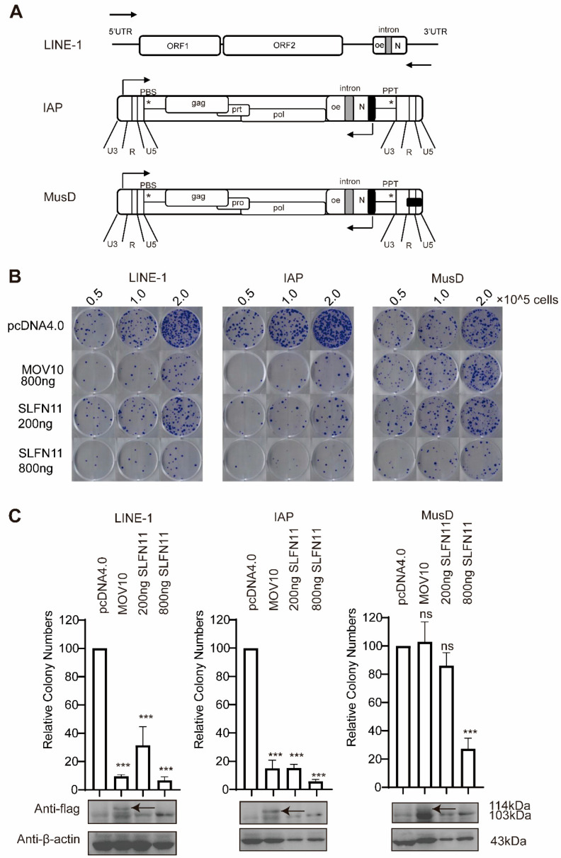
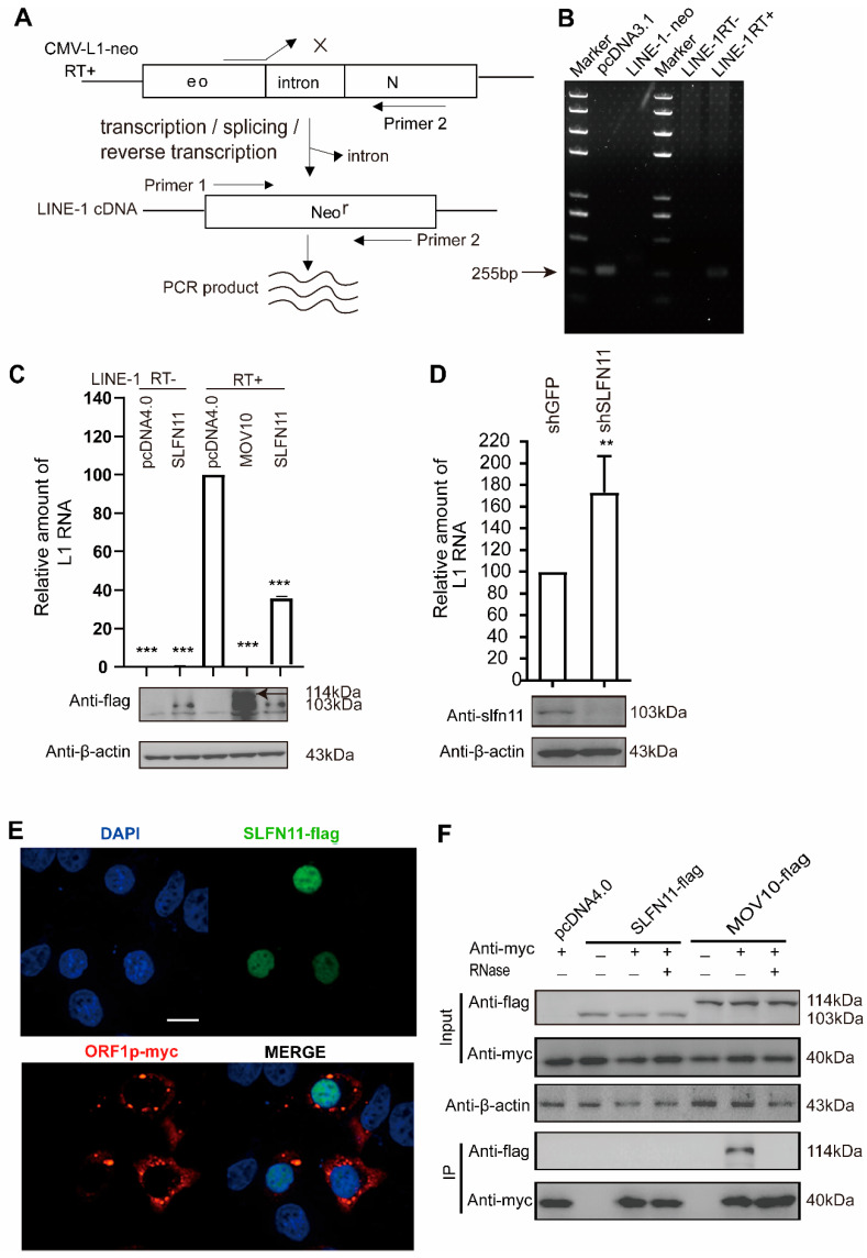
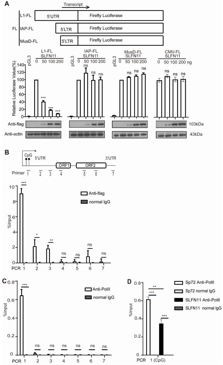
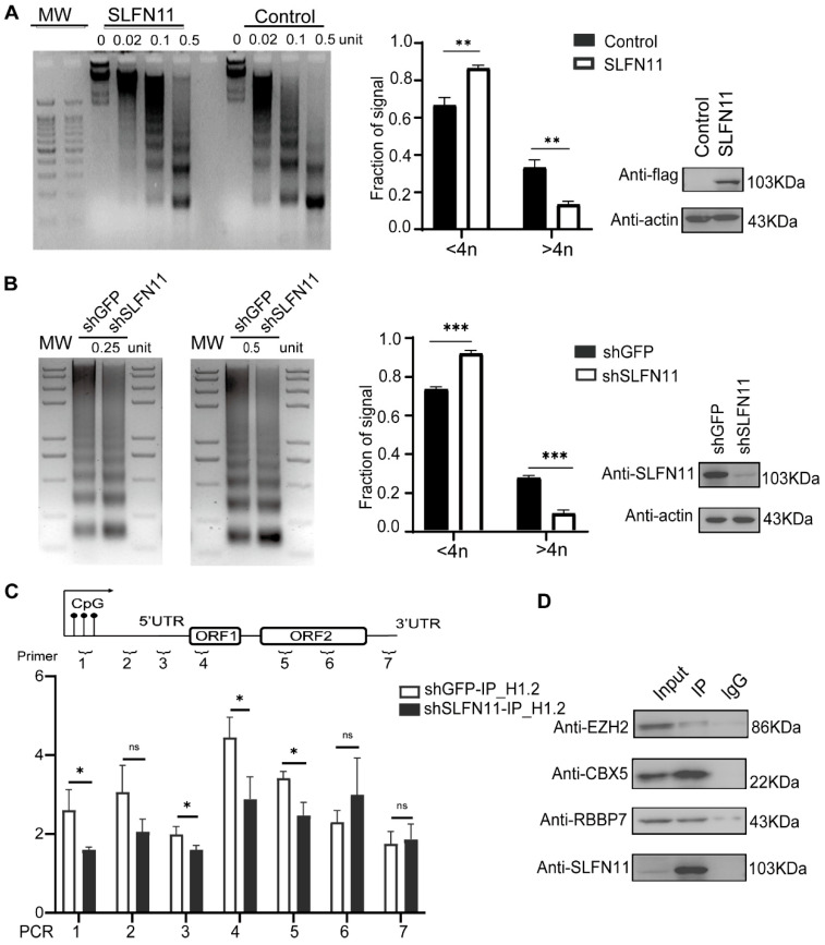
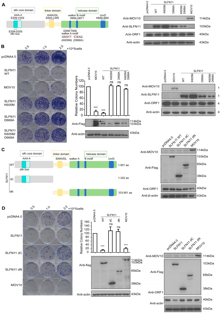
Long interspersed element-1 (LINE-1) is the only active autonomous transposon comprising about 17% of human genomes. LINE-1 transposition can cause the mutation and rearrangement of the host's genomic DNA. The host has, therefore, developed multiple mechanisms to restrict LINE-1 mobility. Here, we report that SLFN11, a member of the Schlafen family, can restrict LINE-1 retrotransposition, and the inhibitory activity requires its helicase domain. Mechanistically, SLFN11 specifically binds to the LINE-1 5' untranslated region (5'UTR) and blocks RNA polymerase II recruitment, thereby suppressing its transcription. Furthermore, SLFN11 promotes heterochromatinization, suggesting an epigenetic inhibition pathway.

Keywords: LINE-1; RNA polymerase II; SLFN11; epigenetics; helicase; heterochromatin; long interspersed element-1; transposon.


Conflict of interest statement

The authors declare no conflicts of interest. The funders had no role in the design of the study.

Figures

Figure 1
Figure 1
SLFN11 inhibits LINE-1 retrotransposition. (A) An illustration of the CMV-L1-neoRT+ reporter plasmid and its transcriptional product conferring G418 resistance. (B) The ectopic expression of SLFN11 suppresses LINE-1 retrotransposition. HeLa cells cotransfected with CMV-L1-NeoRT+ and increasing amounts of the SLFN11 expression plasmid (0, 0.5, 1.0 μg) were subjected to G418 selection. (Left) Representative crystal violet-stained colonies. (Right) Western blot confirming SLFN11 overexpression (lower panel) and the quantitative analysis of relative colony formation (upper panel, normalized to vector control). MOV10 serves as a loading control. Data represent the relative colony numbers normalized to the SLFN11-negative control (set as 100%). (C) SLFN11 expression does not affect basal colony formation efficiency. HeLa cells were transfected with empty pcDNA3.1 and underwent identical G418 selection. Western blot confirmation and colony quantification were performed as described in (B). The black arrow indicates the band for MOV10 protein and the same below. (D) SLFN11 specifically inhibits RT-competent LINE-1 elements. Hela cells were co-transfected with either CMV-L1-neoRT− or CMV-L1-neoRT+ constructs alongside an empty vector, pcDNA4.0 control, or SLFN11 expression plasmid, followed by retrotransposition analysis. (E) Endogeggest that SLFN11 may reduce LINnous SLFN11 depletion enhances LINE-1 retrotransposition. SLFN11-knockdown HeLa cells were transfected with CMV-L1-neoRT+ and subjected to G418 selection. Crystal violet-stained colonies (representative image) and quantitative data (graph) showed increased retrotransposition upon SLFN11 knockdown. All data were representative of at least three independent experiments. The values are expressed as means ± SDs. Statistical analyses were performed with two-tailed, unpaired Student’s t-test (**, p < 0.01; ***, p < 0.001; and ns, not significant).
Figure 2
Figure 2
SLFN11 inhibits IAP and MusD retrotransposition. (A) Schematic diagrams of IAP-neoTNF and MusD-neoTNF reporter constructs. (B,C) The ectopic expression of SLFN11 inhibits IAP and MusD retrotransposition. HeLa cells were co-transfected with CMV-L1-neoRT+, IAP-neoTNF, and MusD-neoTNF along with either 200 ng or 800 ng of SLFN11-Flag, MOV10-Flag (positive control), or equivalent empty vectors of DNA. (B) Retrotransposition efficiency was quantified by the G418-resistant colony formation (crystal violet staining). (C) Upper panel: the quantitative analysis of retrotransposition events normalized to vector control (set as 100%). Lower panel: Western blot analysis confirming SLFN11 and MOV10 expression levels. Data are representative of at least three independent experiments. The values are expressed as means ± SDs. Statistical analyses were performed with two-tailed, unpaired Student’s t-test (*, p < 0.05; ***, p < 0.001; and ns, not significant).
Figure 3
Figure 3
SLFN11 diminishes LINE-1 RNA. (A) A schematic of intron-spanning primer design for specific LINE-1 RNA detection. Primers (arrows) that span the Neo cassette intron in the LINE-1 in DNA cannot amplify LINE-1 DNA, as indicated by the cross symbol. Only spliced LINE-1 RNA can be amplified to eliminate potential DNA contamination from the transfected plasmid. (B) The intron-containing CMV-L1-NeoRT+ plasmid cannot be amplified by PCR using intron-spanning primers targeting the neomycin resistance sequence but successfully amplifies spliced L1 RNA products from successful retrotransposition events. (C) Ectopic SLFN11 expression decreases LINE-1 mRNA. HeLa cells were co-transfected with SLFN11 and CMV-L1-Neo± constructs. LINE-1 RNA levels analyzed by RT-qPCR (72 h post-transfection). (D) Endogenous SLFN11 inhibits LINE-1 mRNA. The figure shows 293FT cells with shSLFN11 knockdown transfected with CMV-L1-neoRT+ and LINE-1 RNA quantified by RT-qPCR (72 h). (E) Subcellular localization analysis of SLFN11 and ORF1p. HeLa cells expressing Flag-SLFN11 and Myc-ORF1p immunostained with anti-Flag (green) and anti-Myc (red). Scale bars, 10 μm. (F) SLFN11 does not interact with ORF1p. Myc-ORF1p co-expressed with Flag-SLFN11 or Flag-MOV10 in 293T cells. RNase-treated lysates immunoprecipitated with anti-Myc; co-precipitated proteins analyzed by immunoblotting. Data are representative of at least three independent experiments. The values are expressed as means ± SDs. Statistical analyses were performed with two-tailed, unpaired Student’s t-test (**, p < 0.01; ***, p < 0.001).
Figure 4
Figure 4
SLFN11 represses the transcription of LINE-1. (A) SLFN11 specifically suppresses the LINE-1 promoter. The 293T cells were co-transfected with SLFN11 DNA and luciferase reporters driven by LINE1-FL, IAP-FL, or MusD-FL promoters (schematic, top). Firefly luciferase activity reflects LINE-1 5′UTR promoter activity, with the promoterless pGL3-basic vector as the negative control. Firefly luciferase activity reflects 5′UTR promoter activity, with the promoterless pGL3-basic vector as the negative control. SLFN11 dose-dependently inhibited the LINE-1 promoter (left) but showed no significant effect on IAP (middle), MusD (right), or CMV-driven luciferase expression. SLFN11 protein levels were verified by Western blot (bottom). Baseline luciferase activity (no SLFN11) was defined as 100%. (B) SLFN11 is associated with endogenous LINE-1 5′UTR regions. The schematic shows primer locations (top) for amplifying genomic LINE-1 elements. DNA co-immunoprecipitated with FLAG-tagged SLFN11 from MCF7 cells was quantified using seven primer pairs spanning LINE-1 loci. (C,D) SLFN11 impairs RNA polymerase II (Ser5-P) recruitment to LINE-1 5′UTR loci. (C) The ChIP analysis of RNA polymerase II (Ser5-P) binding regions on LINE-1 DNA. ChIP assays were conducted in MCF7 cells to investigate RNA polymerase II binding at LINE-1 elements, employing specific antibodies against Ser5-phosphorylated RNA polymerase II (Ser5-P) and control IgG, along with primers targeting defined regions of the LINE-1 locus. (D) SLFN11 impairs the binding of RNA polymerase II (Ser5-P) to LINE-1 retrotransposon DNA. MCF7 cells were transfected with either SLFN11 or the empty pSP72 vector. ChIP was performed using antibodies against RNA polymerase II (Ser5-P) or normal IgG along with primers targeting defined regions of the LINE-1 locus. Data are representative of at least three independent experiments. The values are expressed as means ± SDs. Statistical analyses were performed with two-tailed, unpaired Student’s t-test (*, p < 0.05; **, p < 0.01; ***, p < 0.001; and ns, not significant).
Figure 5
Figure 5
SLFN11 promotes higher-order heterochromatin assembly. (A) Nucleosome organization in SLFN11-overexpressing MCF7 cells. MCF7 cells transfected with pcDNA4.0-SLFN11 or an empty vector were treated with increasing MNase concentrations (0, 0.02, 0.1, 0.5 U). MNase-digested DNA was resolved by agarose gel electrophoresis. DNA fragments corresponding to smaller tetranucleosomes (≤4n) or larger pentanucleosomes (>4n) at 0.5 U MNase were quantified using ImageJ (Version 1.53t, NIH, Bethesda, MD, USA) and normalized to the total lane signal. (B) Nucleosome stability in SLFN11-deficient 293FT cells. Wild-type or SLFN11-knockdown (KD) 293FT cells were treated with MNase (0, 0.02, 0.1, 0.5 U). Fragment size distribution at 0.5 U MNase was analyzed as shown in (A). (C) SLFN11 promotes the binding of the linker histone H1.2 in LINE-1 loci. H1.2 occupancy at LINE-1 regions was assessed by ChIP-qPCR in wild-type versus SLFN11-KD 293FT cells using anti-H1.2 antibodies. (D) SLFN11 interacts with heterochromatin-associated factors. Co-IP assays were used in 293FT cells using anti-SLFN11 antibodies followed by immunoblotting for indicated proteins. Data represent the mean ± SD from ≥3 independent experiments. Statistical significance was determined by two-tailed unpaired Student’s t-test (* p < 0.05, ** p < 0.01, *** p < 0.001; ns, not significant).
Figure 6
Figure 6
The helicase domain of SLFN11 is required for the suppression of LINE-1 retrotransposition. (A) A schematic of the SLFN11 helicase structure. SLFN11 contains conserved motifs, including the SLFN box, AAA-4 domain, SWAVDL motif, Walker A/B motifs, and a UvrD-like helicase domain. Critical residues in Walker A (GSGKT; K605M) and Walker B (IDEAQ; D668A) are highlighted in red. Neither single (K605M, D668A) nor double (K605M/D668A) mutations significantly affect endogenous ORF1p levels. (B) Helicase-inactive SLFN11 mutants fail to suppress LINE-1 activity. Left: A representative colony formation assay following transfection with wild-type (WT) or mutant SLFN11. Right: The quantification of neomycin-resistant colonies normalized to empty vector control (set as 100%). Western blot confirms comparable SLFN11 mutant expression and unchanged ORF1p levels. (C) The schematic illustration of SLFN11 truncation constructs. Neither N-terminal (ΔN) nor C-terminal (ΔC) truncations alter endogenous ORF1p expression. (D) Truncation constructs of SLFN11 lack retrotransposition inhibitory activity. The colony formation assay with CMV-L1-neoRT+ in the presence of SLFN11 truncations is shown in the left panel. Data represent the mean ± SD from ≥3 independent experiments. Statistical significance was determined by two-tailed unpaired t-test (*** p < 0.001; ns, not significant).

Similar articles

References

    1. Brouha B., Schustak J., Badge R.M., Lutz-Prigge S., Farley A.H., Moran J.V., Kazazian H.H.J. Hot L1s account for the bulk of retrotransposition in the human population. Proc. Natl. Acad. Sci. USA. 2003;100:5280–5285. doi: 10.1073/pnas.0831042100. - DOI - PMC - PubMed
    1. Cost G.J., Feng Q., Jacquier A., Boeke J.D. Human L1 element target-primed reverse transcription in vitro. EMBO J. 2002;21:5899–5910. doi: 10.1093/emboj/cdf592. - DOI - PMC - PubMed
    1. Luan D.D., Korman M.H., Jakubczak J.L., Eickbush T.H. Reverse transcription of R2Bm RNA is primed by a nick at the chromosomal target site: A mechanism for non-LTR retrotransposition. Cell. 1993;72:595–605. doi: 10.1016/0092-8674(93)90078-5. - DOI - PubMed
    1. Elbarbary R.A., Lucas B.A., Maquat L.E. Retrotransposons as regulators of gene expression. Science. 2016;351:aac7247. doi: 10.1126/science.aac7247. - DOI - PMC - PubMed
    1. Goodier J.L., Kazazian H.H.J. Retrotransposons revisited: The restraint and rehabilitation of parasites. Cell. 2008;135:23–35. doi: 10.1016/j.cell.2008.09.022. - DOI - PubMed

Publication types

LinkOut - more resources