. 2025 May 28;14(11):792.
doi: 10.3390/cells14110792.

Insulin-Degrading Enzyme Regulates mRNA Processing and May Interact with the CCR4-NOT Complex

Affiliations

Insulin-Degrading Enzyme Regulates mRNA Processing and May Interact with the CCR4-NOT Complex

Barbara Bertocci et al. Cells. .

Abstract

Insulin-degrading enzyme is a zinc metalloprotease that degrades low-molecular-weight substrates, including insulin. Ubiquitous expression, high evolutionary conservation, upregulation of Ide in stress situations, and literature findings suggest a broader function of Ide in cell physiology and protein homeostasis that remains to be elucidated. We used proteomics and transcriptomics approaches to search for leads related to a broader role of Ide in protein homeostasis. We combined an analysis of the proteome and single-cell transcriptome of Ide+/+ and Ide-/- pancreatic islet cells with an examination of the interactome of human cytosolic Ide using proximity biotinylation. We observe an upregulation of pathways related to RNA processing, translation and splicing in Ide+/+ relative to Ide-/- islet cells. Corroborating these results and providing a potential mechanistic explanation, proximity biotinylation reveals interaction of Ide with several subunits of CCR4-NOT, a key mRNA deadenylase regulating gene expression "from birth to death". We propose a speculative model in which human and murine Ide cooperate with CCR4-NOT to control protein expression in proteotoxic and metabolic stress situations through cooperation between their deadenylase and protease functions.

Keywords: CCR4-NOT; RNA processing; beta cell; insulinase; islet of Langerhans; protein homeostasis.


Conflict of interest statement

The authors declare no conflicts of interest. The funders had no role in the design of the study; in the collection, analyses, or interpretation of data; in the writing of the manuscript; or in the decision to publish the results.

Figures
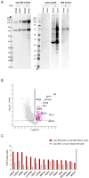
Figure 3
Figure 3
Identification of proteins interacting with IDE. (A) Immunoblot analysis of biotinylated proteins in Cyto-IDE (left), Cyto-TurboID, and untransduced HEK (right) cells incubated for 10 min with 50 μM biotin or buffer control, in total extracts and after enrichment using streptavidin beads. One out of four independent experiments is shown. (B) Scatterplot of biotinylated proteins enriched in Cyto-IDE vs. Cyto-TurboID cells incubated with 50 μM biotin. Data based on four independent replicates. Proteins with FDR=0.05 were analyzed. (C) 15 most-enriched proteins in Cyto-IDE cells relative to both controls (Cyto-IDE without biotin and Cyto-TurboId plus biotin).
Figure 1
Figure 1
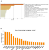
Proteomic analysis of islet proteins from Ide+/+ and Ide−/− NOD mice. (A) Top nonredundant enrichment clusters, as identified by Metascape, among proteins upregulated in Ide+/+ islets. The color scale represents statistical significance levels expressed as -log10. (B) Top 20 enriched proteins in Ide+/+ islets.
Figure 2
Figure 2
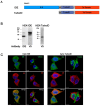
Characterization of TurboID fusion protein. (A) Schema of TurboID constructs for cytosolic expression. Human IDE cDNA starting at Met42 was fused to sequences encoding a V5 tag, the TurboID enzyme, the self-cleaving peptide T2AP2A, and tdTomato. (B) TurboID-fusion protein expression in HEK 293 cells was analyzed by Western blot in 10 μg total protein extracts using antibodies to IDE and V5 tag. (C) The expression of TurboID-fusion proteins in HEK cells was visualized by confocal fluorescent imaging. Cells expressing IDE–TurboID (left panel), or TurboID (right panel) were stained with antibodies to IDE, TOM20, and V5 tag. Merged images show cytosolic localization of each fusion protein.
Figure 4
Figure 4
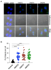
Ide may interact with the CCR4-NOT complex. (A) Proximity of IDE and CCR4-NOT was analyzed by PLA (green dots) using primary mouse antibodies to IDE and rabbit antibodies to CNOT2, CNOT3, and CNOT8. The cell area was outlined in epifluorescence images. Nuclei were counterstained with DAPI (blue). One of two independent experiments is shown. (B) Number of PLA positive signals per cell. The total numbers of cells analyzed were 36 (CNOT2), 26 (CNOT3), 37 (CNOT8), and 36 (isotype). ****, p < 0.001.
Figure 5
Figure 5
Single-cell RNA sequencing of Ide+/+ and Ide−/− C57BL/6 pancreatic islets. (A) Uniform Manifold Approximation and Projection plot (UMAP) depicting clusters of cells from the two genotypes. Clusters are named and color-coded according to the legend shown at the right. (B) Percentage of each cell type among the total numbers of Ide+/+ and Ide−/− cells. (C) Percentage of the two genotypes in each population identified in (A) among pooled sequenced islet cells. (D) Ide RNA expression levels in islet cell subpopulations.
Figure 6
Figure 6
Single-cell RNA-seq analysis of beta and alpha cells indicates enrichment of genes related to translation and RNA processing in Ide+/+ cells. (A,B). Pathways significantly associated with upregulated genes in Ide+/+ compared with Ide−/− cells (FDR < 0.05) were identified with Gene Set Enrichment Analysis (GSEA) with the Molecular Signatures Database (MSigDBr) Reactome gene sets. Gene Ratio refers to the proportion of upregulated genes among each gene set, and Count refers to the number of upregulated genes in the set. (C,D) Dot plots show the genes with the greatest expression changes in Ide+/+ and Ide−/− beta (C) and alpha cells (D). Color indicates the average expression, and the dot size indicates the percentage of cells.

References

    1. Lesire L., Leroux F., Deprez-Poulain R., Deprez B. Insulin-Degrading Enzyme, an Under-Estimated Potential Target to Treat Cancer? Cells. 2022;11:1228. doi: 10.3390/cells11071228. - DOI - PMC - PubMed
    1. Leissring M.A., González-Casimiro C.M., Merino B., Suire C.N., Perdomo G. Targeting Insulin-Degrading Enzyme in Insulin Clearance. Int. J. Mol. Sci. 2021;22:2235. doi: 10.3390/ijms22052235. - DOI - PMC - PubMed
    1. Duckworth W.C. Insulin Degradation: Progress and Potential. Endocr. Rev. 1998;19:608–624. doi: 10.1210/er.19.5.608. - DOI - PubMed
    1. Zhu S., Waeckel-Énée E., Oshima M., Moser A., Bessard M.-A., Gdoura A., Roger K., Mode N., Lipecka J., Yilmaz A., et al. Islet cell stress induced by insulin-degrading enzyme deficiency promotes regeneration and protection from autoimmune diabetes. iScience. 2024;27:109929. doi: 10.1016/j.isci.2024.109929. - DOI - PMC - PubMed
    1. Hamel F.G., Mahoney M.J., Duckworth W.C. Degradation of intraendosomal insulin by insulin-degrading enzyme without acidification. Diabetes. 1991;40:436–443. doi: 10.2337/diab.40.4.436. - DOI - PubMed

Publication types

Associated data

LinkOut - more resources