Review
. 2025 Jun 1;14(11):823.
doi: 10.3390/cells14110823.

Distinct Types of Regulated Cell Death in Melanoma

Affiliations
Review

Distinct Types of Regulated Cell Death in Melanoma

Qi Wu et al. Cells. .

Abstract

Resistance to cell death is one of the core hallmarks of cancer, with regulatory abnormalities particularly pronounced in the malignant progression and therapeutic resistance of melanoma. This review aims to systematically summarize the roles and mechanisms of regulated cell death (RCD) in melanoma. Currently, distinct types of RCD, including apoptosis, autophagy, pyroptosis, immunogenic cell death, necroptosis, and ferroptosis, have all been found to be involved in melanoma. Autophagy promotes the survival of melanoma cells under stress conditions through metabolic adaptation, yet its excessive activation can trigger cell death. Immunogenic cell death has the capacity to elicit adaptive immune responses in immunocompetent syngeneic hosts. Necroptosis, governed by the receptor-interacting protein kinase 1 (RIPK1)/RIPK3 mixed lineage kinase domain-like protein (MLKL) signaling axis, can synergize with immunotherapy to enhance anti-melanoma immune responses when activated. Pyroptosis, mediated by Gasdermin proteins, induces the release of inflammatory factors that reshape the tumor microenvironment and enhance the efficacy of immune checkpoint inhibitors. Ferroptosis, characterized by lipid peroxidation, can overcome melanoma resistance by targeting the solute carrier family 7 member 11 (SLC7A11)/glutathione peroxidase 4 (GPX4) axis. Therapeutic strategies targeting RCD pathways have demonstrated breakthrough potential. Several agents have been developed to target RCD in order to suppress melanoma.

Keywords: apoptosis; autophagy; ferroptosis; immunogenic cell death; melanoma; necroptosis; pyroptosis; regulatory cell death.


Conflict of interest statement

The authors declare no conflicts of interest.

Figures

Figure 1
Figure 1
Potential molecular mechanisms of apoptosis in melanoma. The extrinsic apoptotic pathway is initiated by two types of receptors on the cell membrane: death receptors (DRs) which engage Fas-associated protein with death domain (FADD) and associate with pro-Caspase-8 to form a death-inducing signaling complex (DISC), ultimately leading to the activation of Caspase-8, and pattern recognition receptors (PRRs) which respond to pathogen-associated molecular patterns (PAMPs) to activate Caspases-9. The intrinsic apoptotic pathway is primarily regulated by mitochondrial processes. In response to various stimuli such as DNA damage, hypoxia, oxidative stress, intracellular acidosis and endoplasmic reticulum (ER) stress, the levels of p53 protein rise substantially, which will activate Bcl-2 antagonist/killer (BAK) and Bcl-2-associated X protein (BAX), leading to the release of cytochrome c (Cyto-C) from the mitochondria, which then forms an apoptosome with apoptotic peptidase activating factor 1 (APAF1) and pro-Caspase-9, which activates Caspase-9 and executioner caspases (Caspases-3/7). These executioner caspases facilitate apoptosis by activating DNase and Rho-associated protein kinase 1 (ROCK-1), which are responsible for chromatin condensation, DNA fragmentation, and membrane blebbing.
Figure 2
Figure 2
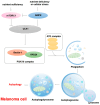
Potential molecular mechanisms of autophagy in melanoma. When intracellular nutrients are sufficient, mammalian target of rapamycin complex 1 (mTORC1) phosphorylates serine 757 of human serine/threonine-protein kinase ULK1 (ULK1) and autophagy-related protein 13 (ATG13), thereby inhibiting the initiation of autophagy. Under conditions of nutrient deficiency or cellular stress, adenosine monophosphate-activated protein kinase (AMPK) becomes activated and phosphorylates serine 317 and serine 777 of ULK1. This phosphorylation by AMPK counteracts the inhibitory effects of mTORC1, thereby promoting the initiation of autophagy. In detail, ULK1 activates the phosphatidylinositol 3-kinase (PI3K-III) complex, which includes Beclin-1 and VPS34, facilitating the formation of phagophores with the assistance of the ATG complex. Ultimately, autophagosomes fuse with lysosomes to form autolysosomes, where their contents are subsequently degraded by hydrolases for recycling.
Figure 3
Figure 3
Potential molecular mechanisms of pyroptosis in melanoma. PAMPs/DAMPs are recognized by PRRs such as NLRs and AIM2, which bind to the ASC adaptor protein to recruit and activate Caspase-1. Activated Caspase-1 cleaves GSDMD, releasing its N-terminal domain (GSDMD-N), which oligomerizes to form pores in the cell membrane, leading to osmotic imbalance and membrane rupture. Concurrently, Caspase-1 processes pro-IL-1β and pro-IL-18 into their active forms, which are released through these membrane pores to trigger robust inflammatory responses. In non-canonical pathways, LPS directly activates Caspase-4/5/11, which cleave GSDMD. Under apoptotic signals, Caspase-3 cleaves GSDME while Caspase-8 cleaves GSDMC, both cleaved gasdermin proteins induce pyroptosis-like cell death. In the granzyme-mediated pathway, granzymes (e.g., GZMA and GZMB) enter target cells via perforin and cleave specific members of the gasdermin family (e.g., GSDMB, GSDME), thereby inducing pyroptosis in cancer cells. All the cleaved gasdermin proteins mentioned above can form gasdermin pores in melanoma cells.
Figure 4
Figure 4
Potential molecular mechanisms of necroptosis in melanoma. Upon binding to TNF-α, TNFR1 recruits TNF receptor type 1-associated death domain protein (TRADD), TRAF2/5, and RIPK1 to form complex I, activating NF-κB pro-survival signals. However, when RIPK1 is deubiquited, it dissociates from complex I and forms complex IIa with FADD and caspase-8, which will then cause apoptosis. If caspase-8 is inhibited, RIPK1 activates RIPK3, which subsequently phosphorylates MLKL to form complex IIb, followed by MLKL oligomerization and translocation to the plasma membrane, resulting in ion imbalance, membrane integrity disruption, and ultimately cell swelling and rupture.
Figure 5
Figure 5
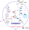
Potential molecular mechanisms of ferroptosis in melanoma. Ferroptosis is regulated by three primary pathways: iron metabolism, the classical GPX4-regulated pathway, and lipid peroxidation metabolism. Ferric/ferrous ions (Fe3+/Fe2+) enter cells via TFR-mediated endocytosis, generating ·OH through the Fenton reaction to trigger lipid peroxidation. The system Xc⁻-mediated cystine uptake and GSH synthesis inhibit lipid peroxidation, while GPX4 maintains cellular redox balance by reducing phospholipid hydroperoxides (PLOOHs). Pro-ferroptotic factors (e.g., ACSL4, lipoxygenases [LOXs]) and anti-ferroptotic factors (e.g., GPX4, NRF2, FSP1) collectively regulate the ferroptotic process in melanoma. The figure also indicates relevant drug targets and inhibitors (e.g., Erastin and Fer-1).

Similar articles

References

    1. Garbe C., Amaral T., Peris K., Hauschild A., Arenberger P., Basset-Seguin N., Bastholt L., Bataille V., Del Marmol V., Dréno B., et al. European consensus-based interdisciplinary guideline for melanoma. Part 1: Diagnostics: Update 2022. Eur. J. Cancer. 2022;170:236–255. doi: 10.1016/j.ejca.2022.03.008. - DOI - PubMed
    1. Vergara I.A., Wilmott J.S., Long G.V., Scolyer R.A. Genetic drivers of non-cutaneous melanomas: Challenges and opportunities in a heterogeneous landscape. Exp. Dermatol. 2022;31:13–30. doi: 10.1111/exd.14287. - DOI - PubMed
    1. Ferlay J., Colombet M., Soerjomataram I., Parkin D.M., Piñeros M., Znaor A., Bray F. Cancer statistics for the year 2020: An overview. Int. J. Cancer. 2021 doi: 10.1002/ijc.33588. in press . - DOI - PubMed
    1. Tribble J.T., Brownell I., Cahoon E.K., Sargen M.R., Shiels M.S., Engels E.A., Volesky-Avellaneda K.D. A Comparative Study of Merkel Cell Carcinoma and Melanoma Incidence and Survival in the United States, 2000–2021. J. Investig. Dermatol. 2025 doi: 10.1016/j.jid.2024.12.014. in press . - DOI - PubMed
    1. Long G.V., Hauschild A., Santinami M., Kirkwood J.M., Atkinson V., Mandala M., Merelli B., Sileni V.C., Nyakas M., Haydon A., et al. Final Results for Adjuvant Dabrafenib plus Trametinib in Stage III Melanoma. N. Engl. J. Med. 2024;391:1709–1720. doi: 10.1056/NEJMoa2404139. - DOI - PubMed

Publication types

LinkOut - more resources