. 2025 Jun 3;14(11):830.
doi: 10.3390/cells14110830.

Isolation and Characterization of Articular Cartilage-Derived Cells Obtained by Arthroscopic Cartilage Biopsy from Non-Osteoarthritic Patients

Affiliations

Isolation and Characterization of Articular Cartilage-Derived Cells Obtained by Arthroscopic Cartilage Biopsy from Non-Osteoarthritic Patients

Pedro Nogueira Giglio et al. Cells. .

Abstract

Cartilage-derived migratory cells show great potential for autologous use in cartilage repair surgery. However, their collection through arthroscopic biopsy has not been previously reported in individuals without osteoarthritis. This study aimed to characterize migratory cartilage cells isolated from arthroscopic biopsies of volunteers without osteoarthritis and compare them with cells obtained by enzymatic digestion. Cell cultures were successfully established using both methods-enzymatic digestion and cell migration-from cartilage explants, with no significant differences observed in stem cell markers or plasticity between the cell lines. Cells derived from both procedures exhibited characteristics of mesenchymal stem cell, including fibroblast-like morphology, expression of CD29, CD90, and CD105 markers, absence of hematopoietic and endothelial cell markers, and the ability to differentiate into adipocytes, chondrocytes, and osteoblasts under appropriate conditions. Cells obtained by migration showed lower expression of collagen I and II, along with reduce collagen II/collagen I ratio, both positively associated with chondral matrix production, as well as lower RUNX2 expression. However, no differences were found in the levels of SOX9, essential for chondrogenic differentiation, or in the expression of perlecan gene. Syndecan-1 expression was lower in cells obtained by migration. In conclusion, this study demonstrates that cartilage-derived migratory cells can be successfully obtained from arthroscopic biopsies of individuals without osteoarthritis, presenting comparable dedifferentiation and plasticity profiles. Furthermore, these cells express essential chondrogenic markers and proteins. Although further in vivo studies are needed to determine their effective regenerative potential, cartilage-derived migratory cells represent a promising avenue for cartilage repair strategies.

Keywords: cartilage explants; cartilage repair; cartilage-derived cells; enzymatic cartilage digestion; mesenchymal stem cells.


Conflict of interest statement

The authors declare no conflicts of interest. The funders had no role in the design of the study; in the collection, analyses, or interpretation of data; in the writing of the manuscript; or in the decision to publish the results.

Figures
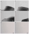
Figure 1
Figure 1
Photomicrograph of migrating cells from cartilage explant and cell culture. (A) Four days of culture; (B) nine days of culture; (C) twelve days of culture; (D) fifteen days of culture; (E) cell culture of explant group and (F) cell culture of collagenase group. Images obtained with an invert microscope Axio A.1 (Carl Zeiss, Oberkochen, Germany). Scale bar, 200 µm.
Figure 2
Figure 2
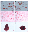
Representative images of differentiation into adipocytes, osteocytes and chondrocytes. (A,B): adipogenic differentiation (Oil Red O); (C,D): osteogenic differentiation (Alizarin Red); (E,F): chondrogenic differentiation (Safranin O). Explant group: (A,C,E); collagenase group: (B,D,F). Images obtained with inverted microscope Zeiss Axio A.1. Scale bar 50 µm in (A,B,E,F) and calibration 200 µm in (C,D).
Figure 3
Figure 3
Analysis of proteins Collagen I, Collagen II. (A,B) Graph of fluorescence intensity of collagen I (A) and collagen II (B). (C) Graph of ratio of collagen II for collagen I. (D,E) Representative images of collagen I (green) and nucleus (blue, stained with Hoechst 33342) in explant cells (D) and in collagenase cells (E). (F,G) Representative images of collagen II (green) and nucleus (blue, stained with Hoechst 33342) in explant cells (F) and in collagenase cells (G). Statistical analysis was performed by Student’s t-test in the GraphPad Prism 8 software. Data are mean ± SEM from three independent experiments in duplicate. * p < 0.05, Scale bar, 100 µM.
Figure 4
Figure 4
Analysis of proteins RUNX2, SOX9 and PPRAγ. (A) Graph of fluorescence intensity of RUNX2; (B,C) representative images of RUNX2 (red) and nucleus (blue, stained with Hoechst 33342 dye) in explant cells (B) and in collagenase cells (C). (D) Graph of fluorescence intensity of SOX9; (E,F) representative images of SOX9 (red) and nucleus (blue, stained with Hoechst 33342 dye) in explant cells (E) and in collagenase cells (F). (G) Graph of nuclear PPRAγ positivity; (H,I) representative images of PPRAγ (red) and nucleus (blue, stained with Hoechst 33342 dye) in explant cells (H) and in collagenase cells (I). Statistical analysis was performed by Student’s t-test in the GraphPad Prism 8 software. Data are mean ± SEM from three independent experiments in duplicate. * p < 0.05, Scale bar, 100 µM.
Figure 5
Figure 5
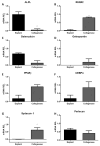
Analysis of mRNA expression for cartilage cells obtained by explantation or collagenase digestion. (A) ALPL, (B) RUNX2, (C) Osteocalcin, (D) Osteopontin; (E) PPARγ; (F) CEBPα; (G) Syndecan-1 and (H) Perlecan gene. Statistical analysis was performed by Student’s t-test in the GraphPad Prism 8 software. Data are expressed by mean ± SEM from three independent experiments in duplicate. * p < 0.05.
Figure 6
Figure 6
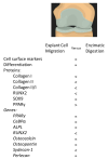
Summary of the effects of isolation methodology in cells derivate from arthroscopic joint biopsies of non-diseased cartilage. = equal expression; <: lower expression; >: higher expression.

References

    1. Hinckel B.B., Thomas D., Vellios E.E., Hancock K.J., Calcei J.G., Sherman S.L., Eliasberg C.D., Fernandes T.L., Farr J., Lattermann C., et al. Algorithm for Treatment of Focal Cartilage Defects of the Knee: Classic and New Procedures. Cartilage. 2021;13:473S–495S. doi: 10.1177/1947603521993219. - DOI - PMC - PubMed
    1. Makarczyk M.J. Cell Therapy Approaches for Articular Cartilage Regeneration. Organogenesis. 2023;19:2278235. doi: 10.1080/15476278.2023.2278235. - DOI - PMC - PubMed
    1. Giglio P.N., Lizier N.F., Levy D., Sobrado M.F., Gobbi R.G., Pécora J.R., Bydlowski D.P., Demange M.K. Autologous chondrocyte implantation in Brazil. Acta Ortop. Bras. 2020;28:131–136. doi: 10.1590/1413-785220202803226503. - DOI - PMC - PubMed
    1. Vinod E., Parameswaran R., Ramasamy B., Kachroo U. Pondering the Potential of Hyaline Cartilage-Derived Chondroprogenitors for Tissue Regeneration: A Systematic Review. Cartilage. 2020;13:34S–52S. doi: 10.1177/1947603520951631. - DOI - PMC - PubMed
    1. Matta C., Boocock D.J., Fellows C.R., Miosge N., Dixon J.E., Liddell S., Smith J., Mobasheri A. Molecular phenotyping of the surfaceome of migratory chondroprogenitors and mesenchymal stem cells using biotinylation, glycocapture and quantitative LC-MS/MS proteomic analysis. Sci. Rep. 2019;9:9018. doi: 10.1038/s41598-019-44957-y. - DOI - PMC - PubMed

Publication types

LinkOut - more resources