. 2025 Jul;26(13):3264-3279.
doi: 10.1038/s44319-025-00466-w. Epub 2025 Jun 11.

AML patient blasts exhibit polarization defects upon interaction with bone marrow stromal cells

Affiliations

AML patient blasts exhibit polarization defects upon interaction with bone marrow stromal cells

Khansa Saadallah et al. EMBO Rep. 2025 Jul.

Abstract

Hematopoietic stem and progenitor cells (HSPCs) polarize in contact with the bone marrow stromal cells constituting their niche. Given the role of cell polarity in protection against tumorigenesis and the importance of the niche in the progression of acute myeloid leukemias (AMLs), we investigated the polarization capacities of leukemic blasts. Using engineered micro-niches and centrosome position with respect to the contact site with stromal cells as a proxy for cell polarization, we show that AML cell lines and primary cells from AML patient blasts are unable to polarize in contact with healthy stromal cells. Exposure to AML patient-derived stromal cells compromises the polarization of healthy adult HSPCs and AML blasts from patients. When cultured in "bone-marrow-on-a-chip", stromal cells from a leukemic niche stimulate the migration of healthy HSPCs and AML blast. These results reveal the detrimental influences of both intrinsic transformation and extrinsic contact with transformed stromal cells on the polarization of AML blasts.

Keywords: Acute Myeloid Leukemia (AML); Artificial Niche; Bone-marrow-on-a-Chip (BMoC); Hematopoietic Stem and Progenitor Cells (HSPCs); Microwell.


Conflict of interest statement

Disclosure and competing interests statement. The authors declare no competing interests.

Figures

Figure 1
Figure 1. AML cell lines failed to polarize when interacting with osteoblasts within microwells.
(A) Schematic drawing representing microwells on a cover slide. The first zoom illustrates the non-adhesive edges (polyacrylamide) and the adhesive bottom (fibronectin coating), allowing the attachment and confining of stromal cells. The second magnification illustrates the position of the centrosome in the hematopoietic cell relative to cell contact with the stromal cell and the measurement of the polarization index (d/D). (B) Immunostainings of the centrosome (red), the nucleus (blue) and the actin cytoskeleton (green) in Cord blood (CB) HSPC and AML cell lines interacting with osteoblasts (hFOB) within microwells. Images were acquired in 3D. The top row shows a top projected view and scale bars correspond to 10 µm. Bottom row shows a side projected view and scale bars correspond to 10 µm. CB HSPCs form a magnupodium in contact with stromal cells. MOLM-14 and NOMO-1 leukemic blasts are both round and do not exhibit a magnupodium upon interaction with the osteoblast. (C) Graphs show the quantification of the polarization index of AML cell lines, MOLM-14 and NOMO-1, in contact with osteoblasts (hFOB) in comparison to CB HSPCs within microwells. Experiments were repeated two or three times and data were pooled (n CB HSPCs = 59, n MOLM-14 = 95; n NOMO-1 = 173). Each spot corresponds to the measurement of the polarity of a single cell. The various shapes of the spots correspond to distinct experiments. Black bars show the median value and the standard deviation of the distribution. Differences between populations were evaluated using non-parametric test Kruskal–Wallis ANOVA with a P value < 0.0001 (****).
Figure 2
Figure 2. AML patient blasts failed to polarize when interacting with various stromal cells from the bone marrow.
(A) Immunostainings of the centrosome (red, pointed at with white arrowheads), the nucleus (blue) and the actin cytoskeleton (green) in HSPCs from healthy adult donor and AML primary leukemic cells from patients #1, #2, #3, and #10 interacting with osteoblasts (hFOB) in microwells. Images were acquired in 3D. The top row shows a top projected view, and scale bars correspond to 10 µm. Bottom row shows a side projected view and scale bars correspond to 5 µm. (B) Same as (A) with adult HSPCs and AML primary leukemic cells from patients #1, #2, and #3 interacting with healthy donor MSCs (HD MSCs) in microwells. Similar to CB HSPCs, adult HSPCs elongate and form magnupodia where can be located their centrosomes in close vicinity to the contact site with stromal cells (osteoblast or HD MSC). AML primary blasts from patients #1, #2, and #3 and AML primary LSCs from patient #10 exhibit large zones of contact with no sign of magnupodia in contact with the stromal cell (osteoblast or HD MSC). (C) Quantification of the polarization index of AML patient blasts (blue dots), compared to HSPCs from healthy adult donor (red dots), in contact with osteoblasts (hFOB) within microwells. Experiments were repeated three times and data were pooled (n adult HSPCs = 132, n patient 1 blasts = 123; n patient 2 blasts = 142, n patient 3 blasts = 61; n patient 10 LSCs = 149). Each spot corresponds to the measurement of the polarity of a single cell. The various shapes of the spots correspond to distinct experiments. Black bars show the median value and the standard deviation of the distribution. Differences between populations were evaluated using a Kruskal–Wallis ANOVA test with P values < 0.0001 (****). (D) Same as (C) for AML patient blasts (blue dots) and HSPCs from healthy adult donor (red dots) in contact with healthy donor (HD) mesenchymal stem cells (MSCs) within microwells. Experiments were repeated three times and data were pooled (n adult HSPCs = 88, n patient 1 blasts = 148; n patient 2 blasts = 29, n patient 3 blasts = 29). Differences between populations were evaluated using a Kruskal–Wallis ANOVA test with P values of 0.03, 0.004 and <0.0001 (*, ** and ****). AML primary blasts from patients #1, #2, #3 and LSCs from patient #10 display a random distribution of their centrosome positioning while interacting with osteoblasts and HD MSC, whereas adult HSPC from HD were polarized in contact with both cell types.
Figure 3
Figure 3. Contact with stromal cells from AML patients impairs the polarization of both adult HSPCs from healthy donors and leukemic blasts from AML patients.
(A) Immunostainings of the centrosome (red, pointed at with white arrowheads), the nucleus (blue) and the actin cytoskeleton (green) of adult HSPCs from healthy donor (HD) interacting with either healthy or AML bone marrow stromal cells. Images were acquired in 3D. Top row shows a top projected view and scale bars correspond to 10 µm. Bottom row shows a side projected view, and scale bars correspond to 5 µm. (B) Quantification of the polarization index of adult HSPCs from HD (red dots) and AML patient blasts (blue dots) upon interaction with AML MSCs in the microwells. Experiments were repeated three times and data were pooled (n adult HSPC / HD MSC = 88, n adult HSPC / Patient 4 MSC = 84; n adult HSPC / Patient 5 MSC = 135, n Patient 1 blasts / Patient 4 MSC = 73). Each spot corresponds to the measurement of the polarity of a single cell. The various shapes of the spots correspond to distinct experiments. Black bars show the median value and the standard deviation of the distribution. Differences between populations were evaluated using a Kruskal–Wallis ANOVA test with P values of 0.0060, 0.0222, and <0.0001 (**, *, ****). In adult HSPCs from HD interacting with AML MSCs derived from patient #5 and patient #4 the centrosome is mispositioned. In AML patient blasts interacting with AML MSCs from patient #4, the centrosome is mispositioned. Furthermore, AML patient blasts display a reverted polarization (polarity index >0.5) in contact with AML MSCs.
Figure 4
Figure 4. AML cell lines and AML patient blast failed to polarize when cultured with stromal cells in a bone marrow-on-a-chip (BMoC).
(A) The microfluidic circuit of the BMOC comprises 6 parallel channels. Channels (1) and (2) (not shown) are committed to medium supplementation. Channels (3), (4) and (5) are dedicated to endosteal (hFOBs), endothelial (HUVECs) and fibroblastic cells (NHLFs), respectively. All stromal cells are cultured in an hydrogel composed of collagen and fibrin. Endosteal compartment harbors central pillars to resist hydrogel contraction due to osteoblasts traction forces. The central channel (6) is devoted to loading hematopoietic cells such as hematopoietic stem and progenitor cells (HSPCs), AML cell lines (MOLM-14 or NOMO-1), or AML patient blasts. The photography on the left shows the BMoC perfused with colorants to illustrate the position of the distinct compartments. Microscopy images on the right show centrosome, actin filaments and nucleus stainings within the endosteal and endothelial compartments. Scale bars correspond to 50 µm. (B) Immunostainings of the centrosome (red, pointed at with white arrowheads), the nucleus (blue) and the actin cytoskeleton (green) of cord blood (CB) HSPCs and AML cell lines, MOLM-14 and NOMO-1, interacting with osteoblasts (hFOBs) in the endosteal compartment. Scale bars correspond to 10 µm. (C) Quantification of the polarization index of CB HSPCs (red dots), MOLM-14 and NOMO-1 (blue dots) in contact with osteoblasts. Experiments were repeated three to five times and data were pooled (n CB HSPCs = 240, n MOLM-14 = 81; n NOMO-1 = 153). Each spot corresponds to the measurement of the polarity of a single cell. The various shapes of the spots correspond to distinct experiments. Black bars show the median value and the standard deviation of the distribution. Differences between populations were evaluated using Kruskal–Wallis ANOVA test with P values of 0.0002 and <0.0001 (***,****). (D) Same as (B) with CB HSPCs, MOLM-14 and NOMO-1 interacting with endothelial cells (HUVECs) in the BMoC endothelial compartment. Scale bars correspond to 10 µm. (E) Same as (C) with CB HSPCs, MOLM-14 and NOMO-1 interacting with endothelial cells (HUVECs) in the BMoC endothelial compartment. Experiments were repeated three times and data were pooled (n CB HSPCs = 106, n MOLM-14 = 167; n NOMO-1 = 215). Differences between populations were evaluated using Kruskal–Wallis ANOVA test with P values < 0.0001 and 0.0008 (****,***).
Figure 5
Figure 5. Adult HSPCs and AML patient blasts migration in a humanized leukemic stromal compartment of a BMoC.
(A) Instead of osteoblast, mesenchymal stem cells (MSCs) were collected from the bone marrow of a either healthy donor (orange in the schematics, left) or AML patient #5 and #6 (blue in the schematics, right) and loaded in the endosteal compartment of the BMoC (channel 3). Hematopoietic cells were collected from healthy adult (red in the schematics, top row) or AML patients #7 (blue in the schematics, bottom row). Images in transmitted light were extracted from time-lapse movies of healthy HSPC or AML patient blasts within healthy or leukemic compartments. Scale bars correspond to 50 µm. (B) Quantification of the mean speed and traveled distance during 20 min of the migration of healthy adult HSPCs (red) and AML blasts from patient #7 (blue) in a healthy (orange) or leukemic (blue) MSCs compartment. Tracks were analyzed at various positions within the stromal compartment (n adult HSPC/ HD MSC = 88, n adult HSPC/ AML MSC = 95, n AML blast / HD MSC = 140, n AML blast / AML MSC = 116). In the violin plots, black bars represent the median and dashed lines the 95% confidence interval. Differences between populations were evaluated using a Mann–Whitney test with P values < 0.0001 (****).
Figure EV1
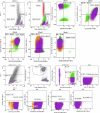
Figure EV1. AML patient n° 10 hematopoietic cell flow cytometry gating strategy (blasts and LSCs).
Flow cytometry analysis of an AML patient with a high LSC fraction.
Figure EV2
Figure EV2. AML patient immunophenotypic profiles of mesenchymal stromal cells (MSCs).
Flow cytometry strategy to characterize from the AML patient MSCs considered as CD45, CD90, CD73, CD105.
Figure EV3
Figure EV3. Leukemic cells originating from AML cell lines lack the motility observed in AML patient blasts within a healthy mesenchymal stromal cell compartment in the BMoC model.
(A) AML cell line (MOLM-14) and AML patient blasts (blue in the schematics) were loaded in the BMOC and thus interacted with MSC from healthy donors (orange in the schematics). Images in transmitted light were extracted from time-lapse movies of AML cell lines and patient blast within healthy compartments. Scale bars correspond to 50 µm. (B) Quantification of the mean speed and traveled distance during 20 min of the migration of leukemic cells (MOLM-14) or AML patient blast in a healthy stromal compartment (HD MSC). Tracks were analyzed at various positions within the stromal compartment (n MOLM-14/ HD MSC = 41, n AML blast/ HD MSC = 77). In the violin plots, black bars represent the median and dashed lines the 95% confidence interval. Differences between populations were evaluated using a Mann–Whitney test with P values < 0.0001 (****).

References

    1. Agarwal A, Bolosky WJ, Wilson DB, Eide CA, Olson SB, Fan G, Druker BJ (2019) Differentiation of leukemic blasts is not completely blocked in acute myeloid leukemia. Proc Natl Acad Sci USA 116:24593–24599 - PMC - PubMed
    1. Althoff MJ, Nayak RC, Hegde S, Wellendorf AM, Bohan B, Filippi M-D, Xin M, Lu QR, Geiger H, Zheng Y et al (2020) Yap1-Scribble polarization is required for hematopoietic stem cell division and fate. Blood 136:1824–1836 - PMC - PubMed
    1. Andrews MG, Subramanian L, Salma J, Kriegstein AR (2022) How mechanisms of stem cell polarity shape the human cerebral cortex. Nat Rev Neurosci 23:711–724 - PMC - PubMed
    1. Bessy T, Candelas A, Souquet B, Saadallah K, Schaeffer A, Vianay B, Cuvelier D, Gobaa S, Nakid-Cordero C, Lion J et al (2021) Hematopoietic progenitors polarize in contact with bone marrow stromal cells in response to SDF1. J Cell Biol 220:e202005085 - PMC - PubMed
    1. Bidlingmaier S, Zhu X, Liu B (2008) The utility and limitations of glycosylated human CD133 epitopes in defining cancer stem cells. J Mol Med 86:1025–1032 - PMC - PubMed