. 2025 Jun 26;129(25):6220-6232.
doi: 10.1021/acs.jpcb.5c01826. Epub 2025 Jun 12.

Triplet-Triplet Annihilation Upconversion Is Impeded in Liposomes that Prevent Sensitizer and Annihilator Co-Confinement

Affiliations

Triplet-Triplet Annihilation Upconversion Is Impeded in Liposomes that Prevent Sensitizer and Annihilator Co-Confinement

Amrutha Prabhakaran et al. J Phys Chem B. .

Abstract

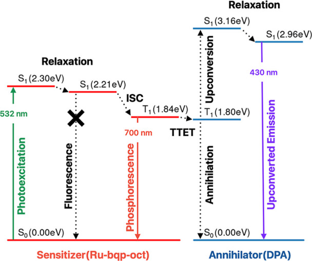
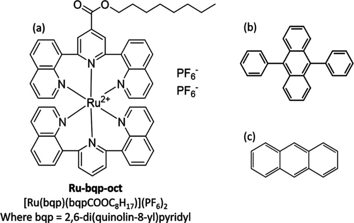
Triplet-triplet annihilation upconversion (TTA-UC) implemented in liposomes may be a promising tool in drug delivery and sensing. Indeed, we recently demonstrated that colocalization of lipophilic reagents to the membrane hydrophobic core improves the TTA-UC efficiency in liposomes compared to solution. Here, we examined if the counter is true, i.e., we evaluate if TTA-UC is inhibited when the sensitizer and annihilator occupy different regions within a single leaflet of a liposome membrane. To test this hypothesis, we used a Ru(II) complex, with tridentate ligand 2,6-di(quinolin-8-yl)pyridyl) (bqp) [Ru(bqp)(bpq-oct)]2+(Ru-bqp-oct) where oct is a C8 alkyl chain appended to facilitate integration into the liposome, as a sensitizer and diphenylanthracene (DPA) as an annihilator. TTA-UC from this pair was evaluated and compared in solution and liposomal nanovesicles. This Ru(II)-bqp complex was selected for its exceptionally long-lived emission and high triplet quantum yield, due to its expanded N-Ru-N bite angles. In solution, TTA-UC was efficient with a quantum yield of 3.11%, but in liposomes, no anti-Stokes shifted emission was observed even with an increased concentration of sensitizer and annihilator in the membrane. Molecular dynamics simulations were used to understand this effect and confirmed poor co-orientation of sensitizer and annihilator in the membrane was responsible for lack of TTA-UC in the membrane. DPA was determined to orient at the hydrophobic core, while the cationic Ru complex is embedded shallowly at the membrane interface, the closest approach of donor and acceptor in the membrane was determined as 0.7 nm. This work highlights the critical importance of colocalization of sensitizers and annihilators, even within a single membrane leaflet to facilitate Dexter energy transfer through collision in membrane-constrained TTA-UC systems and the value of MD simulations in system design.


Figures

1
1. Schematic Illustration of the Jablonski Diagram of the TTA-UC Mechanism for the System Explored Here
1
1
Chemical structures of (a) triplet sensitizer Ru-bqp-oct, and annihilators (b) 9,10-DPA and (c) anthracene.
2
2
Normalized absorption and emission spectra of (a) 10 μM Ru-bqp-oct in acetonitrile and (b) 20 μM 9,10-DPA in acetonitrile. TDDFT vertical energies and oscillator strengths, S1 and T1 0–0 transitions. The emission spectra of Ru-bqp-oct and DPA were collected by exciting at 490 and 395 nm, respectively. Excitation and emission slit width of 5 nm was used.
3
3
Upconverted emission from deaerated acetonitrile containing (a) Ru-bqp-oct and DPA at the 1:20 ratio with three different concentrations of 5 μM:100 μM, 10 μM:200 μM, and 20 μM:400 μM sensitizer/annihilator concentrations including one sample before deaeration showing the absence of TTA-UC and (b) 20 μM Ru-bqp-oct and 400 μM anthracene and 10 μM:200 μM (multiplied by 10). (c) Digital photograph showing the intense upconverted violet emission from deaerated acetonitrile containing 20 μM Ru-bqp-oct and 400 μM DPA. All samples are excited with a 532 nm green laser of 10 mW power, and the emission was collected at 5 nm slit width.
4
4
(a) Double logarithmic plot of integrated upconversion emission intensity measured as a function of the power of incident laser of 532 nm in a mixture of 20 μM Ru-bqp-oct and 400 μM DPA in deaerated acetonitrile. The linear fits with slopes 1 and 2 at high and low power regimes are included. R 2(COD) ≈ 0.98. (b) I th vs DPA concentration.
5
5
Normalized nanosecond transient absorption spectra of (a) Ru-bqp-oct, 20 μM in acetonitrile, inset: decay transient at 380 and 450 nm with monoexponential fits in dashed line, τdec at 380 nm = 4.2 μs; τdec at 450 nm = 4.2 μs. (b) Ru-bqp-oct 20 μM and DPA 100 μM. Inset: decay transient at 380 and 450 nm with monoexponential fits in dashed line. τdec at 380 nm = 1.6 μs; τinc at 450 nm = 1.6 μs.
6
6
(a) Reflectance image of MSLB array showing the aqueous filled cavities, fluorescence lifetime images of the DOPC bilayer labeled with (b) 50 nM Ru-bqp-oct at 532 nm excitation and (c) DOPE-Atto655 at 640 nm excitation. Scale bar indicated 4 μm. (d) Representative FLCS autocorrelation function of DOPC MSLB labeled with 10 nM Ru-bqp-oct (upper leaflet) and the (e) corresponding intensity-time trace. FLCS was measured over 40–50 cavities, and the average is shown. The solid lines are the 2D diffusion fit using eq S1. All measurements were carried out in contact with PBS of pH 7.4.
7
7
Preferred localization of Ru-bqp-oct and DPA molecules within a DOPC membrane. The probability distributions derived from our MD simulations show the positioning of the sensitizer and annihilator molecules along the axis perpendicular to the membrane surface. The brown curve represents the DOPC phosphate groups for reference. The 0.0 position corresponds to the bilayer midplane.

References

    1. Shum J., Leung P. K.-K., Lo K. K.-W.. Luminescent Ruthenium­(II) Polypyridine Complexes for a Wide Variety of Biomolecular and Cellular Applications. Inorg. Chem. 2019;58(4):2231–2247. doi: 10.1021/acs.inorgchem.8b02979. - DOI - PubMed
    1. Yam V. W.-W., Wong K. M.-C.. Luminescent Metal Complexes of D6, D8 and D10 Transition Metal Centres. Chem. Commun. 2011;47(42):11579. doi: 10.1039/c1cc13767k. - DOI - PubMed
    1. Fernández-Moreira V., Thorp-Greenwood F. L., Coogan M. P.. Application of D6 Transition Metal Complexes in Fluorescence Cell Imaging. Chem. Commun. 2010;46(2):186–202. doi: 10.1039/B917757D. - DOI - PubMed
    1. Baggaley E., Weinstein J. A., Williams J. A. G.. Lighting the Way to See inside the Live Cell with Luminescent Transition Metal Complexes. Coord. Chem. Rev. 2012;256(15–16):1762–1785. doi: 10.1016/j.ccr.2012.03.018. - DOI
    1. Arellano-Reyes R. A., Prabhakaran A., Sia R. C. E., Guthmuller J., Jha K. K., Yang T., Dietzek-Ivanšić B., McKee V., Keyes T. E.. BODIPY-Perylene Charge Transfer Compounds; Sensitizers for Triplet-Triplet Annihilation Up-conversion. Chem. - Eur. J. 2023;29(24):e202300239. doi: 10.1002/chem.202300239. - DOI - PubMed

MeSH terms

LinkOut - more resources