[Preprint]. 2025 Jun 2:2025.05.30.656989.
doi: 10.1101/2025.05.30.656989.

Biocompatible Multi-functional Polymeric Material for Mineralized Tissue Adhesion

Affiliations

Biocompatible Multi-functional Polymeric Material for Mineralized Tissue Adhesion

Yan Luo et al. bioRxiv. .

Update in

Abstract

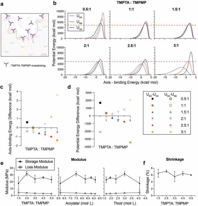
This study developed a biocompatible multifunctional thiol-ene resin system for adhesion to dentin mineralized tissue. Adhesive resins maintain the strength and longevity of dental composite restorations through chemophysical bonding to exposed dentin surfaces after cavity preparations. Dental pulp cells are exposed to residual monomers transported through dentinal tubules. Monomers of conventional adhesive systems may result in inhomogeneous polymer networks and the release of residual monomers that cause cytotoxicity. In this study, we develop a one-step multi-functional polymeric resin system by incorporating trimethylolpropane triacrylate (TMPTA) and bis[2-(methacryloyloxy)ethyl] phosphate (BMEP) to enhance both mechanical properties and adhesion to dentin. Molecular dynamics simulations identified an optimal triacylate:trithiol ratio of 2.5:1, which was consistent with rheological and mechanical tests that yielded a storage modulus of ~30 MPa with or without BMEP. Shear bond tests demonstrated that the addition of BMEP significantly improved dentin adhesion, achieving a shear bond strength of 10.8 MPa, comparable to the commercial primer Clearfil SE Bond. Nanoindentation modulus mapping characterized the hybrid layer and mechanical gradient of the adhesive resin system. Further, the triacrylate-BMEP resin showed biocompatibility with fibroblasts in vitro. These findings suggest the triacrylate-trithiol crosslinking and chemophysical bonding of BMEP provide enhanced bond strength and biocompatibility for dental applications.

Keywords: Triacrylate resin; dentin adhesion; molecular dynamics simulation; nanoindentation; thiol-ene polymerization.


Conflict of interest statement

Conflict of Interest Statement The authors affirm that they do not have any known conflicting financial interests or personal relationships that could have potentially influenced the findings presented in this paper. KHV is a co-inventor of US patent 11224679B2 and European patent 3426182B1 related to the use of TMPTA and TMPMP for dental applications.

Figures

Figure 1.
Figure 1.. Illustration of TMPTA-TMPMP resin-dentin dual adhesion mechanisms.
(a) Overall schematic of resin with Trimethylolpropane triacrylate (TMPTA) and Trimethylolpropane tris(3-mercaptopropionate) (TMPMP) as monomers, Bis[2-(methacryloyloxy)ethyl] phosphate (BMEP) as the primer, and 2,2-Dimethoxy-2-phenylacetophenone (DMPA) as the photoinitiator. (b) Demonstration of both physical and chemophysical interlocks between resin and dentin through crosslinked polymers and chelation reactions involving BMEP.
Figure 2.
Figure 2.. Molecular dynamics simulations and comparative modulus and shrinkage analysis for resin systems with varying compositions of TMPTA and TMPMP.
(a) Illustration of TMPTA-TMPMP crosslinking in resin. (b) Molecular dynamics simulation results of potential energy and axis-binding energy profile for TMPTA and TMPMP reacting under different ratios. U – potential energy; S – screening material, TMPMP; B – base material, TMPTA. Summary plots for (c) axis-binding energy difference and (d) potential energy difference between UBS and USS or UBB for different TMPTA: TMPMP ratios. (e) Storage modulus and loss modulus of resin plotted as a function of: TMPTA: TMPMP ratios, acrylate concentration and thiol concentration. (f) Shrinkage of resin under different TPMTA: TMPMP ratios.
Figure 3.
Figure 3.. Comparative adhesion, storage modulus and shrinkage analysis of the resin-dentin interface and resin systems with and without BMEP.
(a) Illustration of resin-dentin interface. (b) SEM (left) and EDS (right) images of dentin interface with resin tag exposed. (Yellow: sulfur). (c) SEM images of dentin interface without resin tag under 200 μm and 50 μm scale. (d) Shear test demonstration for adhesion situation between resin and dentin sample and (e) corresponding shear strength statistical results for resins and the commercial primer Clearfil SE Bond (Brown-Forsythe and Welch Test, sample size n = 15). (f) Statistical results of two resin systems about storage modulus and shrinkage percentage (unpaired t-test, sample size n = 3).
Figure 4.
Figure 4.. Nanomechanical analysis of the resin-dentin adhesive interface.
(a) Illustration of two interlocking mechanisms on the resin-dentin interface. (b) Nanoindentation mapping array (10 sperate indentation lines * 20 indentation tests), (c) modulus results for resin with (red) and without (black) BMEP, plots for (d) sigmoidal fitting results (blue curves), (e) corresponding summary table, and (f) comparison on HL’s sum of residual (SSres). (g) Weighted k-means clustering results (dentin region – orange, hybrid layer (HL) region – grey, resin region – blue), and (h) corresponding summary table. All the results demonstrated here represent the aggregate outcomes derived from the analysis of all 10 individual indentation line tests.
Figure 5.
Figure 5.. Biocompatibility of resin systems with and without BMEP.
(a) Fluorescence imaging (scale bar 200 μm) of BJ cells after 24 h culture in original condition (100% concentration) media compared to negative control, stained for nuclei (blue) and F-actin (green). (b) Relative cell viability, compared to negative control, and (c) cell counts of BJ cells after 24 h culture in condition media. Condition media are diluted with DMEM to a total concentration of 100%, 50% and 25% of the original condition media. n = 3 biological replicates, error bars represent SD.

References

    1. Farrar D. F. Bone adhesives for trauma surgery: A review of challenges and developments. International Journal of Adhesion and Adhesives 2012, 33, 89–97. DOI: 10.1016/j.ijadhadh.2011.11.009. - DOI
    1. Manuja N.; Nagpal R.; Pandit I. K. Dental adhesion: mechanism, techniques and durability. The Journal of clinical pediatric dentistry 2012, 36 3, 223–234. - PubMed
    1. Bjorndal L.; Fransson H.; Bruun G.; Markvart M.; Kjaeldgaard M.; Nasman P.; Hedenbjork-Lager A.; Dige I.; Thordrup M. Randomized Clinical Trials on Deep Carious Lesions: 5-Year Follow-up. J Dent Res 2017, 96 (7), 747–753. DOI: 10.1177/0022034517702620. - DOI - PubMed
    1. Krifka S.; Spagnuolo G.; Schmalz G.; Schweikl H. A review of adaptive mechanisms in cell responses towards oxidative stress caused by dental resin monomers. Biomaterials 2013, 34 (19), 4555–4563, Review. DOI: 10.1016/j.biomaterials.2013.03.019. - DOI - PubMed
    1. Sisman R.; Aksoy A.; Yalcin M.; Karaoz E. Cytotoxic effects of bulk fill composite resins on human dental pulp stem cells. J. Oral Sci. 2016, 58 (3), 299–305, Article. DOI: 10.2334/josnusd.15-0603. - DOI - PubMed

Publication types

LinkOut - more resources