[Preprint]. 2025 Jun 5:2025.06.05.657996.
doi: 10.1101/2025.06.05.657996.

Eph Receptor Tyrosine Kinases Are Functional Entry Receptors for Murine Gammaherpesvirus 68

Affiliations

Eph Receptor Tyrosine Kinases Are Functional Entry Receptors for Murine Gammaherpesvirus 68

Anna K Grosskopf et al. bioRxiv. .

Abstract

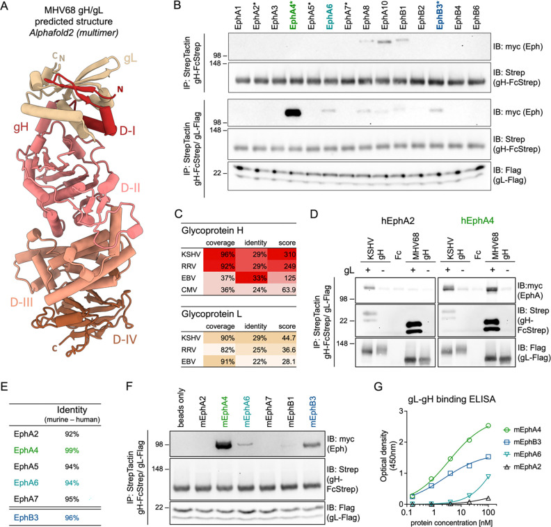
Interactions between viral glycoproteins and cellular receptors determine virus tropism and represent promising targets for vaccines. Eph receptor tyrosine kinases are conserved receptors for the human oncogenic gammaherpesviruses, Kaposi sarcoma herpesvirus (KSHV) and Epstein-Barr virus (EBV), and mediate entry into target cells by interaction with the viral gH/gL glycoprotein complex. To evaluate the use of murine gammaherpesvirus 68 (MHV68), a natural pathogen of rodents, as an in vivo model system for early events in gammaherpesvirus infection, we characterized the interaction of the MHV68 gH/gL complex with Eph receptors. We demonstrate a direct interaction of MHV68 gH/gL with EphA4 and EphB3, that is conserved between human and murine receptors. Pre-incubation of MHV68 inocula with soluble decoy receptors decreased infection of permissive fibroblasts. Ectopic expression of EphA4 and EphB3 enabled MHV68 to infect otherwise non-permissive human B cells, demonstrating EphA4 and EphB3 receptor function. Targeted mutations informed by protein structure predictions demonstrate that the MHV68 gH/gL-Eph interaction is determined by domain I (D-I) and follows structural motifs previously described in the KSHV gH/gL-EphA2 complex. The importance of gH D-I is further highlighted by the analysis of gH-targeting neutralizing antibodies. Antibody adsorption via the full gH ectodomain or gH D-I led to comparable reductions in neutralization capacity of serum from WT infected mice, indicating the Eph-binding domain is a major target for gH/gL-directed neutralizing antibodies. Our study characterizes Eph receptors as novel interaction partners and entry receptors for MHV68. Conservation of entry mechanisms provides the basis for future in vivo analyses of the contribution of Eph receptors to cell-type dependent MHV68 infection, as well as targeted strategies to prevent transmission and diseases associated with chronic infection.


Figures

Fig 1.
Fig 1.. The MHV68 gH/gL complex binds human and murine Eph receptors.
(A) The structure of the MHV68 gH/gL complex was predicted using alphafold2 multimer. MHV68 gH domains (D-I to D-IV) in the top ranked model were annotated based on EBV gH domain structure. Images were generated using ChimeraX. N and C termini of gH and gL are labeled “N” and “C”, respectively. (B) Pairwise precipitation of soluble recombinant MHV68 gH ectodomain in complex with MHV68 gL (gH-FcStrep/gL-Flag) with individual human Eph proteins. MHV68 gH-FcStrep was used as control. Precipitates were analyzed by immunoblot with indicated antibodies. Asterisks indicate known KSHV, EBV or RRV gH/gL interaction partners. (C) Percentage of coverage, identical amino acids in aligned regions and total score of alignments of gH and gL sequences of Eph-interacting herpesviruses were determined by BLAST (Basic Local Alignment Search Tool). MHV68 gH or gL was used as query sequence. No alignment was returned for CMV gL. (D) Comparative precipitation of human EphA2 and EphA4 by soluble recombinant KSHV gH/gL or MHV68 gH/gL. MHV68 gH-FcStrep, KSHV gH-FcStrep and FcStrep alone were used as controls. Precipitates were analyzed by immunoblot with indicated antibodies. (E) Percentage of identical amino acids in aligned regions of human and murine Eph proteins as determined by BLAST. (F) Pairwise precipitation of soluble recombinant MHV68 gH ectodomain in complex with MHV68 gL (gH-FcStrep/gL-Flag) with individual murine Eph proteins. Precipitates were analyzed by immunoblot with indicated antibodies. (G) Binding of dimeric, soluble, murine Eph proteins to an immobilized MHV68 gL-gH fusion protein was measured by enzyme-linked immunosorbent assay. Background corrected optical density at 450 nm is shown. Curve Fitting was performed using a four-parameter dose-response curve, determining an EC50 of 3.6 nM and 2.3 nM for mEphA4 and mEphB3 with a maximum OD of 2.8 and 1.6, respectively. For B, D, F, molecular weight is indicated in kDa.
Fig 2.
Fig 2.. Soluble murine EphA4 and EphB3 inhibit MHV68 infection of endothelial cells and fibroblasts.
(A) Schematic of Eph receptor-dependent block of MHV68 infection of susceptible cell lines. (B) Normalized read counts of the 14 Eph receptor genes in NIH 3T3 cells (GEO dataset series GSE196318 (63)). (C) Dose-dependent inhibition of MHV68 infection by soluble murine EphA4-Fc and EphB3-Fc but not EphA6-Fc on NIH 3T3 murine fibroblasts. MHV68 ORF59-GFP was pre-incubated with murine EphA4-Fc, EphA6-Fc or EphB3-Fc. EphA2-Fc and PBS were used as controls. GFP expression was measured by flow cytometry. Infection is indicated as percentage of GFP+ cells normalized to PBS controls. Mean of three independent experiments, error bars represent SD. (D-G) Cell type-dependent inhibition of MHV68 infection by soluble murine Eph proteins at 100 nM homodimerized protein. EphA2-Fc and PBS were used as controls. GFP expression as indicator of infection was measured by flow cytometry. Infection is shown as percentage of GFP+ cells normalized to PBS controls. Mean and symbols represent three individual experiments, error bars represent SD. Statistical significance was evaluated by ordinary one-way ANOVA, corrected by Holm-Šídák’s multiple comparisons test. *: p-value < 0.05, ***: p-value < 0.001, ****: p-value < 0.0001.
Fig 3.
Fig 3.. Overexpression of human and murine Eph receptors enables MHV68 infection of non-permissive Raji B cells.
(A) Schematic of Eph overexpression-dependent enhancement of MHV68 infection of non-susceptible cell lines. (B) Normalized read counts of the 14 Eph receptor genes in Raji B cells (GEO dataset series GSE111880 (89)). (C) Raji cells were transduced with TwinStrep-tagged murine (m) or human (h) Eph expression constructs or an empty vector (eV) control and selected by antibiotic resistance. Lysates of transduced Raji cell pools were analyzed for Eph-Strep expression by immunoblot. Molecular weight is indicated in kDa. (D) Transduced Raji cells analyzed in (C) were infected with MHV68 ORF59-GFP and GFP expression was measured by flow cytometry. Infection is indicated as fold change of GFP+ cells normalized to empty vector controls. Mean and symbols represent three-four individual experiments, error bars represent SD. Statistical significance was evaluated by ordinary one-way ANOVA, corrected for multiple comparisons by Holm-Šídák’s multiple comparisons test. ****: p-value < 0.0001. (E) Micrographs show representative infection of the indicated cell pools quantified in (D).
Fig 4.
Fig 4.. MHV68 gH/gL shares a structurally conserved Eph-interaction motif with the KSHV gH/gL complex.
(A) Reference structure of KSHV gH/gL in complex with human EphA2 (PDBid: 7B7N). Images were generated using ChimeraX. The core interacting residue pair E52KSHV gH - R103EphA2 is depicted in inset. (B-C) Truncated MHV68 gH (NP_044860.1) and MHV68 gL (NP_044884.3) were predicted in complex with murine EphA4 (NP_031962.2) (B) or murine EphB3 (NP_034273.1) (C) using alphafold2 (multimer, n=15). Resulting models were aligned against the known structure of KSHV gH/gL in complex with human EphA2 (PDBid: 7B7N). Images were generated using ChimeraX. Putative residues involved in Eph interaction or complex stabilization (Y50MHV68 gH, D52MHV68 gH, K24MHV68 gL) and R106EphA4/EphB3 are depicted in insets. Dashed lines represent predicted hydrogen bonds. (D) Pairwise precipitation of soluble recombinant MHV68 gH ectodomain or MHV68 gH D-I in complex with MHV68 gL (gH-FcStrep/gL-Flag), with murine EphA4. Fc and MHV68 gH constructs without gL were used as control. Precipitates were analyzed by immunoblot with indicated antibodies. (E) Putative interaction residues Y50MHV68 gH, D52 MHV68 gH, and K24 MHV68 gL were mutated to alanine in single point mutants or a gH double point mutant. Combinations of MHV68 gH ectodomain and gL point mutants were precipitated with murine EphA4. MHV68 gH-FcStrep was used as control. Precipitates were analyzed by immunoblot with indicated antibodies. (D-E) Molecular weight is indicated in kDa.
Fig 5.
Fig 5.. Neutralizing antibodies in MHV68 infected mice target the MHV68 gH/gL complex.
(A) Schematic of virus neutralization by gH/gL-targeting nAbs. C57BL/6 mice were infected with 1,000 PFU MHV68 WT by intranasal inoculation. Serum was collected at 28 dpi. (B) gH/gL-specific IgG from naïve or MHV68-infected C57BL/6 was measured by MHV68 gL-gH ELISA. Background corrected optical density at 450 nm is shown. (C) Serum of MHV68-infected C57BL/6 mice neutralizes MHV68 infection on NIH 3T3 cells. MHV68 ORF59-GFP was pre-incubated with mouse serum at the indicated dilution. GFP expression was measured by flow cytometry. Infection is indicated as percentage of GFP+ cells normalized to naïve serum. (D-E) Serum neutralization of MHV68 infection on NIH 3T3 cells is mediated by gH/gL-targeting antibodies. Antibodies to gHecto/gL or gHD-I/gL were depleted using soluble complexes pre-coupled to magnetic beads. Fc was used as control. Neutralization was analyzed as in (C) at a serum dilution of 1:80. Micrographs were taken at 16 hpi. Mean and symbols representing individual experiments are shown. Statistical significance was evaluated by ordinary two-way ANOVA corrected for multiple comparisons by Tukey’s multiple comparisons test. ***: p-value < 0.001, ****: p-value < 0.0001.

Similar articles

References

    1. Lurain K, Ramaswami R, Yarchoan R. The role of viruses in HIV-associated lymphomas. Semin Hematol. 2022;59(4):183–91. doi: 10.1053/j.seminhematol.2022.11.002. - DOI - PMC - PubMed
    1. de Martel C, Shiels MS, Franceschi S, Simard EP, Vignat J, Hall HI, et al. Cancers attributable to infections among adults with HIV in the United States. AIDS. 2015;29(16):2173–81. doi: 10.1097/QAD.0000000000000808. - DOI - PMC - PubMed
    1. Lurain KA, Ramaswami R, Krug LT, Whitby D, Ziegelbauer JM, Wang HW, et al. HIV-associated cancers and lymphoproliferative disorders caused by Kaposi sarcoma herpesvirus and Epstein-Barr virus. Clin Microbiol Rev. 2024;37(3):e0002223. doi: 10.1128/cmr.00022-23. - DOI - PMC - PubMed
    1. Grabar S, Costagliola D. Epidemiology of Kaposi’s Sarcoma. Cancers (Basel). 2021;13(22). doi: 10.3390/cancers13225692. - DOI - PMC - PubMed
    1. Losay VA, Damania B. Unraveling the Kaposi Sarcoma-Associated Herpesvirus (KSHV) Lifecycle: An Overview of Latency, Lytic Replication, and KSHV-Associated Diseases. Viruses. 2025;17(2). doi: 10.3390/v17020177. - DOI - PMC - PubMed

Publication types

LinkOut - more resources