[Preprint]. 2025 Jun 3:2025.05.17.654630.
doi: 10.1101/2025.05.17.654630.

Transcriptional Repression of reaper by Stand Still Safeguards Female Germline Development in Drosophila

Affiliations

Transcriptional Repression of reaper by Stand Still Safeguards Female Germline Development in Drosophila

Masaya Matsui et al. bioRxiv. .

Abstract

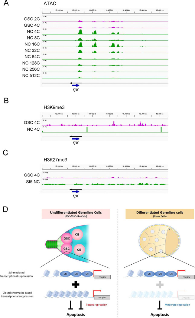
Apoptosis plays a central role in shaping tissues and preserving cellular integrity across developmental stages. In the germline, its precise regulation is critical to ensure both the elimination of aberrant cells and the maintenance of reproductive capacity. However, the molecular mechanisms that control apoptotic susceptibility in germline cells remain poorly defined. Here, we identify stand still (stil) as a female germline-specific regulator of apoptosis in Drosophila. Loss of stil leads to near-complete depletion of germline cells at the time of eclosion, associated with upregulation of the pro-apoptotic gene reaper (rpr) and activation of caspase-dependent cell death. Reporter assays in S2 cells show that Stil directly represses rpr transcription through its N-terminal BED-type zinc finger domain. Despite the absence of stil, undifferentiated germline cells remain resistant to apoptosis. Analysis of publicly available chromatin data reveals that the rpr locus in these cells resides in a closed, H3K9me3-enriched chromatin state, suggesting a Stil-independent mode of transcriptional silencing. Together, our findings uncover two distinct mechanisms that protects the female germline from rpr-dependent apoptosis: Stil-mediated transcriptional repression that operates in both undifferentiated and differentiated germline cells, and an additional chromatin-based silencing mechanism that functions specifically in undifferentiated cells. This work provides new insights into the interplay between transcriptional and chromatin-based regulations that maintain germline cell identity and survival.


Conflict of interest statement

Competing Interest Statement The authors declare that the research was conducted in the absence of any commercial or financial relationships that could be construed as a potential conflict of interest.

Figures

Figure 1.
Figure 1.. Disruption of stil induces female germline cell depletion
(A) Schematic representation of the Drosophila stil gene locus showing the position of P-element insertion, EY16156, mutation site of stil3, and the target site of shRNA for knockdown. Black and blue arrows represent primers for RT-PCR and RT-qPCR, respectively. (B) Expression of stil and the control α-tub transcripts in stilEY16156 mutant and the heterozygous control ovaries or testes analyzed by RT-PCR. (C) Representative images showing entire ovarian morphology (left panels) and ovarioles immunostained with with a germline marker, Vasa (red), and nuclear staining with DAPI (blue) (right panels) from y w, stilEY16156/CyO, stilEY16156, stilEY16156/Df, and stilEY16156 expressing 6×Myc-Stil-FL transgene driven by the germline-specific driver, NGT40; NosGal4-VP16. Scale bar: 1 mm for whole ovaries in the left panels, 50 μm for ovariole in the right panels. (D) Quantification of the percentage of germline cell-containing ovarioles in 2–3-days old females. Genotypes are indicated below the graph and the numbers of ovarioles assessed are shown above each bar. Error bars represent standard deviation (s.d.). (E) Analysis of egg laying and hatching rate. The egg numbers and their hatching rate by three females indicated genotypes corresponding to those in (C) mated with three y w males were measured daily (n=3). Error bars indicate s.d.
Figure 2.
Figure 2.. stil loss induces apoptotic cell death in female germline cells
(A) Quantitative RT-PCR confirming the knockdown efficiency in ovaries of stil-germline knockdown (stil-GLKD) where shRNA targeting stil transcript was driven by NGT40; NosGal4-VP16, and the control with shRNA alone. Expression levels of an internal control, rp49, and stil are normalized to that of α-tub. Error bar indicates s.d. (n=3). (B) Representative images showing entire ovarian morphology (left panels) and ovarioles immunostained with a germline marker, Vasa (red), and nuclear staining with DAPI (blue) (right panels) from stil-GLKD and the control with shRNA alone. Scale bar: 1 mm for whole ovaries and 50 μm for ovarioles, respectively. (C) Quantification of the percentage of germline cell-containing ovarioles in 2–3-days old females. Genotypes are indicated below the graph and the number of germarium assessed is noted above each bar. Error bars represent standard deviation (s.d.). (D) Quantification of the percentage of developed ovarioles defined as ovarioles that contain at least two egg chambers beyond the germarium. Genotypes are indicated below the graph and the number of germarium assessed is noted above each bar. Error bars represent standard deviation (s.d.). (E) Representative images of TUNEL-positive apoptotic cells (green), Vasa (red), and DAPI (blue) in control with shRNA alone and stil-GLKD germaria (white dotted). Scale bar: 20 μm. (F) Quantification of apoptosis in germarium with TUNEL assay in control and stil-GLKD. (G) Representative images of germaria (white dotted) from stilEY16156, stilEY16156; P35 OE (NGT40; NosGal4-VP16> P35), and stilEY16156; DIAP1 OE (NGT40; NosGal4-VP16> DIAP1) immunostained with Vasa (red) with DAPI (blue). Scale bar: 20 μm. (H) Quantification of the percentage of germline cell-containing ovarioles in 2–3-day old females. Genotypes are indicated below the graph and the number of germarium assessed is noted above each bar. Error bars indicate standard deviation (s.d.).
Figure 3.
Figure 3.. stil regulates rpr expression in female germline cells
(A) Top 10 upregulated and downregulated genes in stil-deficient ovaries. Bars indicate fold changes in stil mutant ovaries overexpressing (OE) of the apoptosis inhibitor, P35 in germline cells (stilEY16156, NGT40; NosGal4-VP16> P35), compared to control with P35 OE in a heterozygous background (stilEY16156/CyO, NGT40; NosGal4-VP16> P35). Bars are according to adjusted p-values (p-adj). (B) MA plot of apoptosis-related genes (GO:0006915) showing their mean gene expression (CPM: counts per million) and fold change in stilEY16156; P35 OE comparing stilEY16156/CyO; P35 OE. Each dot represents an individual gene, with dot size inversely correlated with p-value adjusted (p-adj). Larger dots highlight genes with higher statistical significance. The red dots (upregulated genes) and blue dots (downregulated genes) represent the differentially expressed RNAs with fold threshold>2.0, and p-adj<0.01. (C) Top 10 upregulated and downregulated genes related to apoptosis (GO:0006915). Bars represent fold changes and are colored by adjusted p-values (p-adj), as in (A). (D) Quantitative RT-PCR analysis of proapoptotic genes, rpr and hid in stilEY16156; P35 OE and stilEY16156/CyO; P35 OE ovaries. Expression levels were normalized to αtub with rp49 as an internal control. Error bars indicate s.d. (n=3). (E) Immunofluorescence of Vasa (red) and DAPI (blue) in ovarioles from stilEY16156/CyO; rpr87/+, stilEY16156/CyO; rpr87, stilEY16156; rpr87/+, and stilEY16156; rpr87. Scale bar: 50 μm. (F) Quantification of the percentage of germline cell-containing ovarioles in 2–3-days old females. Genotypes of females and the number of germarium assessed are noted above each bar. Error bars represent standard deviation (s.d.). (G) Quantification of the percentage of developed ovarioles defined as ovarioles that contain at least two egg chambers beyond the germarium. Genotypes females and the number of germarium assessed are noted above each bar. Error bars represent standard deviation (s.d.). (H) Analysis of egg laying and hatching rate. Daily egg laying by three females indicated genotypes mated with three y w male and their hatching rate are shown (n=3). Error bar indicates s.d.
Figure 4.
Figure 4.. Stil interacts with rpr-transcriptional control region to repress its expression
(A) Distribution of DamID peaks across entire genome categorized by genomic features including promoter (≤1, 1–2, and 2–3 kb), UTR, exon, intron, distal intergenic, and downstream. (B) The plot showing the log2 ratio of Dam-Stil peaks to Dam-alone control across the rpr genomic region. The blue bar on the bottom indicates the rpr gene body, and the arrow shows the direction of rpr transcription. Three independent biological replicates were performed for both Dam-Stil and Dam-alone and data were merged for visualization. (C) Schematic representation of the Stil full-length (Stil-FL), the truncated variants: Stil-NT (1st to 169th aa) and Stil-CT (169th to the end), and amino acid substitutions of the C2H2 zinc finger CCYC: Stil-AAYA. (D) stilEY16156 ovaries expressing Myc-tagged Stil variants corresponding to C in germline cells. Ovaries are immunostained with a germline marker, Vasa (red), and nuclear staining with DAPI (blue). Scale bar: 50 μm. (E) Quantification of the percentage of germline cell-containing ovarioles in 2–3 days-old females. Genotypes are indicated below the graph and the number of germarium assessed is noted above each bar. Error bars represent standard deviation (s.d.). (F) Quantification of the percentage of developed ovarioles defined as ovarioles that contain at least two egg chambers beyond the germarium. Genotypes are indicated below the graph and the number of germarium assessed is noted above each bar. Error bars indicate standard deviation (s.d.). (G) Schematic representation of rpr-GFP reporter containing the 5 kb upstream region and 5’-UTR of the rpr gene fused to GFP CDS. (H) Reporter assay in S2 cells followed by western blot analysis to detect GFP and α-Tubulin (αTub) as a loading control. Co-transfection of the rpr-GFP reporter and FLAG-tagged Stil variants were performed in S2 cells. The lower panel shows the signal ratio of GFP expression to αTub, normalized to that in cells transfected with the reporter alone. Error bars represent the standard deviation (s.d.) (n = 3).
Figure 5.
Figure 5.. stil mutant germline stem cells resist apoptosis
(A) Fluorescent immunostaining with anti-Engrailed (red), anti-Vasa (green), and DAPI (blue) for terminal filaments of late 3rd instar larval (LL3) ovaries from the heterozygous control, stilEY16156/CyO and stil mutant, stilEY16156. Scale bar: 50 μm. (B) Quantification of surviving germline cells in 3rd instar larval ovaries from stilEY16156/CyO and stilEY16156. (C) Immunostaining of Vasa (red) and DAPI (blue) in control, Tkv.CA (constitutive active Thickveins) OE (NGT40; NosGal4-VP16>Tkv.CA), and bam mutant (bamΔ86) germaria (white dotted) in the control heterozygous (stilEY16156/CyO) or stil mutant (stilEY16156) background. Scale bar: 20 μm. (D) Quantification germline cell-containing germaria per ovary in 2–3 days-old females. Genotypes and the number of germarium assessed are indicated above each bar. Error bars represent standard deviation (s.d.). (E) Representative images of germaria (white dashed lines) showing TUNEL-positive apoptotic cells (green), Vasa (red), and DAPI (blue) in the control tumorous germaria (stilEY16156/CyO; Tkv.CA OE) and tumorous germaria of stil mutant ovaries (stilEY16156; Tkv.CA OE). White arrowheads indicate TUNEL-positive apoptotic cells. Scale bar: 20 μm. (F) Quantification of apoptosis in germaria from the control tumorous ovaries (stilEY16156/CyO; Tkv.CA OE) and stil mutant ovaries (stilEY16156; Tkv.CA OE). (G) MA plot of apoptosis-related genes showing the mean gene expression (CPM: counts per million) and fold change in stil mutant ovaries (stilEY16156; Tkv.CA OE) compared to the control tumorous ovaries (stilEY16156/CyO; Tkv.CA OE). Each dot represents an individual gene, with dot size inversely correlated with p-adj. The blue dots represent the downregulated genes with fold change<−2.0, and p-adj<0.01. (H) Quantitative RT-PCR analysis of rpr in the control tumorous ovaries (stilEY16156/CyO; Tkv.CA OE) and stil mutant ovaries (stilEY16156; Tkv.CA OE). Expression levels of an internal control, rp49, and rpr are normalized to that of α-tub. Error bars indicate s.d. (n=3).
Figure 6.
Figure 6.. chromatin state changes in female germline cells during oogenesis
(A) ATAC-seq signal intensity (RPM-normalized) at the rpr locus in germline stem cells (GSCs: 2N and 4N) and nurse cells (NCs) at different ploidy levels (4C to 512C). The Y-axis is fixed at 0–30 PRM for all tracks. For 64C, 128C, 256C, and 512C NCs, data from three biological replicates are averaged; all other samples are based on single replicates. (B) H3K9me3 ChIP-seq signal at the rpr locus in 4C GSCs and 4C NCs. (C) H3K27me3 ChIP-seq signal at the rpr locus in GSCs and stage 5 (st5) NCs. (D) Schematic model illustrating the rpr repression to inhibit the apoptotic cell death in undifferentiated germline cells (GSCs or GSC-like cells) and differentiated germline cells (nurse cells).

Similar articles

References

    1. Abbott M. K. & Lengyel J. A. (1991). Embryonic head involution and rotation of male terminalia require the Drosophila locus head involution defective. Genetics, 129(3), 783–789. 10.1093/genetics/129.3.783 - DOI - PMC - PubMed
    1. Almeida C., Correia S., Rocha E., Alves Â., Ferraz L., Silva J., Sousa M. & Barros A. (2013). Caspase signalling pathways in human spermatogenesis. Journal of Assisted Reproduction and Genetics, 30(4), 487–495. 10.1007/s10815-013-9938-8 - DOI - PMC - PubMed
    1. Aravind L. (2000). The BED finger, a novel DNA-binding domain in chromatin-boundary-element-binding proteins and transposases. Trends in Biochemical Sciences, 25(9), 421–423. 10.1016/s0968-0004(00)01620-0 - DOI - PubMed
    1. Aughey G. N., Cheetham S. W. & Southall T. D. (2019). DamID as a versatile tool for understanding gene regulation. Development, 146(6), dev173666. 10.1242/dev.173666 - DOI - PMC - PubMed
    1. Baonza A., Tur-Gracia S., Pérez-Aguilera M. & Estella C. (2022). Regulation and coordination of the different DNA damage responses in Drosophila. Frontiers in Cell and Developmental Biology, 10, 993257. 10.3389/fcell.2022.993257 - DOI - PMC - PubMed

Publication types

LinkOut - more resources