[Preprint]. 2025 Jun 5:2025.06.02.657335.
doi: 10.1101/2025.06.02.657335.

Precise MRI-Histology Coregistration of Paraffin-Embedded Tissue with Blockface Imaging

Affiliations

Precise MRI-Histology Coregistration of Paraffin-Embedded Tissue with Blockface Imaging

Yixin Wang et al. bioRxiv. .

Update in

Abstract

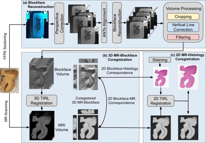
Magnetic resonance imaging (MRI) provides 3D spatial information on tissue, yet it lacks at the molecular level. In contrast, histology provides cellular and molecular information, but it lacks the 3D spatial context and direct in vivo translation. Coregistering the two is key for the 3D-embedding of histological details, validating pathological MRI findings, and finding quantitative imaging biomarkers of neurodegenerative diseases. However, coregistration is challenging due to non-linear distortions of the tissue from histological processing and sectioning leading to microscopic and macroscopic nonlinear 3D deformations between specimen MRI and stained histology sections. To address this, we developed a novel pipeline, named Brewster's Blockface Quantification (BBQ), integrating robust optical approaches with innovative 2D and 3D registration algorithms to achieve precise volumetric alignment of specimen MRI data with histological images. On a variety of brain tissue specimens from distinct anatomical regions and across multiple species, our methodology generated blockface volumes with minimal distortion and artifacts. Using these blockface volumes as an intermediary, we achieve a precise alignment between MRI and histology slides, yielding registration results with an overlapping Dice score of ~90% for whole tissue alignment between MRI and blockface volumes, and >95% for 2D MRI-histology registration. This correlative MRI-histology pipeline with robust 2D and 3D coregistration methods promises to enhance our understanding of neurodegenerative diseases and aid the development of MRI-based disease biomarkers.

Keywords: Biomarkers; Blockface imaging; Coregistration; Neurodegenerative disease; Ultra-high-resolution MRI; Volumetric histology.


Figures

Figure 1:
Figure 1:
Schematic overview of image acquisition and registration pipeline. (a) Reconstruction of block-face volume via distortion correction, 2D alignment, and image filtering. (b) Determination of histology-MR slice correspondence via 3D registration of the MRI to blockface volumes. (c) Alignment of the derived 2D MRI slice with corresponding 2D histology stain images. Note: this 2D registration can be bidirectional.
Figure 2:
Figure 2:
Ex-vivo specimen MRI set-up. Dissected specimens (A) were immersed in fluorinated oil (CHRISTO-LUBE MCG 1065) and enclosed in plastic tubes (B), and then scanned using a 7T Bruker preclinical MRI (C), yielding high-resolution multi-echo gradient echo (MGE) images.
Figure 3:
Figure 3:
Blockface imaging setup, showing both front and top views to illustrate Brewster’s angle (57° to the surface normal) between lightsource, tissue, and camera. A humidifier was applied before each section was cut.
Figure 4:
Figure 4:
Comparison of simultaneously acquired raw and processed blockface images of a neocortical human brain specimen captured under two different lighting setups: (top row) directly at Brewster’s angle (On Brewster’s) and (bottom row) slightly off Brewster (Brewster’s Adjacent). Note the inversion of gray-white contrast between the two positions.
Figure 5:
Figure 5:
Blockface perspective correction. (a) A fixed grid, printed and adhered to the microtome mount, was imaged (first row) and transformed to the correct perspective without distortion (second row). (b) The same transformation matrix was then applied to the blockface images captured from the identical position.
Figure 6:
Figure 6:
3D coregistration between MRI and blockface volumes. (a) Blockface images in axial, sagittal, and coronal planes. (b) The same sections with manually segmented for all specimens: white matter (red), grey matter (green), for hippocampal specimens: the dentate gyrus (blue, arrow), and only for pig: caudate (yellow), putamen (purple), and the anterior commissure (light blue, arrow). Red contours mark segmented region boundaries. (c, d) MR images after deformable (non-linear) transformation into the blockface space. Red and blue outlines trace the boundaries between subregion segmentations performed on the blockface and MRI respectively.
Figure 7:
Figure 7:
Dice similarity coefficient scores (%) of the segmentations of MRI and blockface volumes after coregistration across different specimens. “Whole Tissue” represents the score for the entire composite area. The highest score (best overlap) is indicated in Red.
Figure 8:
Figure 8:
Qualitative evaluation of 2D coregistration between histology and MRI correspondences. (i) displays H&E-stained sections with manually labeled outlines of white matter (red), grey matter (green), dentate gyrus (light blue), caudate (yellow), putamen (purple), and the anterior commissure (black). (ii) and (iii) present MRI images transformed into the histology space. Manually annotated regions of interest within the circles and pointed by arrows on the histology slides correspond precisely to MRI images transformed using TIRL Nonlinear registration.
Figure 9:
Figure 9:
Dice similarity coefficient (%) analysis for 2D registration of MRI and histology across multiple hippocampal head, tail, 4-mm cortex and pig coronal slab slides with H&E staining. TIRL shows a superior performance in the dentate gyrus (DG) and Anterior Commissure (AC).
Figure 10:
Figure 10:
3D coregistration between MRI and blockface volumes of Human 2-mm Cortex. A(i). Original blockface images in 3D planes with manually segmented white matter (red), grey matter (green), and red contours marking segmented region boundaries. A(ii, iii). MR images after undergoing deformable (non-linear) transformation into the blockface space using ANTs Nonlinear (SyN) and TIRL Nonlinear. B presents Dice coefficient across subregions using different registration algorithms.
Figure 11:
Figure 11:. 2D coregistration between MR and histology correspondences of Human 2-mm Cortex.
A(i) displays manual segmentation of white matter and grey matter on MRI slices, and A(ii) presents the corresponding histology sections transformed into MRI space using TIRL Nonlinear. B shows the quantitative Dice coefficient for each coregistered histology slide to its corresponding MR slice using TIRL Nonlinear and ANTs Nonlinear (***: p < 0.001). Each dot represents a single slice.
Figure 12:
Figure 12:
Reconstruction of volumetric histology from Human 2-mm Cortex. A shows the reconstructed volumes, while B illustrates rendering using 3D Slicer. The manually segmented vessels in both MRI (red) and histology volumes (green) are highlighted in C and overlaid on B, with two continuous vessels (blue and yellow) selected to assess voxel overlap in D and mean Euclidean distances in E.

Similar articles

References

    1. Adler D. H., Pluta J., Kadivar S., Craige C., Gee J. C., Avants B. B., & Yushkevich P. A. (2014). Histology-derived volumetric annotation of the human hippocampal subfields in postmortem MRI. NeuroImage, 84, 505–523. 10.1016/j.neuroimage.2013.08.067 - DOI - PMC - PubMed
    1. Ali S., Wörz S., Amunts K., Eils R., Axer M., & Rohr K. (2018). Rigid and non-rigid registration of polarized light imaging data for 3D reconstruction of the temporal lobe of the human brain at micrometer resolution. NeuroImage, 181, 235–251. 10.1016/j.neuroimage.2018.06.084 - DOI - PubMed
    1. Alkemade A., Bazin P.-L., Balesar R., Pine K., Kirilina E., Möller H. E., Trampel R., Kros J. M., Keuken M. C., Bleys R. L. A. W., Swaab D. F., Herrler A., Weiskopf N., & Forstmann B. U. (2022). A unified 3D map of microscopic architecture and MRI of the human brain. Science Advances. 10.1126/sciadv.abj7892 - DOI - PMC - PubMed
    1. Amunts K., Kedo O., Kindler M., Pieperhoff P., Mohlberg H., Shah N. J., Habel U., Schneider F., & Zilles K. (2005). Cytoarchitectonic mapping of the human amygdala, hippocampal region and entorhinal cortex: Intersubject variability and probability maps. Anatomy and Embryology, 210(5), 343–352. 10.1007/s00429-005-0025-5 - DOI - PubMed
    1. Avants B., Tustison N. J., & Song G. (2009). Advanced Normalization Tools: V1.0. The Insight Journal. 10.54294/uvnhin - DOI

Publication types

LinkOut - more resources